CLINICAL SIGNIFICANCE OF BLOOD COLLECTION FOR TESTING
An important aspect of nursing care is the collection of blood samples for laboratory testing. This chapter details the most commonly performed laboratory tests—their purpose and normal values—and the collection and proper handling of blood samples. An overview of point-of-care testing as well as laboratory and nursing roles is included. Laboratory procedures are not a focus and thus are not explained.
Role of the Infusion Nurse
An IV department with responsibility for venous sampling is in a unique position to ensure adequate venous access and quality outcomes. First, the nurse, understanding the importance
of preserving veins for infusion therapy, is cautious when choosing veins and in applying blood-drawing techniques. Second, the infusion nurse is skilled at using a single venipuncture to permit both the withdrawal of blood and the initiation of an infusion, thereby preserving veins, reducing discomfort, and avoiding undue patient distress. Third, patient-blood identification is of paramount importance in preventing the error of infusing incompatible blood. Because the IV department assumes responsibility for patient-blood identification before transfusion and is aware of existing hazards, its personnel are well qualified and educated in the collection of samples for typing and crossmatching. Another advantage of having infusion nurses collect blood samples is advocacy for patients to reduce blood loss due to phlebotomy: reduce unwarranted repeat testing and consolidate blood sampling.
The infusion nurse in laboratory testing adheres to the Infusion Nursing Standards of Practice for blood sampling (
Infusion Nurses Society, 2011). The phlebotomy standard outlines evidence-based practices to promote patient safety, prevent infection, and reduce blood loss. All nurses should be knowledgeable and skillful in blood sampling. For example, withdrawing blood for specimen collection and safely maintaining central venous catheter patency should be a demonstrated competency outlined in institutional policy and procedure.
Laboratory Language
Clinical laboratories may be a separate business entity within a medical center or a private business contracting with an infusion center to provide services. They balance their own budget, maintain a professional environment, and aim for high customer satisfaction. In the case of the laboratory, both patients and ordering professionals are the consumers. Laboratories often operate on the premise that in order to provide a quality “product” (results), it is critical that an accurate sample is received. This begins with the correct selection and ordering of tests, proper identification of the patient, and superior sample collection. These steps enable the laboratory to provide reliable results to its customers. Understanding this information promotes a beneficial working relationship among all departments and the sharing of vital information.
PRACTICE STANDARDS IN TESTING
The nurse is responsible for maintaining the practice standard in diagnostic testing (
Table 7-1). The collection of blood samples for certain tests must meet special requirements. Some tests call for whole blood, whereas others require components such as plasma, serum, or cells. The requirements must be met correctly to prevent erroneous or misleading laboratory analysis. Practice standards cover a wide range of test procedures, techniques, and circumstances.
Safety
Safety is the first priority in obtaining, handling, and processing blood samples. In the past, infusion departmental responsibilities included the collection of venous blood samples. Although collection of venous blood is usually delegated now to a team of laboratory phlebotomists or technologists, in many instances, the IV nurse will be involved in the following situations:
When veins have become exhausted;
When the patient is to receive an IV infusion;
When the specimen is to be obtained from a central venous catheter that is to be removed, from a multilumen central venous catheter, or from an implanted vascular access device.
A critical practice in blood sampling is specimen labeling. Errors can occur along the continuum from patient identification to results reporting. Use two patient identifiers when meeting the patient to collect the sample. Verify the accuracy of the labels and ensure that they are fully completed, easily read, and applied before leaving the patient and that they reflect the correct patient information.
Point-of-Care Testing
Point-of-care testing is synonymous with bedside testing or ancillary testing and is defined as medical testing at or near the site of patient care (
Kost, 2002). The function of this testing is to provide rapid turnaround of test results that may directly affect patient care. Compact and mobile instruments or testing kits are employed. It is generally under the direction of the central laboratory and must adhere to the same expectations of quality involving the total testing process, covering the pre-, intra-, and postanalytic phases of testing (
College of American Pathologists—Point of Care Testing Committee, 2009). Tests that may fall under this category include blood glucose, pregnancy dipstick, and fecal occult blood.
NURSING ROLE
The complexity of these tests generally falls under the
waived testing category dictated by the Clinical Laboratory Improvement Amendments of 1988 (CLIA) law (
U.S. Food and Drug Administration, 2009). Testing personnel must meet facility-defined minimum requirements and have documented training of each testing methodology. A small sample (whole blood, urine, stool, etc.) is collected and tested immediately, according to a specific procedure. In some cases, software interface may allow for integration directly into the patient’s electronic medical record.
BENEFITS
Point-of-care testing can allow for swift, on-the-spot decisions and may facilitate goaldirected therapy. It generally requires a small volume sample, which is desirable for patients by reducing blood loss. This testing can increase efficiency for nurses by providing immediate information rather than waiting for clinical laboratory results. With proper monitoring and training, point-of-care testing can be a valuable nursing tool.
COLLECTING BLOOD AND BLOOD COMPONENTS
Serum consists of plasma minus fibrinogen and is obtained by drawing blood in a dry tube and allowing the blood to coagulate. Serum is required for most of the laboratory tests in common use.
Plasma consists of the stable components of blood minus the cells and is obtained by using an anticoagulant to prevent the blood from clotting. Several anticoagulants are available in color-coded tubes. Choice of the anticoagulant depends on the test to be performed. Most of the anticoagulants, including sodium or potassium oxalate, citrate, and ethylenediaminetetraacetic acid (EDTA), prevent coagulation by binding the serum calcium. Other anticoagulants, such as heparin, are valuable in specific tests but are not commonly used. Heparin prevents coagulation for only limited periods.
Whole blood is required for many tests, including blood counts and bleeding time. Potassium oxalate is commonly used to preserve whole blood.
Carbon dioxide and pH are examples of tests in which results are affected by hemoconcentration. If the tourniquet is required to withdraw the blood, it should be noted on the requisition that the blood was drawn with stasis.
Hemolysis causes serious errors in many tests in which lysis of the red blood cells (RBCs) permits the substance being measured to escape into the serum. When erythrocytes, which are rich in potassium, rupture, the serum potassium level rises, giving a false measurement. See
Box 7-1 for a summary of special precautions for avoiding hemolysis.
EFFECT OF INFUSION FLUIDS ON BLOOD SAMPLE
Infusion fluids may contribute to misleading laboratory interpretations. Blood samples should never be drawn proximal to an infusion but preferably from the contralateral extremity. If the fluid contains a substance that may affect the analysis, an indication of its presence should be made on the requisition—for example, “potassium determination during infusion of electrolyte solution.”
SPECIAL HANDLING
Some samples require special handling when a delay is unavoidable. Some determinations, such as the pH, must be done within 10 minutes after the blood is drawn. When a delay is inevitable, the sample is placed in ice, which partially inhibits glycolysis—the production of lactic acid by the glycolytic enzymes of the blood cells—resulting in a rapid lowering of pH on standing.
Samples for blood gas analyses also require special handling and must be analyzed as soon as blood is collected. When the carbon dioxide content of serum is to be determined, the blood must completely fill the tube or carbon dioxide will escape. Several procedures are currently in use; in each, the escape of carbon dioxide must be prevented.
FASTING
Because absorption of food may alter blood composition, test results often depend on the patient fasting. Blood glucose and serum lipid levels are increased by ingestion of food. Serum inorganic phosphorus values are depressed after meals.
INFECTED SAMPLES
Special caution must be observed in the care of all blood samples because blood from all patients is considered infective. Blood samples should not be allowed to spill on the outside of the containers. Contaminated material should be placed in bags and treated according to institutional policies for disposal of infectious material. Catheters should not be recapped or broken but disposed of intact in catheter-proof containers. All personnel handling blood should wear gloves.
EMERGENCY TESTS
Blood for tests ordered on an emergency basis must be sent directly to the laboratory. Special markings or labels, according to the facility’s procedures, may be used to indicate results are needed as soon as possible, or STAT, due to an emergency or crisis situation. Tests most likely to be designated as emergency tests include amylase, blood urea nitrogen (BUN), carbon dioxide, potassium, prothrombin, sodium, glucose, and blood typing.
EFFECTIVE EQUIPMENT: VACUUM SYSTEM
Infusion nurses are often involved in venous sampling from peripheral or long-term vascular access devices. The vacuum system, which has increased the efficiency of the blood sampling process, consists of a plastic holder into which screws a sterile, disposable, doubleended needle. A rubber-stoppered vacuum tube slips into the barrel. The barrel has a measured line denoting the distance the tube is inserted into the barrel; at this point, the needle becomes embedded in the stopper. The stopper is not punctured until the needle has been introduced into the vein. Advancing technology has made this process relatively bloodless and consistent with Occupational Safety and Health Administration (OSHA) and Centers for Disease Control and Prevention (CDC) safety standards.
After entry into the vein, the rubber-stoppered tube is pushed the remaining distance into the barrel. As the needle is pushed into the vacuum tube, a rubber sheath covering the shaft is forced back, allowing the blood to flow. The tourniquet is released, and several blood specimens may be obtained by simply removing the tube containing the sample and replacing it with another tube. As the tube is removed, the rubber sheath slips back over the needle, preventing blood from dripping into the holder (
Figure 7-1).
If the vein is not located, removing the tube before the needle is withdrawn preserves the vacuum in the tube.
It sometimes is necessary to draw blood from small veins. If suction from the vacuum tube collapses the vein, drawing the blood becomes difficult. By pressing a finger against the vein beyond the point of the needle or by placing the bevel of the needle lightly against the wall of the vein, suction is reduced and the vein is allowed to fill. In the latter process, the nurse should exercise particular caution to prevent injury to the endothelial lining of the vein. The pressure is intermittently applied and released, filling and emptying the vein. A small-gauge winged infusion needle with vacuum adapter may also be used; the smaller needle reduces the amount of suction and may prevent collapse of the vein. A syringe is often used to draw blood from small veins because the amount of suction can be more easily controlled.
Skillful Technique
PERIPHERAL VENIPUNCTURES
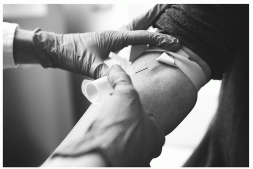
When skillfully executed, a venipuncture causes the patient little discomfort. The numerous blood determinations necessary for diagnosis and treatment make good technique imperative. The one-step entry technique should be avoided because too often it results in through-and-through punctures, contributing to hematoma formation. The needle should be inserted under the skin and then, after relocation of the vein, into the vessel.
The veins most commonly used are those in the antecubital fossa. The median antecubital vein, although not always visible, is usually large and palpable. Because it is well supported by subcutaneous tissue and least likely to roll, it is often the best choice for venipuncture. Second choice is the cephalic vein. The basilic vein, although often the most prominent, is likely to be the least desirable. This vein rolls easily, making the venipuncture difficult, and a hematoma may readily occur if the patient is allowed to flex the arm, which squeezes the blood from the engorged vein into the tissues.
Sufficient time should be spent in locating the vein before attempting venipuncture. Whenever the veins are difficult to see or palpate, the patient should lie down. If the patient is seated, the arm should be well supported on a pillow.
CENTRAL ACCESS CATHETER BLOOD WITHDRAWAL
Venous samples are often drawn from a central line, especially because these catheters are commonly placed in patients with limited venous access. A dual-lumen catheter may facilitate this process. When multiple-lumen catheters are used for central venous blood
sampling, the proximal lumen is the preferred site from which to draw the specimen. Aseptic technique is vital in preventing the introduction of bacteria into the catheter. A sterile IV catheter cap (Luer locking) reduces the risk of bacterial invasion. A recommended method is highlighted in
Box 7-2. Manufacturers have kept pace with our demands for technologically advanced products that enhance the sampling process, maintain the integrity of the sample, and ensure safety for both patient and caregiver.
Complications Resulting From Venipuncture and Blood Sampling
Complications related to venipuncture include hematomas, clotting, blood-borne diseases, syncope, and other problems.
HEMATOMAS
Hematomas are the most common complication of routine venipuncture for withdrawing blood, and they contribute more to the limitation of available veins than any other complication. They may result from through-and-through puncture to the vein or from incomplete insertion of the needle into the lumen of the vein, which allows the blood to leak into the tissues through the bevel of the needle. In the latter case, advancing the needle into the vein corrects the situation. At the first sign of uncontrolled bleeding, the tourniquet should be released and the needle withdrawn.
Hematomas also result from the application of the tourniquet after an unsuccessful attempt to draw blood. The tourniquet should never be applied to the extremity immediately after a venipuncture.
Hematomas most frequently result from insufficient time spent in applying pressure and from the bad habit of flexing the arm to stop the bleeding. Once the venipuncture is completed, the patient should be instructed to elevate the arm; elevation causes a negative pressure in the vein, collapsing it and facilitating clotting. With patients who have cardiac disease, elevation of the arm should be avoided. Constant pressure is maintained until the bleeding stops. The pressure is applied with a dry, sterile sponge because a wet sponge encourages bleeding. Adhesive strips do not take the place of pressure and, if ordered, are not applied until bleeding stops. Ecchymoses on the arm indicate poor or haphazard technique.
BLOOD-BORNE DISEASE
Special caution must be exercised to prevent blood-borne exposure from needle puncture or blood splash. Contaminated needles should be placed immediately in a separate container for disposal, and standard precautions should be followed. A vacuum tube with stopper provides adequate protection against accidental puncture from the contaminated needle until proper disposal can be made. Any needle puncture should be reported at once.
 PATIENT SAFETY
 PATIENT SAFETY


