The management and understanding of hereditary hemochromatosis have evolved with recent advances in iron biology and the associated discovery of numerous genes involved in iron metabolism. HFE -related (type 1) hemochromatosis remains the most frequent form, characterized by C282Y mutation homozygosity. Rare forms of hereditary hemochromatosis include type 2 (A and B, juvenile hemochromatosis caused by HJV and HAMP mutation), type 3 (related to TFR2 mutation), and type 4 (A and B, ferroportin disease). The diagnostic evaluation relies on comprehension of the involved pathophysiologic defect, and careful characterization of the phenotype, which gives clues to guide appropriate genetic testing.
Key points
- •
Hereditary hemochromatosis can damage liver, heart, pancreas, endocrine glands, bones, and joints. However clinical expression spans from a few biologic anomalies to full-blown multiorgan involvement.
- •
Hereditary hemochromatosis is mainly related to inappropriately deficient hepcidin secretion related to mutations in genes involved in hepcidin regulation.
- •
Ferroportin disease is associated with normal hepcidin secretion, but mutations in ferroportin induce loss of iron export function or resistance to hepcidin regulation.
- •
HFE -related hemochromatosis is the most frequent form in the white population. Genetic testing for rare forms of hereditary hemochromatosis must be guided by the clinical phenotype.
Introduction
To efficiently assess patients with suspected hereditary hemochromatosis, clinicians must understand the normal physiology of iron metabolism and the pathophysiologic mechanisms leading to iron overload.
Iron Metabolism
Iron absorption and export
Iron absorption occurs in the proximal duodenum. Nonheme iron is transported across the luminal membrane of the duodenal enterocyte into the cytoplasm by divalent metal transporter 1. Transport of heme iron into the enterocyte membrane is performed by a pathway that remains controversial.
Ferroportin ( SLCA40A1 ) is located at the basolateral membrane of the enterocyte and at the membrane of macrophages. It is the only known cellular iron exporter, allowing iron egress from the cytoplasm into the bloodstream.
Hepcidin
Hepcidin ( HAMP ) is synthesized primarily by hepatocytes, but is also produced at lower levels by adipocytes and macrophages. This small peptide, initially identified as an antimicrobial peptide, was later shown to be the key hormone regulator of iron metabolism.
Hepcidin regulates iron availability through the modulation of iron export by ferroportin: hepcidin binds to ferroportin at the membrane of enterocytes or macrophages, and causes subsequent ferroportin endocytosis and degradation, thus impairing iron export.
Hepcidin regulation
Because of its central role in iron metabolism, hepcidin is tightly regulated by several pathways including inflammation, erythropoiesis, and body iron stores. The regulation of hepcidin according to body iron stores involves distinct pathways for long- and short-term regulation. Basal expression is regulated through a bone morphogenetic protein/Son of Mother Against Decapentaplegic (SMAD) pathway. Bone morphogenetic protein 6 plays a major role in association with its coreceptor hemojuvelin (HJV), and is also involved in the response of hepcidin expression to iron stores over the long term.
Regarding the short-term regulation, it is proposed to be mediated through serum transferrin saturation. Although the molecular mechanisms remain debated, it is proposed that HFE, TFR1, TFR2, and HJV form an iron-sensing complex at the hepatocyte membrane, with subsequent regulation of hepcidin expression.
Iron Overload
Iron overload in hereditary hemochromatosis occurs through two main distinct mechanisms.
Hepcidin deficiency
Hepcidin deficiency is the key mechanism of iron overload in HFE , HJV , HAMP , and TFR2 related hemochromatosis. Mutations in these genes lead to defective or inappropriately low hepatic synthesis of hepcidin for the degree of iron burden. In HFE hemochromatosis it has been demonstrated that correction of liver hepcidin secretion normalized iron metabolism, confirming the predominant role of liver.
Relative hepcidin deficiency leads to a sustained and unregulated activity of ferroportin with two consequences: increased duodenal iron absorption, and enhanced release of iron from reticuloendothelial macrophages into the bloodstream originating from erythrophagocytosis.
The overall result is increased plasma iron concentration and increased saturation of transferrin. If the capacity of transferrin is exceeded, an abnormal physiologic form of iron appears, called nontransferrin bound iron. Nontransferrin bound iron is rapidly taken up by the liver, pancreas, and heart, and leads to pathologic parenchymal iron deposition and organ dysfunction. Moreover, if transferrin saturation exceeds 75%, labile plasma iron appears, which has a high propensity for generating reactive oxygen species that can directly damage tissues.
Ferroportin disease
There are two types of ferroportin disease that are distinguished by the molecular mechanism involved. Type A is characterized by a loss of ferroportin activity caused by mutations in the SLC40A1 gene. As a consequence of the functional deficiency, macrophage iron overload results from decreased iron export. The pathologic consequence is reticuloendothelial macrophage iron loading. Type B is characterized by mutations in ferroportin, which confer “resistance” to hepcidin. Thus, despite an increased serum hepcidin level, ferroportin regulation is defective, resulting in constitutive ferroportin activity and a “functional” hepcidin deficiency, as in HFE hemochromatosis. The pathologic consequence is reticuloendothelial macrophage iron sparing and parenchymal iron loading in target organs.
Penetrance
Clinical expression in hereditary hemochromatosis is variable, especially regarding HFE hemochromatosis. This variable phenotype makes it difficult to define hereditary hemochromatosis: does it correspond only to the identification of genetic mutations, to the association of biologic iron overload with genetic mutations, or only to iron-related clinical expression? Moreover, for the latter two conditions, the clinically significant level of iron overload remains to be determined.
HFE hemochromatosis is primarily associated with homozygosity for the C282Y HFE mutation. However, depending on the diagnostic criteria used, clinically significant iron overload is observed in only 5% to 75% of homozygotes patients. A recent meta-analysis showed an overall penetrance of 13.5%, and longitudinal studies showed iron overload in up to 50% of patients.
This variable penetrance is caused by numerous other acquired and genetic factors affecting iron metabolism regulation. Male sex and alcohol consumption increase the severity of iron overload, whereas obesity, through higher hepcidin expression, has been proposed to exert a protective effect. Furthermore, the occurrence of mutations in other genes involved in iron metabolism has been showed to modify the iron burden; however, the search for other genetic factors has shown variable results.
Because of the relatively few cases of type 2 and 3 hemochromatosis described, data regarding their penetrance and expression are scarce. If the penetrance is nearly complete, the severity of iron burden and age of presentation seems to be more variable than initially thought. This may be caused by the same reasons as seen in HFE hemochromatosis, but likely also related to the nature and consequences of the specific mutations involved.
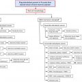
Diagnostic Work-up
We must consider that hereditary hemochromatosis encompasses a wide continuum of hepcidin deficiency states, whose clinical expression varies according to the main genes involved and the subsequent impaired molecular mechanisms. Moreover, clinical expression can be further modified by other acquired and genetics factors.
Therefore, the diagnostic evaluation requires a careful characterization of the patient’s phenotype, and an accurate assessment of iron overload and potential disease modifiers. Then, the phenotype guides the clinician through the appropriate genetic testing for determination of the genes and mutations involved.
Introduction
To efficiently assess patients with suspected hereditary hemochromatosis, clinicians must understand the normal physiology of iron metabolism and the pathophysiologic mechanisms leading to iron overload.
Iron Metabolism
Iron absorption and export
Iron absorption occurs in the proximal duodenum. Nonheme iron is transported across the luminal membrane of the duodenal enterocyte into the cytoplasm by divalent metal transporter 1. Transport of heme iron into the enterocyte membrane is performed by a pathway that remains controversial.
Ferroportin ( SLCA40A1 ) is located at the basolateral membrane of the enterocyte and at the membrane of macrophages. It is the only known cellular iron exporter, allowing iron egress from the cytoplasm into the bloodstream.
Hepcidin
Hepcidin ( HAMP ) is synthesized primarily by hepatocytes, but is also produced at lower levels by adipocytes and macrophages. This small peptide, initially identified as an antimicrobial peptide, was later shown to be the key hormone regulator of iron metabolism.
Hepcidin regulates iron availability through the modulation of iron export by ferroportin: hepcidin binds to ferroportin at the membrane of enterocytes or macrophages, and causes subsequent ferroportin endocytosis and degradation, thus impairing iron export.
Hepcidin regulation
Because of its central role in iron metabolism, hepcidin is tightly regulated by several pathways including inflammation, erythropoiesis, and body iron stores. The regulation of hepcidin according to body iron stores involves distinct pathways for long- and short-term regulation. Basal expression is regulated through a bone morphogenetic protein/Son of Mother Against Decapentaplegic (SMAD) pathway. Bone morphogenetic protein 6 plays a major role in association with its coreceptor hemojuvelin (HJV), and is also involved in the response of hepcidin expression to iron stores over the long term.
Regarding the short-term regulation, it is proposed to be mediated through serum transferrin saturation. Although the molecular mechanisms remain debated, it is proposed that HFE, TFR1, TFR2, and HJV form an iron-sensing complex at the hepatocyte membrane, with subsequent regulation of hepcidin expression.
Iron Overload
Iron overload in hereditary hemochromatosis occurs through two main distinct mechanisms.
Hepcidin deficiency
Hepcidin deficiency is the key mechanism of iron overload in HFE , HJV , HAMP , and TFR2 related hemochromatosis. Mutations in these genes lead to defective or inappropriately low hepatic synthesis of hepcidin for the degree of iron burden. In HFE hemochromatosis it has been demonstrated that correction of liver hepcidin secretion normalized iron metabolism, confirming the predominant role of liver.
Relative hepcidin deficiency leads to a sustained and unregulated activity of ferroportin with two consequences: increased duodenal iron absorption, and enhanced release of iron from reticuloendothelial macrophages into the bloodstream originating from erythrophagocytosis.
The overall result is increased plasma iron concentration and increased saturation of transferrin. If the capacity of transferrin is exceeded, an abnormal physiologic form of iron appears, called nontransferrin bound iron. Nontransferrin bound iron is rapidly taken up by the liver, pancreas, and heart, and leads to pathologic parenchymal iron deposition and organ dysfunction. Moreover, if transferrin saturation exceeds 75%, labile plasma iron appears, which has a high propensity for generating reactive oxygen species that can directly damage tissues.
Ferroportin disease
There are two types of ferroportin disease that are distinguished by the molecular mechanism involved. Type A is characterized by a loss of ferroportin activity caused by mutations in the SLC40A1 gene. As a consequence of the functional deficiency, macrophage iron overload results from decreased iron export. The pathologic consequence is reticuloendothelial macrophage iron loading. Type B is characterized by mutations in ferroportin, which confer “resistance” to hepcidin. Thus, despite an increased serum hepcidin level, ferroportin regulation is defective, resulting in constitutive ferroportin activity and a “functional” hepcidin deficiency, as in HFE hemochromatosis. The pathologic consequence is reticuloendothelial macrophage iron sparing and parenchymal iron loading in target organs.
Penetrance
Clinical expression in hereditary hemochromatosis is variable, especially regarding HFE hemochromatosis. This variable phenotype makes it difficult to define hereditary hemochromatosis: does it correspond only to the identification of genetic mutations, to the association of biologic iron overload with genetic mutations, or only to iron-related clinical expression? Moreover, for the latter two conditions, the clinically significant level of iron overload remains to be determined.
HFE hemochromatosis is primarily associated with homozygosity for the C282Y HFE mutation. However, depending on the diagnostic criteria used, clinically significant iron overload is observed in only 5% to 75% of homozygotes patients. A recent meta-analysis showed an overall penetrance of 13.5%, and longitudinal studies showed iron overload in up to 50% of patients.
This variable penetrance is caused by numerous other acquired and genetic factors affecting iron metabolism regulation. Male sex and alcohol consumption increase the severity of iron overload, whereas obesity, through higher hepcidin expression, has been proposed to exert a protective effect. Furthermore, the occurrence of mutations in other genes involved in iron metabolism has been showed to modify the iron burden; however, the search for other genetic factors has shown variable results.
Because of the relatively few cases of type 2 and 3 hemochromatosis described, data regarding their penetrance and expression are scarce. If the penetrance is nearly complete, the severity of iron burden and age of presentation seems to be more variable than initially thought. This may be caused by the same reasons as seen in HFE hemochromatosis, but likely also related to the nature and consequences of the specific mutations involved.
Diagnostic Work-up
We must consider that hereditary hemochromatosis encompasses a wide continuum of hepcidin deficiency states, whose clinical expression varies according to the main genes involved and the subsequent impaired molecular mechanisms. Moreover, clinical expression can be further modified by other acquired and genetics factors.
Therefore, the diagnostic evaluation requires a careful characterization of the patient’s phenotype, and an accurate assessment of iron overload and potential disease modifiers. Then, the phenotype guides the clinician through the appropriate genetic testing for determination of the genes and mutations involved.
Patient history
The patient history is a crucial element for accurate diagnosis, avoidance of being misled by confounding factors, and to optimize resource use. Regarding the laboratory evaluation, it should be emphasized that because of significant variability of biochemical iron parameters (transferrin saturation) related to diurnal variation and sensitivity to dietary iron, repeated early morning and fasting measurements should be performed.
Family history is also a cornerstone of the diagnostic evaluation. Search for putative diagnosis of iron overload in relatives can suggest a dominant or recessive inheritance pattern and strengthen the need for genetic evaluation of an individual or family.
Careful search for possible causes of secondary iron overload ( Box 1 ) and potential confounding factors or acquired modifiers of iron burden ( Box 2 ) help the clinician in the assessment of iron overload.
- •
Parenteral iron supplementation
Ensure that the patient has not undergone prolonged iron supplementation, especially in a patient with chronic anemia or seeking enhanced sport performance.
- •
Oral iron supplementation
Has been described as a potential cause of iron overload, but is controversial.
- •
Hematologic conditions
Chronic or rare anemias can be associated with iron overload through repeated transfusions and/or hepcidin deficiency (ineffective erythropoiesis leads to inhibition of hepcidin expression) possibly through erythroferrone.
- •
History of chemotherapy
Growth factors or transfusions used in this context can also lead to iron overload.
Related posts:


Stay updated, free articles. Join our Telegram channel

Full access? Get Clinical Tree


