Iron deficiency anemia (IDA) is a common hematologic condition, affecting a substantial proportion of the world’s women and young children. Optimal management of IDA requires an accurate diagnosis, identification and correction of the underlying cause, provision of medicinal iron therapy, and confirmation of treatment success. There are limited data to support current treatment approaches regarding oral iron preparation, dosing, monitoring, and duration of therapy. New intravenous iron agents have improved safety profiles, which may foster their increased utilization in the treatment of patients with IDA. Clinical trials focused on improving current treatment standards for IDA are sorely needed.
Key points
- •
Iron deficiency anemia (IDA) affects approximately 3% of young children in the United States, whereas iron deficiency without anemia occurs in 8% to 10% of infants and children. Globally, approximately half of the world’s children may have IDA, and millions of adults are affected as well.
- •
IDA in young children is primarily nutritional, resulting from poor iron intake due to prolonged breast-feeding and/or excessive cow milk intake, whereas in female adolescents and adults, it is usually secondary to heavy menstrual bleeding (or menorrhagia) and pregnancy. In men and postmenopausal women, its cause is primarily occult gastrointestinal blood loss.
- •
Manifestations of IDA in adolescents and adults include pallor, fatigue, diminished work performance, exercise intolerance, and dizziness. In children of all ages, anemia is often identified incidentally by screening or by accident when blood counts are performed for another reason.
- •
Initial therapy for IDA is an oral iron medication for a minimum of 3 months, while attempting to identify and correct the underlying cause. Nevertheless, firm data regarding optimal dose, duration of therapy, and monitoring of the hematologic response are unavailable.
- •
The administration of intravenous iron to patients who fail oral iron treatment warrants further investigation and strong consideration as a potentially safe and effective option to oral iron dosing.
History of iron therapy
In ancient Greece, iron was thought to be imparted from the mythological figure Mars and therefore associated with “force” and “strength.” It was thus primarily used therapeutically in war wounds. In the seventeenth century, Syndenham used a medicinal syrup of iron filings steeped in cold wine to reduce symptoms of chlorosis or “green sickness” in young women. The treatment improved their headache, greenish nonicteric skin color, and consumption of stones and dirt. He keenly observed in 1661 that “when one gives mars in the pale color the pulse becomes at once fuller and slower, the pallor disappears and once again the face is rosy and ruddy,” clearly documenting the success of a therapeutic trial of iron. In the nineteenth century IDA was confirmed as the cause of chlorosis based on identification of reduced iron concentration in the blood. Use of the microscope allowed for a formal description of the corresponding hypochromic, microcytic erythrocytes on peripheral smear. In 1831, Blaud described iron treatment of chlorosis and developed the first modern-day pill—a combination of ferrous sulfate and potassium carbonate—along with specific recommendations on daily dose and duration of therapy. To date, the concept of a therapeutic trial of iron, and specifically, the use of ferrous sulfate remains widely accepted by the medical community.
Problem of IDA
Incidence/Prevalence
Iron deficiency is the world’s most common nutrient deficiency. It affects a substantial population of women and children in both lower and higher income nations. For centuries, confounders such as malnutrition and infection often masked the underlying anemia due to iron deficiency. Improved nutrition and infection control later fostered better recognition of the condition, although additional confounders, such as malaria, inflammation, sickle cell disease, thalassemia, and other nutritional deficiencies, persist. Despite prevention efforts, iron deficiency and IDA are estimated to affect between 2 to 3 billion persons globally. The highest risk groups are young women, especially those who are pregnant or postpartum, and young children.
In the United States, the widespread switch from breast-feeding to cow milk–based formulas in the 1940s resulted in a high prevalence (up to 15%) of “cow milk anemia” due to iron deficiency in young children. In 1969, the American Academy of Pediatrics (AAP) recommended the use of iron-fortified formula, which was subsequently adopted by the Women, Infants, and Children Program in the 1970s. Along with increased breast-feeding rates, the result was a decline in iron deficiency in some high-risk populations. Nevertheless, IDA still occurs in approximately 3% to 7% of young children and iron deficiency without anemia has an estimated prevalence of 8% to 10%, with peak incidence from age 12 to 48 months. Adolescent women are at risk for IDA because of heavy menstrual bleeding after onset of menarche, with an incidence of approximately 9%, and another 15% to 20% have iron deficiency without anemia. IDA also affects 2% to 5% of pregnant women because of the expansion of red blood cell mass, which can persist postpartum due to blood loss associated with delivery. The prevalence is quite low in men and postmenopausal women but increases again in the elderly.
Lower income nations experience a higher prevalence of IDA due to a lack of access to meat, vegetarian diets, chronic gastrointestinal blood loss from parasitic infections, and scarcity of oral iron supplements.
In this review, the optimal treatment approaches for IDA in developed countries that result from iron poor diet or blood loss are focused on, with an emphasis on young children and adolescents. However, general principles of diagnosis and therapy are applicable to patients of all ages with IDA.
Clinical features and sequelae of IDA
Iron deficiency is a multisystem condition with a wide range of clinical features and sequelae, including effects on neurologic, cardiac, and immunologic functioning ( Box 1 ). Anemia, the extreme manifestation of iron deficiency, becomes apparent after tissue iron stores are depleted. In addition to pallor, fatigue, headache, and dizziness, patients with iron deficiency often develop pica, an irresistible craving for nonfood items (ice, dirt, clay, paper, or cornstarch), which typically resolves within days to weeks after starting iron therapy.
Nutritional
Pica, pagophagia
Neurocognitive
Reduced mental and motor function
Poorer outcomes in executive function and recognition memory
Visual and auditory systems’ functioning
Neurologic
Restless leg syndrome
Neuronal hypomyelination
Decreased neurotransmitter production
Cardiac
Impaired myocyte function
Immunologic
Impaired resistance to bacterial infection
Gastrointestinal
Epithelial tissue injury (glossitis; angular stomatitis)
Esophageal web or stricture, gastric atrophy
Microvillus damage; protein-losing enteropathy
Hematologic
Anemia (fatigue; diminished exercise tolerance and productivity)
A driving force behind efforts to identify and prevent iron deficiency in young children is its association with lower scores on tests of mental and motor development during infancy and later at school entry, even after controlling for lower socioeconomic status. Long-term studies have demonstrated poorer developmental outcome for children with IDA more than 10 years after treatment during infancy as well as identifying deficits in executive function and recognition memory almost 2 decades later.
Iron deficiency is associated with elevated fatigue scores in young women with menorrhagia. Adolescent girls with iron deficiency without anemia have improved verbal learning and memory after taking ferrous sulfate for 8 weeks compared with those receiving placebo. Studies in developing countries have demonstrated increased productivity in workers with IDA who receive iron therapy versus placebo. Thus, extensive evidence supports the concept that iron deficiency has a diversity of undesirable consequences.
Prevention and early diagnosis are the ideal
Although IDA in children and adolescents is usually preventable, prevention strategies are often unsuccessful, as evidenced by only modest decreases in the prevalence of iron deficiency in the United States during recent decades. Once it occurs, however, prompt identification and institution of effective therapy are critical to minimize its sequelae.
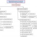
Recommendations for IDA screening are summarized in Table 1 . Screening of infants at 1 year of age is suggested but only identifies patients who are already anemic. Infants who are primarily formula fed are at low risk of IDA at 1 year of age. The transition to cow milk places them at risk for IDA during their second year of life, yet no formal screening recommendations exist in this age group. Screening for IDA in adolescent women, another high-risk population, is not recommended by the AAP, so identification of these patients relies on the astute clinician to recognize risk factors for iron deficiency and to diagnose and treat those girls with heavy menstrual bleeding and IDA.
| Source of Recommendation | Children | Women |
|---|---|---|
| American Academy of Pediatrics (AAP) | Universal screening at 9–12 mo; selective screening at any age for patients with risk factors | |
| American Academy of Family Physicians (AAFP) | Follow USPSTF Guidelines | Follow USPSTF Guidelines |
| American College of Obstetrics & Gynecology (ACOG) | All pregnant women | |
| US Preventive Services Task Force (USPSTF) Guidelines | Evidence insufficient to recommend for or against routine screening | All pregnant women |
| Centers for Disease Control (CDC) | High-risk infants and high-risk preschool children | All nonpregnant women of childbearing age every 5–10 y |
Management of IDA
Regardless of patient age or underlying cause, 5 principles guide IDA management ( Box 2 ).
- 1.
Confirm the diagnosis
- 2.
Identify its cause
- 3.
Correct or manage the primary cause
- 4.
Provide iron therapy, either orally or parenterally
- 5.
Confirm the success of such therapy
Confirmation of the Diagnosis
Although a complete blood count (CBC) or a measurement of the hemoglobin concentration alone is the primary screening test used for IDA, iron deficiency progresses through several phases, with frank anemia manifested only after erythropoeisis has become markedly impaired ( Table 2 ).
| Iron Depletion | Iron-Restricted Erythropoiesis | Iron Deficiency Anemia | |
|---|---|---|---|
| Hemoglobin concentration | Normal | Normal | Reduced |
| Mean corpuscular volume | Normal | Normal-Reduced | Reduced |
| Reticulocyte hemoglobin content a | Normal | Reduced | Reduced |
| Serum iron concentration | Normal | Reduced | Reduced |
| Serum ferritin concentration | Reduced | Reduced | Reduced |
| Total iron binding capacity | Normal | Increased | Increased |
| Soluble transferrin receptor | Normal | Increased | Increased |
a Ret-He or CHr, the first peripheral blood count marker that becomes abnormal in iron deficiency.
No specific test confirms the diagnosis of IDA in all patients, because serum ferritin may be falsely normalized/elevated and transferrin saturation may be reduced in inflammatory states. The reticulocyte hemoglobin content (CHr or Ret-He) is reduced in both IDA and thalassemia trait. All of the clinical information and laboratory data must be taken into account to establish the correct diagnosis. Review of the combination of CBC, reticulocyte count, CHr or Ret-He, and peripheral smear along with serum iron, serum ferritin, and total iron binding capacity often provides evidence to support the diagnosis of IDA. However, the most convincing evidence of IDA is an increase in the hemoglobin concentration after a therapeutic trial of medicinal iron.
Other Causes of Microcytic Anemia
In patients with limited response to iron therapy and/or some normal iron measurements, other causes should be considered, such as thalassemia trait, other hemoglobinopathy, or anemia of inflammation. Iron refractory iron deficiency anemia (IRIDA), a rare form of inherited IDA described in the article by Heeney and Finberg elsewhere in this issue, should be considered in patients with lifelong IDA that is minimally responsive to oral supplementation despite good adherence to treatment and transiently responsive to parenteral iron.
Identification and Management of the Primary Cause: Children and Adolescents
Iron deficiency can result from insufficient nutritional iron intake, increased requirements, malabsorption, or external bleeding. In young children, the cause is primarily nutritional. Risk factors include prematurity (as most iron transfer from mother occurs during the third trimester), exclusive breast-feeding for greater than 6 months without supplemental iron (as breast milk contains little iron), prolonged bottle-feeding, obesity, low socioeconomic status, and early, excessive, and/or prolonged intake of cow milk. Iron in cow milk has lower bioavailability (5%–10% absorption) than human breast milk (50% absorption) and potentially interferes with duodenal absorption of iron in other foods. When consumed in excessive quantities, cow milk proteins can also damage the intestinal mucosa, leading to microvascular gastrointestinal blood loss and occasionally protein-losing enteropathy.
In adolescent women, IDA primarily results from excessive acute and/or chronic menstrual bleeding. Given that 0.4 to 0.5 mg of iron is lost along with every 1 mL of blood, menorrhagia results in IDA that often persists or recurs. In patients with IDA not clearly caused by poor intake or heavy menstrual bleeding, other causes must be evaluated and addressed appropriately. Specifically, gastrointestinal blood loss and/or malabsorption should be considered as well as chronic intrapulmonary bleeding or intravascular hemolysis resulting in urinary iron loss.
Identification and Management of Primary Cause: Adults
Total body iron in men is 4 g versus 3 g in women. Because most women begin pregnancy with low iron reserves, the combined average iron loss during pregnancy of approximately 900 mg that results from the diversion of iron to the fetus, blood loss at delivery, and lactation often results in IDA. In men and postmenopausal women, IDA is most often secondary to gastrointestinal blood loss, so it is imperative to identify and correct its source as well as provide adequate medicinal iron therapy. In adults, there is a higher incidence than in children of iron-restricted erythropoiesis during use of erythropoiesis-stimulating agents (ESAs), in which the iron supply cannot meet the increased requirements. Moreover, adults have a higher incidence of “anemia of chronic disease” resulting from an elevation in the regulatory peptide hepcidin that inhibits duodenal iron absorption and renders iron stores inaccessible, as seen in chronic inflammatory conditions.
Providing Iron Therapy
Regarding the first 3 management principles (see Box 2 ), there is little debate. However, recommendations in the literature about the specifics of iron therapy vary widely with regard to preparation, dosing, frequency and timing, duration, and monitoring. Recent suggestions regarding iron deficiency in young children from the AAP emphasize prevention and early diagnosis but give virtually no attention to its treatment. So-called iron supplements are often prescribed at a dose that is too low and therefore ineffective, or too high, with the risk of adverse effects and poor adherence. In all patients, misguided therapy and/or poor patient education regarding IDA can result in a subpar adherence and response to therapy.
Reasons for Near Absence of Data to Inform Treatment
Many clinicians dismiss mild IDA as a benign condition with inconsequential effects, creating a false perception that research aimed to improve IDA treatment is of low priority, which has in turn resulted in a paucity of published data informing its management. The scarcity of IDA clinical trials supported by governmental funding agencies, including the National Heart, Lung, and Blood Institute and National Institute of Diabetes and Digestive and Kidney Disease, is noteworthy. Moreover, limited funding from industry impedes the development of well-structured and rigorous trials aimed at answering important therapeutic questions. For example, at present, only one randomized clinical trial in the United States is comparing different oral iron medications in the treatment of IDA.
Oral iron therapy
Iron Preparation
There are myriad preparations and formulations of iron, many of them available over-the-counter, causing much confusion for patients and physicians alike. These agents, labeled “supplements,” undergo a separate approval process by the Food and Drug Administration (FDA) and are often marketed based on slight differences involving other added vitamins and minerals (eg, B12, folate), with or without extended release properties. A 1958 review by Nathan Smith indicated that among 170 iron preparations then used in the prevention and treatment of IDA, ferrous sulfate was the least expensive and thought at the time to be the most effective. To date, ferrous sulfate remains the most frequently used treatment of IDA, a testimonial to the lack of progress in the management of this condition.
Ferrous gluconate and ferrous fumarate, 2 other iron salts, have demonstrated efficacy as iron supplements for prevention and treatment of IDA. Carbonyl iron (eg, Feosol) has also been demonstrated as a safe, effective, and inexpensive iron therapy, but it is now less frequently used. Iron polysaccharide combinations (eg, NuIron, Niferex, NovaFerrum) are formulated as well hydrated microspheres remaining in solution over a wide range of pH values, theoretically allowing for improved absorption and tolerability.
Dosing
After a specific agent is chosen, the total daily dose and divided dose schedule are based more on clinical experience than scientific evidence, but 2 basic tenets are essential: administering an adequate daily dose for a sufficient duration. In persons with normal iron status, approximately 10% of dietary iron is absorbed by the duodenum, with a total daily absorptive capacity of approximately 25 mg of elemental iron. Few data seem to support this estimate, and it is unclear whether it refers to the absorptive capacity following a single meal or a total daily value. However, it serves as the basis for the frequent recommendations of divided daily doses of iron therapy and refutes the concept that high single-daily doses of iron result in improved absorption and faster resolution of anemia. Not surprisingly, recommended dosing in children ranges widely from 2 to 6 mg/kg/d, administered either once, twice, or 3 times daily ( Table 3 ). Adult dosing suggestions vary as well from 60 to 300 mg/d of elemental iron in 2 to 4 divided doses.
Related posts:


Stay updated, free articles. Join our Telegram channel

Full access? Get Clinical Tree


