Diagnosis and Management of Effusions


Diagnosis and Management of Effusions


Gary T. Buckholz

Charles F. von Gunten


DIAGNOSIS AND MANAGEMENT OF ASCITES

Ascites, the accumulation of fluid in the abdomen, is common. Its formation may be a direct result of a malignant process or secondary to liver cirrhosis or other comorbidities. Because the pathophysiology of fluid collection varies, treatment strategies differ. Clinical distinction between the causes of ascites is therefore important.

Of all patients with ascites, approximately 80% have cirrhosis (1). Other causes of nonmalignant ascites include the following: heart failure, 3%; tuberculosis, 2%; nephrogenic ascites related to hemodialysis, 1%; pancreatic disease, 1%; and miscellaneous entities such as hepatic vein thrombosis (Budd-Chiari syndrome), pericardial disease, and the nephrotic syndrome account for approximately 2% (1). Only 10% of patients who have ascites have malignancy as the primary cause (1). In these patients, epithelial malignancies, particularly ovarian, endometrial, breast, colon, gastric, and pancreatic carcinomas, cause over 80% of malignant ascites. The remaining 20% are due to malignancies of unknown origin (2). In one study, Runyon has shown that 53.3% of malignant ascites is associated with peritoneal carcinomatosis, 13.3% is associated with massive liver metastases, 13.3% is associated with peritoneal carcinomatosis and massive liver metastases, 13.3% is associated with hepatocellular carcinoma with portal hypertension, and 6.7% is associated with chylous ascites (3).

In general, the presence of ascites portends a poor prognosis, regardless of the cause. Patients with nonmalignant ascites related to cirrhosis have a survival rate of approximately 50% at 2 years (1). The mean survival in patients with malignant ascites is generally <4 months (4). However, with ascites due to a malignancy that is relatively sensitive to chemotherapy, such as newly diagnosed ovarian cancer or lymphoma, the mean survival may improve significantly (4).


PATHOPHYSIOLOGY


Nonmalignant Ascites

The mechanisms that lead to the development of ascites are many, and controversy still exists regarding which factors are most important. The most common cause of nonmalignant ascites is cirrhosis of the liver. In cirrhotic ascites, abnormal sodium retention is mediated by various hormonal and neural mechanisms, similar to those responsible for excess fluid retention in congestive heart failure (CHF). A hemodynamic state exists where total blood volume is increased, cardiac output is increased, and systemic vascular resistance is low. Studies have implicated nitric oxide as one of the potential mediators of this arterial vasodilation (5). In response, the vasoconstrictors of the renin-angiotensin-aldosterone system and the sympathetic nervous system are activated. Although atrial natriuretic peptide levels are increased, there is reduced renal responsiveness (6). In addition, arginine vasopressin, a potent vasoconstrictor, is activated in a manner independent of the osmotic state (7). The net result is an increase in total body sodium and water. In conjunction with cirrhosis, which has caused increased hepatic venous and lymphatic resistance, severe portal hypertension ensues. The increase in hepatic venous sinusoidal and portal pressures causes the excess fluid volume to localize to the peritoneal cavity secondary to fluid transudation from the splanchnic capillary bed. Ascites accumulation is also exacerbated by diminished intravascular oncotic pressure, resulting from hypoalbuminemia due to decreased synthetic capacity of the cirrhotic liver.


Malignant Ascites

Malignant ascites arises through pathophysiologic mechanisms different from those of nonmalignant ascites. First, in peritoneal carcinomatosis, neovascularization and subsequent “leak” from vessels is thought to play a prime role in ascites development. Researchers have identified a vascular growth and permeability factor that increases fluid leak from peritoneal vasculature; vascular endothelial growth factor (VEGF) is a prime candidate for this activity (8). Compared with cirrhotic ascites, high levels of VEGF are present in malignant ascites from gastric, colon, and ovarian cancers (9). In animal models, inhibiting the tyrosine kinase activity of VEGF receptors reduced ascites formation (10). Matrix metalloproteinases (MMPs) also appear to be involved in this process. Breaking down the extracellular matrix is an important step in neovascularization and metastatic spread. In animal models, MMP inhibitors significantly reduced malignant ascites (11). Second, portal pressures may be raised by direct tumor invasion of the liver with resultant hepatic venous obstruction. The resultant portal hypertension leads to transudation of fluid across the splanchnic bed into the abdominal cavity similar to cirrhotic ascites. A final mechanism of ascites formation is due to lymphatic obstruction, commonly caused by lymphoma, resulting in chylous ascites.


MANAGEMENT

Overall goals for patient care should be considered before specific choices for managing ascites are made. The prognosis, expected response to management of the underlying conditions, and preferences for treatment should be established with the patient and family before any treatment plan is instituted. Each ascites treatment modality has associated burdens and benefits that deserve to be considered and discussed.

Whether ascites has a low or a high SAAG is critical in determining the overall management plan. Ascites due to portal hypertension is in equilibrium with total body fluid. The most common cause of nonmalignant ascites, cirrhosis, falls within this category. Efforts to restrict salt and to affect fluid balance with diuretics are often successful. Malignant ascites may or may not be responsive to these efforts, depending on its cause. In peritoneal carcinomatosis, the SAAG is low, there is no portal hypertension, and the ascites is not in equilibrium with total body fluid (18). Consequently, salt and fluid restriction and diuretics may be of little use. Their injudicious use may result in intravascular volume depletion, diminished renal perfusion, azotemia, hypotension, and fatigue (2). However, there are high SAAG forms of malignant ascites that are responsive to salt restriction and diuretics. For example, in cases of massive hepatic metastasis, portal hypertension is present and salt restriction and diuretics are indicated (18). One exception to this rule is nephrotic syndrome in which the SAAG is low but the ascites is diuretic responsive (19). The total protein is also low (<25 g/L) in nephrotic syndrome and thus is helpful in identifying this form of ascites (see Table 16.1 for a summary).

Interventions for ascites management in the supportive or palliative care setting should generally be reserved for patients who are symptomatic. The following ascites-related symptoms may spur intervention:


  • Dyspnea


  • Fatigue


  • Anorexia or early satiety


  • Nausea/vomiting


  • Pain


  • Diminished exercise tolerance


Dietary Management

The dietary management of ascites with a high SAAG begins with sodium restriction. Patients with cirrhosis may excrete as little as 5 to 10 mEq of sodium/d in their urine. Limiting sodium intake to 88 mmol or 2 g/d (equivalent to 5 g of
sodium chloride/d) is an attainable goal for a motivated patient, but it does make food less palatable. Considering a patient’s goals of care, it may be better to liberalize the sodium intake and control ascites through other methods.


TABLE 16.1 Causes of ascites and diuretic responsiveness


Cause of Ascites


Serum-Ascites Albumin Gradient


Typical Diuretic Response


Cirrhosis


High (≥1.1 g/dL)


Yes


Alcoholic hepatitis


High (≥1.1 g/dL)


Yes


Cardiac ascites


High (≥1.1 g/dL)


Yes


Fulminant hepatic failure


High (≥1.1 g/dL)


Yes


Budd-Chiari syndrome


High (≥1.1 g/dL)


Yes


Portal vein thrombosis


High (≥1.1 g/dL)


Yes


Venoocclusive disease


High (≥1.1 g/dL)


Yes


Acute fatty liver of pregnancy


High (≥1.1 g/dL)


Yes


Myxedema


High (≥1.1 g/dL)


Yes


Tuberculosis (without cirrhosis)


Low (≤1.1 g/dL)


No


Pancreatic ascites (without cirrhosis)


Low (≤1.1 g/dL)


No


Biliary ascites (without cirrhosis)


Low (≤1.1 g/dL)


No


Nephrotic syndrome


Low (≤1.1 g/dL)


Yes


Serositis from connective tissue disease


Low (≤1.1 g/dL)


No


Bowel obstruction/infarction


Low (≤1.1 g/dL)


No


Mixed ascites (i.e., cirrhosis plus infection or cancer)


High (≥1.1 g/dL)


Yes


Peritoneal carcinomatosis


Low (≤1.1 g/dL)


No


Massive hepatic metastasis


High (≥1.1 g/dL)


Yes


Patients are also prone to develop dilutional hyponatremia. The management of this condition has typically been by fluid restriction to 1 L/d. In the patient with advanced disease, when treatment goals are purely palliative, fluid restriction is usually intolerably burdensome. Judicious medical management may be less burdensome. For patients with cirrhotic ascites, serum sodium levels as low as 120 mmol/L are well tolerated and rarely dictate intervention (1).


Pharmacologic Management

For the majority of patients, the pharmacologic management of ascites is palliative. That is, the goal of therapy is to minimize symptoms and optimize quality of life without the expectation that the underlying cause can be reversed.

Systemic chemotherapy may be an effective management strategy for patients with malignant ascites due to a responsive cancer (e.g., lymphoma, breast, or ovarian cancer). In addition to systemic chemotherapy, intraperitoneal chemotherapy is an option. In theory, intraperitoneal chemotherapy can deliver high doses to peritoneal sites with minimal systemic side effects. In practice, intraperitoneal chemotherapy is often limited by uneven distribution and poor tissue penetration. Hyperthermic intracavitary chemotherapy after surgical debulking may overcome some of these limitations and enhance the cytotoxicity of chemotherapy (20,21). Biologically active agents have also been used intraperitoneally to treat malignant ascites. Early clinical trials have used interferon (IFN)-α, IFN-β, and IFN-γ, tumor necrosis factor, interleukin-2, anti-VEGF antibodies, anti-VEGF receptor antibodies, VEGF receptor tyrosine kinase inhibitors, and metalloproteinase inhibitors. To date, no phase III clinical trials have been performed. Overall, the efficacy and role of intraperitoneal chemotherapeutic and biologic agents in both curative and palliative care remain to be determined.

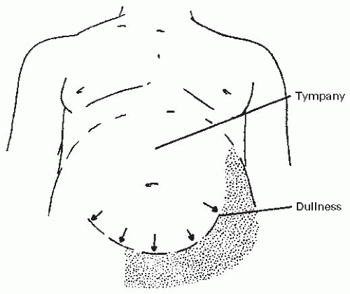
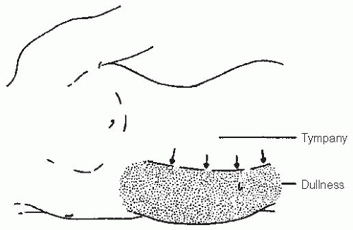
Diuretic therapy could be useful in patients whose ascites has a high SAAG, as opposed to low SAAG ascites that is typically diuretic unresponsive. As with any drug therapy in the supportive care setting, the patient’s symptoms should first be ascertained and the benefit versus the burden of therapy considered. The goal of diuretic therapy is to reduce extravascular fluid accumulation. Diuretic therapy should be directed to achieve a slow and gradual diuresis that does not exceed the capacity for mobilization of ascitic fluid. In the patient with ascites and edema, edema acts as a fluid
reservoir to buffer the effects of a rapid contraction of plasma volume. Approximately 1 L/d (net) can safely be diuresed. In patients with ascites but without edema, diuresis may be achieved at the expense of the intravascular volume, leading to symptomatic orthostatic hypotension. In these patients, a more modest goal is to achieve net diuresis of 500 mL/d. Diuretics should not be administered with the goal to render the patient free of edema and ascites. Rather, only enough fluid should be mobilized to promote the patient’s comfort. Overly aggressive diuretic therapy for ascites in a patient with high SAAG ascites has been associated with the hepatorenal syndrome and death (22).

For patients with high SAAG ascites in whom diuretics may be helpful, the renin-angiotensin-aldosterone system is activated. Therefore, the initial diuretic of choice for management is one that acts at the distal nephron to block the effect of increased aldosterone activity (23,24). Spironolactone, an aldosterone receptor antagonist, is a first-line therapy. Dosing begins at 100 mg/d and can be titrated up to effect or a maximum of 400 mg/d (Table 16.2). Given spironolactone’s long half-life, daily dosing is sufficient. Spironolactone may cause painful gynecomastia (25). Amiloride hydrochloride, 10 mg/d, is an alternative. It acts faster and does not cause gynecomastia. It can be titrated up to a dose of 40 mg/d. Because these diuretics are relatively potassium sparing, patients should be advised not to use salt substitutes, as these are usually preparations of potassium chloride. If patients have a suboptimal response despite maximal use of the distal diuretics, a loop diuretic may be added, beginning at low doses (e.g., furosemide, 40 mg orally daily). There is evidence to support the combined use of a distal tubule diuretic and a loop diuretic at the beginning of therapy (24). This combination may effect a more rapid diuresis while maintaining potassium homeostasis. A ratio of 100 mg of spironolactone to 40 mg of furosemide is recommended as a starting point (1). The ratio can be adjusted to maintain normokalemia. The dosages can be increased in parallel until the goals of therapy have been attained, up to a maximum of spironolactone, 400 mg/d, and furosemide, 160 mg/d, or until therapy is limited by side effects (1). If there is no response to this level of therapy, the ascites is considered refractory to diuretic therapy if the following are true: (a) salt intake is appropriately limited and (b) nonsteroidal anti-inflammatory medications, which can affect glomerular filtration, are not being used.


TABLE 16.2 Diuretics


Diuretics


Major Site of Action


Dosage Range (mg/d)


Comments


Spironolactone


Distal tubule


100-400


Long half-life and gynecomastia


Amiloride hydrochloride


Distal tubule


10-40


Triamterene


Distal tubule


100-300


Furosemide


Loop of Henle


40-160


Ethacrynic acid


Loop of Henle


50-200


Can be used for sulfa allergy

Only gold members can continue reading. Log In or Register to continue

Stay updated, free articles. Join our Telegram channel

Aug 25, 2016 | Posted by in ONCOLOGY | Comments Off on Diagnosis and Management of Effusions

Full access? Get Clinical Tree

Get Clinical Tree app for offline access