Primary
Genetic disorders
Congenital adrenal hyperplasia
Adrenoleukodystrophy
ACTH resistance syndromes
ACTH insensitivity: familial glucocorticoid deficiency, triple-A syndrome
Adrenal hypoplasia congenita
Other transcription factors: SF1 (NR5A1)
Drug induced
Adrenostatic/adrenolytic: etomidate, ketoconazole, metyrapone, mitotane, osilodrostat, abiraterone
Glucocorticoid receptor antagonist: mifepristone
Autoimmune
Destructive
Hemorrhage
Central
Genetic disorders
Septo-optic dysplasia
Isolated ACTH deficiency
Drug induced
Exogenous corticosteroids (any route of administration), medroxyprogesterone
Opioids
Immune-checkpoint inhibitor-induced hypophysitis: ipilimumab
Benzodiazepines: alprazolam
Atypical antipsychotics: olanzapine, quetiapine
Acquired hypothalamic-pituitary disease
Pituitary tum ors
Granulomatous diseases
Hypophysitis: granulomatous or lymphocytic
Surgery
Radiation
Today, the prevalence of secondary and tertiary adrenal insufficiencies is 150–280 per million, which is much more common than PAI. Because it is often impossible and of little clinical utility to distinguish pituitary and hypothalamic sites of dysfunction, the term central adrenal insufficiency (CAI) is often used to encompass secondary and tertiary adrenal insufficiencies. Etiologies include primarily pituitary pathologies: tumors, congenital or developmental causes, and hypophysitis (Table 9.1). Brain trauma, radiation damage, and granulomatous diseases primarily affect the neuroendocrine components of the hypothalamic-pituitary-adrenal (HPA) axis.
Of course, the most common cause of CAI is the administration of exogenous glucocorticoids. The biochemical findings are a low or normal plasma ACTH in the presence of a subnormal cortisol or impaired cortisol response to stimulation. Any route of glucocorticoid administration (oral, parenteral, inhaled, or topical) will suppress corticotropin-releasing hormone (CRH) and the HPA axis if the dose and duration of use are sufficient. Insoluble suspensions, particularly when injected into a relatively avascular space, such as paraspinous injections of triamcinolone acetonide, can suppress the HPA axis for months. In addition, high doses of synthetic progestins with partial glucocorticoid agonist activity, such as megestrol acetate [4], will suppress the HPA axis, and many types of opiates also potently suppress the HPA axis [5–9]. Although the exogenous glucocorticoids compensate for the reduction in cortisol production, the atrophied zona fasciculata and zona reticularis of the adrenal glands are unable to produce cortisol when the glucocorticoids are withdrawn, which can precipitate clinical features of AI if abruptly discontinued.
As a general guide, HPA axis suppression from opiates is rapid in onset and, although variable among individuals, can occur with even small doses when taken regularly. There is some evidence that axis suppression reverses quickly upon discontinuation, even with chronic opiate use, and that acute stress such as insulin-induced hypoglycemia can overcome the suppression; however, data characterizing opiate-induced HPA axis suppression are limited. For exogenous glucocorticoids, HPA axis suppression persists starting about 3–4 weeks after a supraphysiologic dose and persists roughly for a period of time equal to the duration of treatment. Duration of use is more important than dose in determining the probable duration of axis suppression, and drugs with the longest the duration of action are the most suppressive. For example, HPA axis suppression from 4 mg dexamethasone daily for 6 months will typically last longer than from 40 mg prednisone daily for 3 months. In addition, the clinical state during treatment is important, as the HPA axis becomes resistant to suppression during critical illness.
Clinical Presentation
The presentation of chronic PAI encompasses vague and nonspecific symptoms, but some signs and symptoms are more specific than others, and the presence of certain symptoms can virtually rule out the diagnosis. For example, weight loss, hypotension, skin hyperpigmentation, and auricular cartilage calcification are more specific for AI than abdominal pain, nausea, vomiting, fatigue, general malaise, and myalgias. Of note, weight gain, in the absence of comorbidities such as heart failure, liver failure, or renal failure, essentially rules out PAI. On laboratory studies, hyponatremia , hyperkalemia , and eosinophilia are notable findings in PAI.
The presentations of PAI and CAI are similar, with a few notable differences. Patients with CAI do not experience skin hyperpigmentation, because elevated levels of pro-opiomelanocortin, a prohormone precursor of ACTH and melanocyte stimulation hormone, are absent. Due to intact mineralocorticoid production in CAI, hyperkalemia is absent, and hypotension is less prominent. However, hyponatremia may still be present due to the absence of glucocorticoid negative feedback on vasopressin secretion as well as the diminished glomerular filtration rate that may accompany cortisol deficiency. Consequently, the presence of unexplained hyponatremia should always raise the possibility of hypocortisolemia. Gastrointestinal manifestations are less common in CAI than in PAI; it is possible that these symptoms are more prominent with mineralocorticoid deficiency . CAI may present with headaches, visual deficits, and symptoms of deficiencies in other pituitary hormones.
Acute AI, or adrenal crisis , presents predominantly with weakness, malaise, nausea, vomiting, and hypotension. Without appropriate therapy, shock and multisystem organ failure may ensue. Well-done longitudinal studies have shown that 6–8% of patients with a known diagnosis of either PAI or CAI will experience an episode of adrenal crisis annually, and the annual death rate may be as high as 0.5% [10]. Various situations can precipitate adrenal crisis: for example, patients with partial AI may be asymptomatic until they encounter a stressor, such as acute illness or surgery. Adrenal crisis is also seen in patients treated with glucocorticoids chronically, who do not take extra doses during acute stressors. Bilateral adrenal infarcts, adrenal hemorrhage, and pituitary infarcts can all precipitate adrenal crises. Table 9.2 lists the various signs and symptoms of PAI and CAI and their utility in supporting the diagnosis.
Table 9.2
Clinical presentation of adrenal insufficiency
Primary | Central | ||
|---|---|---|---|
Sign and symptoms | Constitutional | Hypotension, weight loss, myalgias, arthralgias, malaise | Hypotension less prominent. Symptoms of other pituitary hormone deficiencies may be present |
Gastrointestinal | Abdominal pain, nausea, vomiting, constipation, diarrhea, splenomegaly | Less common | |
Neuropsychiatric | Impaired memory, confusion, depression, psychosis | Other neurological deficits may also be present: headaches, visual field deficits | |
Reproductive | Typically in women: decreased axillary and pubic hair, decreased libido, amenorrhea | Present | |
Dermatologic | Hyperpigmentation, vitiligo | Absent | |
Other | Auricular cartilage calcification | Present | |
Laboratory studies | Basic metabolic panel | Hyponatremia, hyperkalemia, hypoglycemia | Hyperkalemia absent, hypoglycemia more common |
Complete blood count | Leukocytosis, eosinophilia | Present | |
As alluded to above, in any patient suspected of having AI, it is important to take a thorough medication history. Glucocorticoids taken via various routes, including inhaled, intra-articular, transdermal, ocular, and rectal, can suppress the HPA axis. It should be emphasized that other commonly prescribed medications like opiates [5, 6] and benzodiazepines [11] are also associated with CAI. Because CAI of organic etiology is quite rare in the absence of other pituitary deficits, isolated CAI should be presumed due to exogenous substances until proven otherwise (Table 9.3).
Table 9.3
Medical management of adrenal insufficiency
Setting | Recommended treatment | Comments |
|---|---|---|
Chronic therapy | ||
Adult | Hydrocortisone 12 mg/m2/day in two or three divided doses, 15 to 25 mg total daily dose; prednisone 4–8 mg every morning; prednisolone 5–7.5 mg every morning; methylprednisolone 4–8 mg every morning | Titrate dosing every 3–6 months based on signs, symptoms, and serum sodium |
9?-fludrocortisone acetate 0.05–0.4 mg/day | Only for primary; titrate dosing every 6–12 weeks based on volume status, blood pressure, and serum potassium | |
Pregnancy | In third trimester, increase glucocorticoid by up to 50% Titrate fludrocortisone based on blood pressure and serum potassium | Avoid dexamethasone |
Children | Hydrocortisone 12–17 mg/m2/day divided in two or three doses | Hydrocortisone preferred over other glucocorticoids |
9?-fludrocortisone acetate 0.05–0.4 mg/day | Titration is similar to that for adults | |
Acute stressors | ||
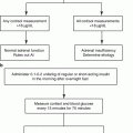
Acute illness with fluid loss and poor PO intake | Double or triple glucocorticoid dose and continue 3 days or until feeling well for 1 day | Monitor blood pressure and heart rate |
Acute illness with inability to take PO glucocorticoid or maintain blood pressure | Hydrocortisone 100 mg intramuscular or subcutaneous (act-O-vial) once | Paramedics cannot administer glucocorticoids, so administer prior to transport to acute care facility |
Minor surgery | 25 mg hydrocortisone once prior to surgery and then resume normal therapy | Overtreatment can impair recovery |
Major surgery, including childbirth | 50 mg once, followed by 25–50 mg every 6 h for 2 days | Extend intensified therapy if complications |
Acute adrenal crisis | Hydrocortisone 50 mg every 6 h Fluid resuscitation Electrolyte repletion | Monitor volume status and electrolytes; evaluate etiology of adrenal crisis |
Approach to the Patient
There are basal and dynamic methods of testing for adrenal insufficiency. Basal testing measures hormone levels at specific times of the day, while dynamic testing involves measuring the hormonal response to a stimulus. As an initial evaluation in the outpatient setting in the absence of an acute illness, basal testing alone can be used to exclude all forms of adrenal insufficiency in the majority of patients. Because of the strong diurnal rhythm for these hormones, it is important to test hormone levels when they are high; serum cortisol and plasma ACTH values are most useful when drawn before 0900. A serum cortisol of >14 μg/dL (400 nmol/L) obviates the need for dynamic testing [12]. Although ACTH values cannot be used to diagnose AI per se, the ACTH is used to distinguish primary (>100 pg/mL, >20 pmol/L) from secondary (<10 pg/mL, <2 pmol/L) AI if the cortisol is diagnostically low (<5 μg/dL, <140 nmol/L). Basal cortisol values 5–14 μg/dL, however, neither exclude nor establish the diagnosis and therefore prompt additional testing.
When performing basal testing, additional information can be obtained with the measurement of other ACTH-dependent products of the adrenal cortex, particularly dehydroepiandrosterone sulfate (DHEAS). Unlike cortisol and ACTH, serum DHEAS concentrations vary only slightly throughout the day due to its extensive protein binding; consequently, values need not be obtained in the early morning. Unless the patient is taking DHEA supplements, a serum DHEAS value >60 μg/dL (1500 nmol/L) at any time of day indicates normal adrenal function [13]. DHEAS measurements are generally used not as a primary test but as an ancillary criterion, typically when the basal serum cortisol is 10–14 μg/dL or when the cortisol is drawn after 0900 [14]. DHEAS production declines with age and is usually easily suppressed from any exogenous glucocorticoid use, even when cortisol secretion may remain normal; these properties limit its utility in the elderly (age > 65 years) and for patients who are already treated with glucocorticoids. Consequently, a low DHEAS level by itself is certainly not diagnostic of adrenal hypofunction. Serum aldosterone may also be useful in distinguishing PAI from CAI, since patients with PAI have a subnormal aldosterone and elevation of plasma renin activity.
When Is Dynamic Testing Necessary?
In an outpatient setting, when evaluating a patient with suspicion of chronic AI, basal morning testing often provides enough information to exclude the diagnosis, which obviates the need for dynamic testing . Difficulties in establishing or excluding a diagnosis of AI arise in a variety of settings, including patients who have already received treatment with glucocorticoids, patients seen in an afternoon clinic, patients who are taking certain medications, and patients with known pituitary disease. In general, the diagnosis of PAI is much easier than the diagnosis of CAI, particularly partial forms. Given the low prevalence of AI and the profound implications of the diagnosis, the primary objective should be to exclude the diagnosis in the chronic setting and to establish the diagnosis in the acute setting. As in all testing, the positive predictive value is based on the estimated pretest probability of disease, which is based on the aforementioned specific signs and symptoms. Considerable controversy has arisen for patients with critical illness, derived from the anesthesia and critical care literature. Criteria for critically ill patients are no different than for well outpatients, although hypoproteinemia should be taking into account as discussed below. Here we review the four most commonly used dynamic tests for AI.
To definitively exclude PAI and long-standing CAI, the standard test remains the conventional 250 μg cosyntropin stimulation test, called the short Synacthen test in Europe. The cosyntropin is administered as an intravenous bolus or intramuscular injection, and 1–3 serum cortisol samples are obtained after 30–60 min. The only criterion used for a normal test is a peak serum cortisol value of >18 μg/dL (500 nmol/L) anytime during the test [15]. The advantage to this test is that it can be performed any time of the day to elicit maximal cortisol production from the adrenals. The increase in serum cortisol is highly dependent on the basal value, which varies during the day, and the increase is no longer used as a diagnostic criterion. For example, a test is performed at 0900, and the cortisol rises from 16 to 19 μg/dL. This result should be interpreted as normal adrenal function. The normal change of serum aldosterone in response to cosyntropin is a doubling in value [16], and this change in aldosterone is also helpful for confirming normal adrenal function when cortisol testing is borderline normal and/or confounded.
The poorly named insulin tolerance test (ITT) is the gold standard test to diagnose any form of AI [15, 17]; however, only patients with suspected CAI and equivocal basal testing need to undergo an ITT. Furthermore, the logistical limitations render the study impractical in most circumstances, but the details of this test are nevertheless included for historical context. Insulin-induced hypoglycemia might not be a physiologically meaningful stimulus for most adults, but unlike other stimuli for the HPA axis, such as exercise [18, 19] or acute illness, the ITT follows a standardized protocol with normative criteria. The ITT should not be performed in patients who have seizure disorders or significant cardiovascular disease, in elderly and frail patients, or in patients who lack the ability to verbalize symptoms of hypoglycemia. The test is performed in the early morning after an overnight fast. Regular insulin or short-acting analog is given as an intravenous bolus of 0.1–0.2 unit/kg, and serum cortisol samples are drawn every 15 min for 75 min. In order for a negative test to be valid, the glucose should drop to <40 mg/dL (2.2 mmol/L), which typically occurs between 30 and 45 min after the dose. The normal response is a cortisol rise to >18 μg/dL at any time, including the basal value; the measurement of plasma ACTH during ITT provides no additional diagnostic information [20].
The low-dose 1 μg cosyntropin stimulation test has been proposed as a test to diagnose CAI of recent onset, to avoid false-positive results with the standard 250 μg test. This low-dose test, however, requires sampling precisely 30 min after bolus administration of diluted cosyntropin directly into an intravenous access without a significant length of tubing. To exclude CAI, the serum cortisol should rise to 18 μg/dL, although other criteria are found in the literature derived from results in specific patient cohorts. This test is most helpful when a diagnosis is urgently needed and/or appropriately timed basal samples cannot be obtained. In patients with partial CAI, the serum DHEA (not DHEAS) response to low-dose cosyntropin is lost before the serum cortisol response. Although strict cutoff values are not known, a cortisol/DHEA molar ratio >85 before or after low-dose cosyntropin is good evidence of impaired HPA axis integrity [21].
Related posts:
 Pheochromocytomas and Paragangliomas: Genetics and Pathophysiology
 Adrenal Zonation and Development
 Diagnosis and Management of Adrenal Insufficiency
Pheochromocytomas and Paragangliomas: Genetics and Pathophysiology
Diagnosis and Management of Adrenal Insufficiency
Pheochromocytomas and Paragangliomas: Genetics and Pathophysiology
 Genetics and Pathophysiology of Congenital Adrenal Hyperplasia
 Adrenal Cushing’s Syndrome: Updates on Overt and Mild Hypercortisolism
Stay updated, free articles. Join our Telegram channel

Full access? Get Clinical Tree


