High-risk
Allopurinol
Carbamazepine
Lamotrigine
Nevirapine
Oxicam nonsteroidal anti-inflammatory drugs (e.g., piroxicam)
Phenobarbital
Phenytoin
Sulfasalazine
Sulfonamide antibiotics
Significant but lower risk
Acetic acid nonsteroidal anti-inflammatory drugs (e.g., diclofenac)
Other antibiotics (aminopenicillins, cephalosporins, macrolides, quinolones, tetracyclines)
In general, physicians’ knowledge of SJS-TEN is derived from cases caused by conventional medications. However, chemotherapeutic agents can also rarely cause SJS-TEN. Table 16.2 lists chemotherapeutic agents that were identified as causes of SJS-TEN in case reports from 1978 to 2003. Each agent has only one or two reports of associated SJS-TEN. SJS-TEN that occurs in the setting of chemotherapy seems to be very idiosyncratic.
Table 16.2
Chemotherapeutic agents that have caused 1 or more episodes of SJS-TEN
Asparaginase | Gemcitabine with radiation therapy |
Bleomycin | Imatinib mesylate |
Chlorambucil | Interleukin-2 |
Cytarabine | Methotrexate |
Docetaxel | Mithramycin |
Doxorubicin | Rituximab |
Etoposide | Suramin |
Fludarabine | Thalidomide |
Clinical Features
The onset of SJS-TEN typically occurs 7–21 days after the first exposure to the causative medicatio n. In a review of 69 cases of TEN, 78 % occurred within 3 weeks after exposure (Stern and Chan 1989). For certain drugs, such as phenytoin, the onset may take 8 weeks or more. However, re-exposure to a causative medication may result in the onset of SJS-TEN within 48 h.
A prodrome with high fever, sore throat, myalgia, and malaise precedes the skin disease by up to 3 days. Afterward, painful red patches suddenly appear on the trunk and subsequently on the face and proximal extremities. The palms and soles but usually not the distal extremities may be involved. The presence of many tender red patches on the trunk and proximal extremities is highly suggestive of incipient TEN. The patches become grey and start to form fragile blisters; the blister roofs then slide off the skin, leaving behind large, moist erosions (Fig. 16.1).
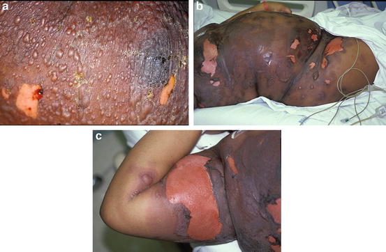

Fig. 16.1
(a) Tense blisters and erosions in a patient with TEN. (b) Large erosions in a patient with TEN. (c) Close-up of a large erosion and intact blister
Classification of the patient’s condition depends upon measurement of the detached and detachable epidermis.
SJS: <10 % of body surface area (BSA)
TEN: >30 % of BSA
SJS-TEN overlap: 10–30 % of BSA
Most patients with SJS-TEN have oral mucosal disease . The initial symptoms are burning of the lips and buccal mucosa. Blisters then appear on the lips and buccal mucosa and sometimes on the palate, gingiva, tongue, and pharynx. These blisters rupture quickly, leaving painful erosions that prevent the patient from eating or drinking. The lips become covered in hemorrhagic crusts (Fig. 16.2). The onset of oral mucosal disease may occur concurrently with the skin disease or precede it by a few days. Patients with SJS-TEN may also develop painful erosions at almost any mucosal site, including the eyes, nostrils, esophagus, bronchi, genitals, and anus.
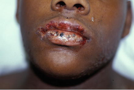

Fig. 16.2
Erosion and crusting of the lips in a patient with TEN
During the initial active phase of SJS-TEN , the patient has persistent fever, formation of mucocutaneous blisters, and partial shedding of the skin and oral mucosa. The active phase is complete when no new blisters appear. At about this time, the extent of skin detachment reaches its peak. If the patient is no longer receiving the causative medication, the active phase lasts 5 days. Barring other complications, the eroded skin then slowly re-epithelializes over the next 2–3 weeks. Mucosal sites take longer to heal than skin sites.
Complications
Patients with TEN have very large cutaneous erosions that may persist for 2–3 weeks. As a result of a compromised skin barrier, these patients may develop bacteremia, especially with Staphylococcus aureus or Pseudomonas species. This is the main cause of death in patients with TEN. Candida fungemia also may occur. The extensive loss of the skin barrier also predisposes patients to dehydration, electrolyte abnormalities, and hypothermia. These complications are much less common in patients with SJS.
Other possible complications of SJS-TEN are pneumonia, infected catheters, and partial or complete loss of vision owing to ocular erosions and scarring.
Prognosis
The overall mortality rate for SJS is 5 %, whereas that for TEN is 30 %. The severity-of-illness score for TEN (SCORTEN) is a predictor of disease severity and mortality based on a 7-point checklist. Patients are evaluated for clinical/laboratory parameters such as age greater than 40 years and extent of epidermal detachment (Table 16.3). The more adverse prognostic factors present, the worse the predicted mortality rate.
Table 16.3
Severity-of-illness score for TEN variables
Variable | Weight |
|---|---|
Age ≥40 years | 1 |
Malignancy | 1 |
BSA detached ≥10 % | 1 |
Tachycardia ≥120/min | 1 |
Serum urea level ≥27 mmol/L | 1 |
Serum glucose level ≥250 mmol/L | 1 |
Serum bicarbonate level <20 mmol/L | 1 |
Prompt discontinuation of a causative drug reduces the risk of death in SJS-TEN patients by about 30 % per day. An episode of SJS-TEN caused by a drug with a long half-life is more likely to end in death than one caused by a drug with a short half-life.
Diagnosis
The clinical presentation of an established case of SJS-TEN is characteristic. The patient has fever; painful oral mucositis; hemorrhagic crusts on the lips; painful red patches, especially on the trunk; fragile blisters; and cutaneous erosions. This presentation often occurs within 7–21 days of receiving a drug associated with SJS-TEN. Incipient SJS-TEN may be suspected in a patient who presents with an early morbilliform eruption and any tenderness of the oral mucosa or skin.
Skin biopsy for routine histology reveals substantial or full-thickness necrosis of the epidermis, a subepidermal split, and a sparse inflammatory infiltrate in the dermis. These findings are not specific.
Once SJS-TEN is diagnosed, the next step is to identify the causative medication. We have found that using a drug calendar can be very helpful. The culprit is usually not one of the patient’s chronic medications but rather a drug that has been given for the first time in the 3-week period prior to the onset of SJS-TEN. However, if a patient is re-exposed to a causative drug, the reaction may occur within 48 h. A review of the literature also helps to identify those drugs that are commonly associated with SJS-TEN.
A medication is probably not the trigger for SJS-TEN if it was first given within the previous 24 h or if the duration of treatment exceeds 3 weeks. An exception to this is with anticonvulsant medications (especially phenytoin), which may take up to 8 weeks to cause SJS-TEN.
Differential Diagnosis
Staphylococcal scalded skin syndrome (SSSS) may clinically resemble SJS-TEN. Patients with both disorders are febrile and have widespread tender erythema and erosions. The erosions of SSSS are more superficial than those of SJS-TEN. In addition, patients with SSSS do not have oral mucosal disease. When we suspect either of these diseases, we perform a skin biopsy on the edge of a blister for rapid (frozen) section. A pathology department can report the results in only a few hours. Frozen sections reliably differentiate between SJS-TEN (subepidermal split) and SSSS (intraepidermal split).
Grade IV (severe) acute cutaneous GVHD often resembles TEN both clinically and histologically. Differentiating between the two disorders can be nearly impossible. Also included in the differential diagnosis are paraneoplastic pemphigus and linear IgA disease.
Therapy
An important initial step in treating SJS-TEN is discontinuation of the offending drug. If the offending drug cannot be reliably identified, then all nonessential medications should be withdrawn.
TEN patients have extensive skin detachment with water, electrolyte, and protein loss: they physiologically resemble burn patients. There is some evidence that admission to a burn unit (not to a general intensive care unit) leads to improved outcomes (Endorf et al. 2008).
Treatment of TEN is primarily supportive: the goal is to protect the skin while it heals and correct any fluid and nutritional deficiencies. Intravenous fluid and electrolyte replacement in TEN patients is important, as insensible water loss may be significant. TEN patients need nutritional support because of a hypermetabolic state, wound exudative losses, and their inability to eat. Pain management and thermoregulation are also important. An ophthalmologist should evaluate patients with any evidence of ocular involvement.
TEN patients are at high risk for skin colonization by pathogenic bacteria with subsequent bacteremia owing to the compromised skin barrier. Patient care is therefore performed in a sterile fashion, and the patient may receive daily hydrotherapy with chlorhexidine gluconate or another antiseptic agent.
Another goal of skin care is to protect the large cutaneous erosions while re-epithelialization occurs. Some centers use synthetic skin substitutes such as silicone film embedded in nylon mesh (Biobrane), whereas others use porcine xenografts or human skin allografts. Physicians also take different approaches to the care of the patches of necrotic epidermis: some centers debride them surgically or with hydrotherapy, whereas others leave them in place.
TEN patients usually remain in the burn unit until re-epithelialization is almost complete and until they are otherwise stable. The median burn unit stay in one series of 19 patients was 14 days (Gerdts et al. 2007). Practical guidelines for the management of patients with TEN are available (Fromowitz et al. 2007).
Investigators have evaluated various pharmacologic agents in an attempt to halt progression of TEN. These agents include systemic steroids, cyclosporine, and intravenous immunoglobulins (IVIGs). With one exception (thalidomide, which produced a negative result), investigators have not performed randomized controlled trials of these agents. A large retrospective study (n = 281) did not demonstrate a clear benefit of treatment of SJS-TEN with either systemic steroids or IVIG (Schneck et al. 2008).
The most intensively studied of these pharmacologic agents is IVIG. In SJS-TEN patients, the destruction of the epidermis is caused by massive apoptotic cell death of keratinocytes via intracellular signaling proteins. One implicated pathway involves binding of the FAS ligand to the keratinocyte death receptor FAS. In vitro, pooled IVIG (which contains anti-FAS ligand antibodies) halted subsequent keratinocyte necrosis. Later, some clinical case studies demonstrated an improved mortality rate in TEN patients given IVIG, whereas others found no improvement (Abood et al. 2008).
The literature contains individual case reports in which patients with SJS-TEN almost certainly benefitted from receiving IVIG (Hebert and Bogle 2004). IVIG is less toxic than systemic steroids; some clinicians therefore feel that the current clinical and laboratory evidence is sufficiently compelling to justify its use, especially given the morbidity and mortality associated with TEN. The purpose of any medical therapy for TEN is to halt progression of the blistering process before it becomes extensive. Therefore, early initiation of this therapy is important.
Drug Reaction with Eosinophilia and Systemic Symptoms
DRESS is a severe nonblistering drug reaction . The typical findings are fever, a maculopapular skin eruption, lymphadenopathy, hepatitis, and a peripheral eosinophilia. The hepatitis may be severe and may be the cause of death; other internal organs are sometimes affected as well. In the early stage of DRESS, patients may simply appear to have an uncomplicated maculopapular drug eruption.
Another name for DRESS is drug hypersensitivity syndrome . Phenytoin (Dilantin) hypersensitivity syndrome, anticonvulsant hypersensitivity syndrome, and dapsone hypersensitivity syndrome are all probably subtypes of DRESS.
In general, the most common causes of DRESS are anticonvulsants, sulfonamide antibiotics, minocycline, allopurinol, and certain antiretroviral medications (Table 16.4). The incidence of DRESS caused by anticonvulsants and sulfonamides ranges from 1 in 1000 to 1 in 10,000 exposures.
Table 16.4
Drugs associated with DRESS
Abacavir |
Allopurinol |
Carbamazepine |
Dapsone |
Lamotrigine |
Minocycline |
Nevirapine |
Phenobarbital |
Phenytoin |
Sulfasalazine |
Trimethoprim-sulfamethoxazole |
Clinical Features
The onset of DRESS typically occurs 2–6 weeks after the first exposure to the causative medication. Patients may experience a prodrome of high fever, malaise, and sore throat that precedes the onset of the rash by a few days. At least 70 % of patients with DRESS have fever (temperature greater than 37.5 °C) during the disease course. The most clinically striking feature of DRESS is the rash, a pink or red maculopapular eruption that starts on the face and upper trunk (Fig. 16.3). The rash then progresses caudally to involve the entire trunk and thighs. This eruption occasionally progresses to erythroderma or TEN.
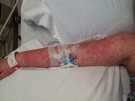

Fig. 16.3
Morbilliform eruption on the arm in a patient with DRESS
Some patients with DRESS have very characteristic periorbital or lip edema that is not associated with laryngeal edema (Fig. 16.4). The facial edema may distort facial features and occurs in 30–50 % of patients with DRESS. The majority of patients develop mild mucosal involvement such as small lip erosions or redness of the conjunctiva and oral mucosa. Many also have tender lymphadenopathy of the neck, axillary, or inguinal lymph node basins.
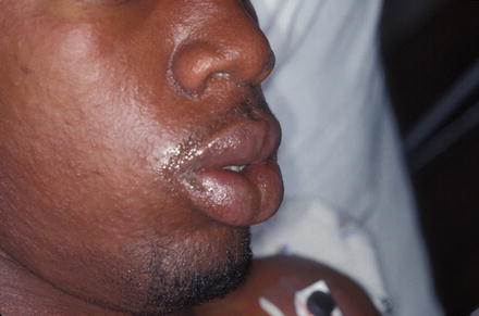

Fig. 16.4
Lip edema in a patient with DRESS
Most patients with DRESS develop drug-induced hepatitis of variable severity. Some have elevated transaminase levels without any clinical sequelae. In one series of 27 patients, 96 % had elevated transaminase levels, with 50 % having marked elevation (greater than 10 times normal) (Ang et al. 2010). Patients may have palpable hepatomegaly. Some patients develop hepatic necrosis and liver failure, the most common cause of death related to DRESS. The degree of hepatitis is partially related to the interval between the onset of the syndrome and discontinuation of the drug.
Stay updated, free articles. Join our Telegram channel

Full access? Get Clinical Tree


