A2. Deficiency anaemias
Iron Deficiency Anaemia
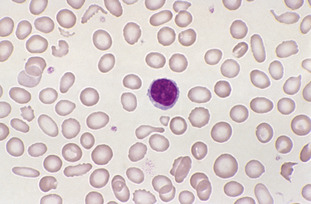
A major cause of iron deficiency anaemia is blood loss. It may also result from an inadequate diet and rarely from malabsorption. Pregnancy and growth are associated with greater requirements for iron; thus the risk of development of iron deficiency is high at these times. Microcytic hypochromic red cells are characterised by an MCV less than 80 fL and an MCH less than 27 pg. Red cell size may be assessed by comparing the red cell with a small lymphocyte.
The classical features found on the blood film in iron deficiency include anisocytosis, microcytes, hypochromasia, elliptocytes, and pencil cells and fragmented cells. Thrombocytosis is often present. When iron-deficiency anaemia is treated, a dimorphic blood film will result, that is, one in which there are two distinct populations of red cells: microcytic and hypochromic as well as normocytic and normochromic.
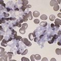
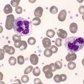
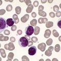
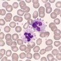
Severe cases of iron deficiency anaemia may also be detected in the bone marrow by the presence of smaller than normal erythroblasts with ragged and incompletely haemoglobinised cytoplasm. Iron stores may be assessed from the bone marrow by performing a Perl’s Prussian blue stain. Haemosiderin, which is present in the marrow fragments, will stain a turquoise colour in the presence of iron; decreased or absent haemosiderin is characteristic of iron deficiency. Refer to Figure A2-1, Figure A2-2, Figure A2-3 and Figure A2-4.
Stay updated, free articles. Join our Telegram channel

Full access? Get Clinical Tree


