© Springer International Publishing Switzerland 2016
Celestino Pio Lombardi and Rocco Bellantone (eds.)Minimally Invasive Therapies for Endocrine Neck Diseases10.1007/978-3-319-20065-1_2121. Critical Appraisal
(1)
Department of Surgery, Duke University, Durham, NC, USA
Keywords
Intraoperative nerve monitoringRecurrent laryngeal nerveExternal branch superior laryngeal nerveLigaSureHarmonic scalpel21.1 Introduction
Over the last 20 years, new technologies have been developed which have dramatically changed the field of surgery. While meticulous surgical technique and attention to detail remain the hallmarks of thyroid surgery, many surgeons have incorporated several technologies to enable them to perform thyroidectomy safely and faster. The previous chapters have reviewed the most common technologies used by thyroid surgeons, including intraoperative stimulation and monitoring of the external branch of the superior laryngeal nerve and the recurrent laryngeal nerve, as well as utilization of hemostatic devices and agents. This chapter will critically appraise the literature regarding these new technologies and summarize our recommendations.
21.2 External Branch Superior Laryngeal Nerve Intraoperative Nerve Monitoring
Injury to the external branch of the superior laryngeal nerve (EBSLN) can occur during thyroidectomy, resulting in alterations in the patient’s voice quality, projection, and ability to produce pitch in the highest ranges [1, 2]. These changes may be subtle, but are particularly significant for singers and other voice professionals. The standard techniques to minimize the risk of injury to the EBSLN are either direct visualization of the nerve before ligation of the superior thyroid vessels or ligation of superior thyroid vessels on the thyroid capsule without attempts to identify the nerve [3]. Nerve injury may still occur in up to 58 % of patients [4]. Identification and preservation of the EBSLN is often difficult because of its small caliber and variable anatomic pathway, particularly for patients with short necks, large goiters, and advanced cancers, where the anatomy is altered and the nerve is more intimately associated with the superior thyroid vessels [5]. Furthermore, the EBSLN cannot be identified in approximately 20 % of patients by direct visualization; this is generally the case when the nerve travels within the inferior constrictor muscle [6]. Because of the high rate of injury (as frequent as 58 %) [4] and increased difficulty identifying the EBSLN, intraoperative nerve stimulation and monitoring (IONM) have been employed as an adjunct to identify and minimize injury to the EBSLN.
There have been several studies that have evaluated IONM for EBSLN; however, the majority of these studies have been small, retrospective, nonrandomized, or single armed. To date, three randomized controlled trials [7–9] have evaluated the effect of IONM on EBSLN injury. Cernea et al. assessed EBSLN function with stimulation of the nerve followed by visual inspection for contraction of the cricothyroid muscle and a subsequent twitch. Seventy-six patients were randomized to three groups: IONM, dissection by a surgical resident without nerve identification, and dissection by a senior surgeon without nerve identification. Voice function was evaluated 1 week after surgery, and cricothyroid muscle function measured by percutaneous electromyograph 1 month after surgery. Both voice function and cricothyroid muscle electromyography were repeated at 6 months for patients with early EBSLN paralysis. The nerve identification rate was 93 % in the IONM group. There were no EBSLN injuries in the IONM group. However, 28 % of patients in the resident group and 12 % of patients who had surgery by the senior surgeon group had EBSLN injury (p = 0.01) on early assessment. Voice function correlated with nerve injuries. At 6 months follow-up, 57 % of patients in the resident group and no patients in the senior surgeon group had persistent nerve injury. Due to the small number of patients, a statistical analysis between these groups was not performed. The study had several limitations. Many patients were lost to follow-up, and 26 % of patients were excluded during the course of the study for reasons not described. Accordingly, an intention to treat analysis was not performed, so potential bias in the results cannot be excluded.
Lifante et al. [8] assessed EBSLN function under regional anesthesia using electrodes placed directly into the cricothyroid muscle. Forty-seven patients were randomized either to visual identification of the EBSLN plus IONM, or to direct visual identification alone. Following surgery, patients were assessed with the Voice Handicap Index-10 [10] and the Reflux Symptom Index [11]. The nerve identification rate was 66 % in the IONM group compared to 21 % in the control group (p = 0.003). Voice function 3 months after surgery was impaired in the control group compared to the IONM group (p = 0.034). Long-term follow-up of voice function was not performed. Unfortunately, a blinded, objective assessment of EBSLN function was not performed with either laryngoscopy or cricothyroid muscle electromyography, thereby preventing a correlation between patients’ perceived voice function and EBSLN function.
Barczynski et al. [9] performed the largest randomized controlled trial to date evaluating IONM of the EBSLN. Two hundred ten patients were randomized either to meticulous exploration for, and identification of, the EBSLN, or to fastidious exploration for, and identification of, the EBSLN with the aid of IONM. EBSLN function was assessed using endotracheal tube electrodes and direct visual contraction of a cricothyroid muscle twitch. The nerve identification rate was 84 % for the IONM compared to 34 % for the control group (p < 0.001). The rate of temporary EBSLN palsy was 1 % for the IONM group compared to 5 % for the control group (p = 0.02). Voice impairment and abnormal stroboscopy were more common in the control group than the IONM group 3 weeks after surgery (p = 0.03), but after 3 months, these differences resolved (p = 0.57). There was no difference in permanent EBSLN palsy rates (p = 0.57).
In summary, studies evaluating IONM for EBSLN have been small and have employed different methodologies to identify the nerve. Data appear to support the concept that IONM is associated with increased identification of the EBSLN, with subsequent reduction in transient voice disturbance, and decreased frequency of temporary dysfunction in the EBSLN. Compared to visualization alone, IONM does not appear to be associated with alteration in long-term voice function or the rate of permanent EBSLN injury. The American Academy of Otolaryngology and Head and Neck Surgery guidelines for improving voice outcomes after thyroid surgery [12] evaluated the published evidence addressing protection of the EBSLN. Based on an intermediate level of evidence and a preponderance of benefit over harm, the guidelines recommended that surgeons should take steps to preserve the EBSLN when performing thyroid surgery. The role of IONM was not specifically addressed.
We feel that the evidence demonstrating the potential benefit of IONM of the EBSLN is still lacking; therefore, the technology cannot be recommended routinely, although the data to date are promising. The available studies are small, and the techniques used for nerve identification and measurement of patient outcomes have been inconsistent between studies. Further, there are to date no data to definitively demonstrate that IONM is associated with superior long-term voice function. While cost-effectiveness was not specifically evaluated in any of these studies, it is likely lower with IONM because long-term patient quality of life is not significantly compromised when IONM is not employed. We feel that additional studies are needed to provide adequate data to assess the value of EBSLN IONM; in particular, they should include evaluation of cost and cost-effectiveness.
21.3 Recurrent Laryngeal Nerve Intraoperative Nerve Monitoring
Recurrent laryngeal nerve (RLN) IONM was introduced in the 1990s [13] as an adjunct to the gold standard of RLN visualization. Recent studies have revealed that 53 % of general surgeons [14] and 65 % of otolaryngologists [15] in the United States employ IONM during thyroid surgery. The prevalence of IONM is even more common in Germany, where a recent survey of surgery departments was performed. The survey response rate was 53 %, and the respondents accounted for 75 % of all thyroid surgeries done in Germany. The survey revealed that IONM was used for 92 % of thyroidectomies in 2010 [16]. Despite the widespread use of IONM, there is considerable debate regarding its benefit in improving outcomes for thyroid surgery, and about whether the technology is cost-effective.
Numerous studies have evaluated the role of RLN IONM; however, the vast majority of studies have been observational in nature. In large series (greater than 100 nerves at risk), the average rate of nerve paralysis with IONM was 4.7 %, compared to 5.7 % without IONM. These two injury rates were not significantly different in any of the studies [12]. A study by Dralle et al. [17] reviewed 16,448 patients with 29,998 nerves at risk that underwent thyroid surgery recorded in a German registry of data from 63 hospitals. There was no significant difference (p = 0.97) in the permanent RLN injury rate for IONM (0.80 %) compared to nerve visualization alone (0.89 %). Risk factors for permanent RLN injury were recurrent malignant goiter, recurrent benign goiter, and thyroid malignancy (Odds Ratio 6.66, p < 0.0001, 4.67, p < 0.0001, and 2.0, p = 0.002, respectively). A nonrandomized study by Chan et al. [17] evaluating 1,000 nerves at risk revealed no difference in RLN injury rate as measured by laryngoscopy with IONM in primary surgery, but did show a significant benefit in the setting of remedial surgery (19 % vs. 7.8 %, p = 0.017). There was also a trend towards lower RLN injury rates for patients with cancer and retrosternal goiters. A recent meta-analysis by Higgins et al. [18] of 44 studies (34 case series, seven comparative trials, and one randomized controlled trial) evaluated 64,699 nerves at risk and showed that the rate of transient and permanent RLN injury for visualization alone (3.12 % and 0.59 %, respectively) compared to IONM (3.52 % and 0.75 %, respectively) was not significantly different (p = 0.44). This study did not specifically evaluate the potential value of IONM for remedial surgery. A significant weakness of this meta-analysis was the heterogeneity of the studies included, making a comparison of the data challenging. The methodology for IONM was different and included electrodes placed directly in the vocalis muscle as well as endotracheal tube electrodes. The method for IONM allocation included surgeon preference, consecutive allocation, and equipment availability. Surgeon experience and resident involvement were not evaluated. The timing of initial laryngoscopy ranged from immediately postoperative to 2 weeks after surgery. Persistent vocal cord paralysis was assessed at times ranging from 3 to 24 months after surgery.
Only two randomized controlled trials have compared RLN visualization and IONM. Dionigi et al. [19] assessed IONM for video-assisted thyroidectomy. Seventy-two patients were randomized to IONM or visualization alone. The incidence of RLN injury identified by direct laryngoscopy was 2.7 % (1 patient) for the IONM group compared to 8.3 % (3 patients) for the nerve visualization only group (p = NS). There were no permanent injuries to the RLN in either group. However, the study was small and underpowered to identify a significant difference between the study arms. A larger study by Barczynski et al. [20] randomized 1,000 patients to visualization alone or IONM. Nerve monitoring was performed using the Neurosign® system, where an electrode is placed directly into the vocalis muscle. Transient RLN injury was significantly higher in the visualization group compared to the IONM group (3.8 % versus 1.9 %, p = 0.011). The permanent RLN injury rate was similar for both groups (1.2 % versus 0.8 %, p = 0.368). A subgroup analysis of patients stratified by surgical risk revealed that for low-risk patients, there was no difference in transient RLN injury rates (2.8 % vs. 1.8 %), but injury to the RLN was more frequent in the high risk group (4.9 % vs. 2.0 %, p = 0.011). There was no difference in permanent RLN injury rates based on risk stratification.
In addition to minimizing RLN injury, proponents of IONM have advocated that its use facilitates nerve identification, aids in nerve dissection, prognosticates postoperative nerve function, and can identify the site of nerve injury [21]. RLN identification rates using IONM and nerve mapping are between 98 and 100 %. In a study by Snyder et al. [22], IONM assisted with identification for 3 % of nerves with aberrant anatomy and 7 % of nerves during difficult dissections. Another study by Sari et al. [23] showed that time needed to identify the RLN was significantly shortened by 7 min (p < 0.005) using IONM compared to nerve visualization alone, leading to shorter total operating room times with IONM (p = 0.001). The experience of the participating surgeons was not stated for the study, bringing into question the generalizability of the findings.
After RLN identification, advocates of IONM suggest that it can aid in dissection of the nerve and determine the mechanism and site of injuries. In a study by Snyder et al. [24] of 666 RLNs at risk during thyroid and parathyroid surgery, IONM was helpful during the dissection of 7.5 % of patients, including patients with a bifurcated RLN, extensive scar tissue, and medial nerve displacement. The location and mechanism of nerve injury can also be determined with IONM. In the same study, the most common mechanism of RLN injury was a traction injury at the ligament of Berry. The injury location can be precisely identified using nerve mapping along the length of the nerve. If a ligature or clip is identified at the site of RLN injury, it can be removed, which theoretically should facilitate eventual nerve recovery.
Another perceived advantage of IONM is the ability to prognosticate nerve function at the conclusion of surgery. This is particularly important for bilateral surgery where injury to both RLNs is devastating and may lead to tracheostomy. While a RLN transection is readily apparent, blunt injury from stretch or compression is frequently not visibly detectable. Several studies have shown that surgeons are very poor visually judging RLN function intraoperatively and detect only 10–14 % of injuries [25, 26]. Furthermore, nerve injury was only suspected in one of six (16 %) patients with bilateral RLN injury [27]. In contrast, modern IONM using electromyography waveforms is highly specific and associated with a high negative predictive value, ranging from 92 to 100 % [28]. In a study by Goretzki et al. [29], 48 patients with planned total thyroidectomy using IONM lost signal during dissection of the first lobe. In 26 (55 %) patients, the surgical strategy was changed and the operation terminated after the first lobectomy, with completion thyroidectomy performed at a later date. For the remaining patients, either a total thyroidectomy with the assistance of a senior surgeon or a smaller bilateral surgery was performed. No patients in the delayed group had bilateral nerve injuries compared to 17 % in the nondelayed group.
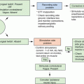
While IONM intermittently assesses the integrity of the RLN, a new method using continuous IONM with vagal stimulation has been developed. This could potentially allow identification of an impending nerve injury and alert the surgeon to alter surgical technique to avoid injury. Early studies have revealed no adverse side effects from continuous vagal stimulation [30, 31]. To date, the studies evaluating continuous IONM are small, but do appear to identify neural changes prior to loss of nerve signal [32, 33]. We eagerly anticipate larger studies and further development of this technology to determine if this may be associated with decreased frequency of RLN injury. Investigations to date have largely consisted of adult patients, so application in the pediatric population is still under-studied.
Related posts:
 High-Intensity Focused Ultrasound Ablation (HI-FU) in Endocrine Neck Diseases
Minimally Invasive Surgical Techniques: Critical Appraisal and Future Perspectives
Topical Hemostatic Agents
Patient Counselling and Patients’ Involvement in Health Policy
High-Intensity Focused Ultrasound Ablation (HI-FU) in Endocrine Neck Diseases
Minimally Invasive Surgical Techniques: Critical Appraisal and Future Perspectives
Topical Hemostatic Agents
Patient Counselling and Patients’ Involvement in Health Policy
 Minimally Invasive Video-Assisted Neck Dissection
 Recurrent Laryngeal Nerve Monitoring
Stay updated, free articles. Join our Telegram channel

Full access? Get Clinical Tree


