Stage
Description
Tumor extent
Surgical category
IA
Tumor is <2 cm and limited to the pancreas
Localized
Resectable
IB
Tumor is >2 cm and limited to the pancreas
IIA
Tumor extends directly beyond the pancreas
No arterial involvement. LN−
Locally advanced
Resectable or borderline resectable
IIB
Tumor may or may not extend beyond the pancreas
No arterial involvement. LN+
III
Tumor involves major local arteries (SMA, celiac). LN− or LN+
Locally advanced
Unresectable
IV
Primary tumor may be any size. Metastatic disease is present
Metastatic
Unresectable
Surgical resection followed by adjuvant therapy is the main stay of treatment for patients with Stage I/II disease. The use of neoadjuvant therapy for these patients is controversial. Patients with Stage II borderline resectable disease should receive neoadjuvant therapy preoperatively [28]. Patients with stage III locally advanced disease should be treated with chemotherapy and/or chemoradiotherapy. A great number of these patients tend to develop metastatic disease later on; however, a few select cases can still be considered suitable for surgical resection. Systemic therapy is administered to patients with Stage IV disease with a good performance status while those with poor overall health are given supportive and palliative therapy [2, 27].
Controversies
The Role of Diagnostic Laparoscopy
Radiological diagnosis of pancreatic cancer has improved greatly due to advancement in imaging technology leading to increased sensitivity of available modalities. The first step in the diagnosis of suspected pancreatic adenocarcinoma is a pancreas protocol CT and a chest CT [27]. The pancreas protocol CT is a detailed examination of the pancreas. It includes both a non-contrast scan and contrast enhancement of arterial, pancreatic parenchymal, and portovenous phases. The difference between the tumor and the pancreatic parenchyma is most prominent during the late arterial phase during contrast enhancement. Post processing three-dimensional reconstructions of images assist in determining the relationship of the tumor to its surrounding vasculature, thus helping to determine tumor resectability. MRI scans are helpful in finding small metastatic deposits especially in the liver and peritoneum. It can also be used in patients who cannot tolerate contrast for CT. CT scans and MRIs have comparable sensitivities [27]. Endoscopic ultrasound can be used to visualize the tumor and also to obtain tissue for diagnosis. PET scanning has a role in select cases [29] and retrospective studies have shown that it has increased sensitivity to identifying metastatic disease compared to CT scan alone [30]. It can also be used to determine the extent of soft tissue invasion before planning radiotherapy [31]. Evidence of extrapancreatic spread, involvement of celiac axis and the SMA, and the patency of the SMV-PV confluence are all factors that can be identified on imaging and help determine tumor resectability [32].
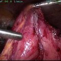
Although CT remains the main tool for staging, the major variability in false negatives, is small peritoneal disease. One older study reported that 20–30 % of patients who appear to have resectable disease on CT were found to have undetected local spread or hepatic and peritoneal implants [33]. For this reason, some centers advocate the routine use of diagnostic laparoscopy. Laparoscopy has the potential to better establish tumor resectability by allowing direct visualization of the tumor and detecting hepatic and peritoneal metastases, particularly when coupled with laparoscopic ultrasound. Doppler techniques can also be used to assess vascular involvement [34, 35]. Moreover, in-depth laparoscopic staging in which celiac, periportal, and peripancreatic lymph nodes are examined has been reported [36]. Thus, laparoscopy potentially prevents the morbidity associated with a non-therapeutic laparotomy in patients with unresectable tumors. These patients can then receive chemotherapy without delay [37]. In patients who are found to have resectable disease using frozen section evaluation of specimens during laparoscopy, the surgeon can proceed with a laparotomy during the same operation [34, 38].
However, diagnostic laparoscopy is time consuming and costly. In addition, data demonstrating benefit to diagnostic laparoscopy are dated. In the contemporary period with high-quality cross-sectional imaging, the finding of unexpected metastatic disease is low. More recent reports demonstrate radiographic failure to detect metastatic disease in only approximately 15 % of all cases [39, 40]. Fisher et al. have recommended the use of laparoscopy in cases where there is a large primary tumor, substantial weight loss, a tumor located in the body or tail of the pancreas, hypoalbuminemia or considerable elevated CA 19-9 levels [41]. The use of diagnostic laparoscopy, at large well-established centers for pancreatic surgery, given the availability of powerful diagnostic modalities and the expertise of the treating physicians, is probably best applied in select cases [37].
Surgery-First Versus Neoadjuvant Approach
Approximately 15–20 % of patients with PDAC have resectable disease at the time of diagnosis. The goal of a potentially curative operation in this cohort is to achieve a margin free of cancer (R0) leaving the patient in a condition that allows systemic treatment for micrometastatic disease [42–44]. There is still no definite consensus on whether patients who initially have resectable tumors should undergo a neoadjuvant approach or a surgery-first approach. Advocates of the surgery-first approach believe that the single most important treatment for a chance of cure is resection of the tumor and this should be accomplished prior to systemic therapy. On the other hand, potential advantages of the neoadjuvant approach include earlier control of micrometastatic disease with systemic therapy and an opportunity for better selecting patients for pancreatectomy by excluding those with more aggressive disease that progresses on therapy. Based on current studies, it is still unclear whether a neoadjuvant approach improves survival as a better treatment modality or whether tumors with favorable tumor biology are simply selected. Designing a randomized clinical trial to make this differentiation is challenging.
Several well-done trials have evaluated the outcomes of the neoadjuvant approach (Table 2). In summary, when taking into account intention to treat, it appears that that both initial surgery and neoadjuvant therapy have comparable survival rates and that the major advantage of neoadjuvant approach is with patient selection. In this regard, several studies reported that patients who receive neoadjuvant therapy have a better median survival than what has traditionally been reported for the surgery-first approach, but 10.71–36.58 % do not make it to surgery [28, 45–48]. The reasons for not making it to surgery included rapid progression in 20 % (n = 17) of the patients of which 9.4 % (n = 8) were deemed unresectable at the time of restaging while 10.6 % (n = 9) were found to have metastatic disease [28].
Table 2
Neoadjuvant therapy for resectable pancreatic cancer
Study | Treatment | Patients | Median survival (months) | Median survival mo. (resected) | Median survival mo.(unresected) |
|---|---|---|---|---|---|
Desai [69] | Gem/Ox + XRT> Gem/Ox | 12(44)a | 12.5 (44 pts.) | NR | NR |
Varadhachary [45] | Gem + Cis > Gem + XRT | 79 | 17.4 | 31 (52 pts.) | 10.5 (27 pts.) |
Evans [28] | Gem + XRT | 86 | 23 | 34 (64 pts.) | 7.1 (22 pts.) |
Heinrich [70] | Gem + Cis | 28 | 26.5 | 19.1 (25 pts.) | NR (3 pts.) |
Le Scodan [46] | 5FU/Cis + XRT | 41 | 9.4 | 9.5 (26 pts.) | 5.6 (15 pts.) |
Golcher [47] | Gem + Cis | 33 | 17.4 | 25 (19 pts.) | NR |
A retrospective study identified 199 patients out of a total of 1562 patients who received neoadjuvant therapy. The percentage of patients who underwent preoperative biliary stenting (57.9 vs. 44.7 %, p = 0.0005), vascular resection (41.5 vs. 17.3 %, p < 0.0001), and open resections (94.0 vs. 91.4 %, p = 0.008) was higher in the neoadjuvant group. However, there was no significant difference between the 30 days mortality (2.0 vs. 1.5 %, p = 0.56) and postoperative morbidity (56.3 vs. 52.8 %, p = 0.35) between the two groups [49].
Another recent retrospective study evaluated patients with resectable disease who received neoadjuvant therapy and provided a classification by dividing them into three groups: those who had clinically resectable cancer with no extrapancreatic disease, those for whom there was a suspicion of extra pancreatic disease, and those who had poor performance status or significant co-morbidities. Resection rates in the three groups were 75, 46, and 37 % respectively (p < 0.001). Old age, poor performance status, pain, and treatment complications (p < 0.05) were factors which were identified for selection against surgery. The overall median survival was 21 months for all patients. Moreover, survival rates in resected and unresected patients who had suspicion of extra pancreatic disease and those who had poor performance status or co-morbidities were similar to those resected and unresected patients who did not have these adverse factors, respectively (p > 0.22) [50].
The standard paradigm in PDAC treatment has involved bias toward a surgery-first approach in resectable tumors, driven by the relatively low efficacy of standard systemic therapies such as gemcitabine. With the advent of more potent chemotherapy regimens, such as FOLFIRINOX and gemcitabine/abraxane, the bias is shifting toward the increased use of neoadjuvant therapy. PDAC is a highly systemic disease and thus aggressive systemic therapy must be the goal in all patients [51].
In contrast to patients with resectable disease and no major vessel involvement, neoadjuvant chemotherapy with chemoradiation is given to all patients with borderline resectable disease as there is a general consensus that such patients will benefit from this therapy. There is a greater probability of positive margin resection if these patients are not given neoadjuvant therapy [52]. Patients who receive neoadjuvant therapy have an 80–90 % rate of margin-negative resection with comparable or sometimes even better survival outcomes than patients who have tumors that are initially classified as resectable [52].
Definition of Borderline Resectable Disease
The treatment of pancreatic cancer is dependent on the stage and resectability of the disease, therefore different institutions have proposed staging systems that determine whether the disease is resectable, borderline resectable , unresectable, or metastatic [27]. These institutions include the M.D. Anderson Cancer Center (MDACC) and the National Comprehensive Cancer Network (NCCN). Moreover a recent consensus guideline which modified the M.D. Anderson staging criteria was recommended by a joint committee of American Hepato-Pancreato-Biliary Association (AHPBA), Society of Surgical Oncology (SSO), and the Society for the Surgery of the Alimentary Tract (SSAT) [53].
Based on these guidelines, resectability is determined by the presence or absence of metastatic disease and involvement of major blood vessels. Both portovenous and arterial vessels are evaluated separately. A tumor involving the portovenous vessels is considered resectable as long as the vessels can be reconstructed after resection. On the other hand, assessing the involvement of arterial vasculature, most importantly the superior mesenteric, hepatic and celiac arteries, is crucial as the degree of involvement determines resectability of the tumor. The degree of involvement is assessed on axial planes of high-quality cross-sectional imaging [54–57]. Encasement (>180° involvement) by the tumor of any arterial vessel is considered unresectable. Abutment (<180° involvement) of the celiac arteries or the superior mesenteric artery is considered to be borderline resectable but is associated with margin-positive resections, high rates of recurrence, and a lower survival [27].
Although these guidelines have proven to be useful in the classifying resectability in the majority of patients, several controversies exist on what should be considered borderline resectable versus locally advanced. With regards to venous involvement, the term “technically reconstructable” varies depending on the experience of the surgeon and variations of anatomy in each patient. For example, involvement of the inferior superior mesenteric vein at the level of the confluence of ileal and jejunal branches is often considered to be locally advanced. However, examples exist in which one or more of these branches can be sacrificed or reconstructed based on the size of remaining branches and location to the tumor. Another example is encasement of the hepatic artery and celiac axis which is strictly considered to be locally advanced. However, in some cases of local involvement of these vessels resection is possible. This may include focal involvement of the hepatic artery that is amenable to reconstruction or in some cases a patient with an accessory right or left hepatic artery may make resection of the hepatic artery possible. In addition, focal encasement of the celiac artery and proximal common hepatic artery with preservation of the gastroduodenal artery may be resected through an Appleby’s procedure [26]. Therefore, the definition of what is borderline resectable versus locally advanced unresectable may not be the same at all institutions. Ultimately, we believe that it is up to the surgeon’s judgment to determine whether successful resection of the tumor is feasible
Ablative Therapy for Locally Advanced Disease
Management of locally advanced disease is difficult and is best managed through a multidisciplinary approach. The literature available to us is limited and as new data become available to us our approach to this stage of the disease will improve. A great deal of emphasis is given to the role of systemic chemotherapy in these patients given that there is a high probability of presence of microscopic systemic disease at the time of diagnosis and/or the risk of development of metastases during the course of radiation. It has been reported that in rare cases that the use of currently available regimens may result in a significant downstaging of the disease.
Moertel et al., in the largest randomized study in this field, demonstrated that concurrent use of radiation with 5-fluorouracil (5-FU) significantly prolonged the median overall survival of these patients, which now is the standard of care for this subset of our patient population [58]. Subsequent research studies support the use of systemic chemotherapy followed by chemoradiation. The team at Johns Hopkins in a retrospective analysis of data from these patients demonstrated better survival outcomes in patients who had a longer course of induction chemotherapy prior to the chemoradiation [59]. Unfortunately, a large-scale study comparing the multiple chemotherapeutic agents available is not available.
A new emerging frontier in the therapy of pancreatic cancer is irreversible electroporation (IRE) [60]. The use of IRE in pancreatic surgery is controversial given that it is a new technique, with only few centers having trained professionals who can use this modality. The current focus lies on the use of IRE to treat locally advanced pancreatic cancer. Careful selection of patients is vital. Experts usually treat only stage III disease using this method. It is a method that employees the use of short pulses of high voltage low energy electric current, to create nanoscale micropores in the lipid bilayer of cell membranes. This leads to an increased permeability to ions and macromolecules and subsequently, leads to cell swelling and subsequent apoptosis. Amplitude of the pulse, its duration, frequency, and number of pulses applied all determine the extent of cell damage and death [61, 62].
Stay updated, free articles. Join our Telegram channel

Full access? Get Clinical Tree


