Common Gastrointestinal Disorders in the Elderly


Common Gastrointestinal Disorders in the Elderly


Kristen M. Robson

Anthony Lembo


CLINICAL PEARLS


  • The elderly may not present with typical symptoms of peptic ulcer disease, such as abdominal pain and dyspepsia.


  • The mainstay of therapy for cholangitis are broad-spectrum antibiotics, relief of biliary obstruction and biliary drainage.


  • Choledocholithiasis in elderly patients can be adequately treated by endoscopy with clearance of the bile duct and does not necessarily need to be followed by cholecystectomy.


  • In elderly patients with nonacute dysphagia, it is often helpful to obtain a barium esophagram as the first imaging study.


  • Patients should be directly asked about fecal incontinence, as many patients are reluctant to bring it up with their caregivers.


  • The hallmark symptoms of gastroesophageal reflux disease (GERD) are heartburn and regurgitation.


  • An upper endoscopy should be considered in patients with suspected GERD who have had symptoms longer than 5 years to exclude erosive esophagitis, esophageal stricture, or Barrett’s esophagus.


  • Among the elderly who complain of constipation, the most common symptom is that of excessive straining and hard stools.


  • Increasing dietary fiber to 20 to 25 g per day is often effective in treating mild to moderate constipation.


  • Patients are often reluctant to discuss this problem with their physician due to social embarrassment.


  • Anorectal biofeedback therapy, which involves retraining the pelvic floor and the abdominal wall musculature, has been shown to improve symptoms in two thirds of patients with fecal incontinence.

The physiologic function of the gastrointestinal (GI) tract is generally maintained with aging. Nevertheless, GI symptoms and diseases are particularly common in the elderly. New GI symptoms in elderly patients should be investigated to determine if pathology exists. The evaluation of symptoms in an elderly patient should be performed in a timely fashion. Elderly patients may not be able to compensate or withstand the effects of a GI illness as compared to their younger counterparts.


PEPTIC ULCER DISEASE

Peptic ulcer disease (PUD) can affect patients of all ages. Although the approach to the management of PUD is similar in both younger and older patients, there may be an increased risk of morbidity and mortality in elderly patients.1 In addition, data from the National Health Interview Survey suggest that increasing age is a risk factor for PUD.2 There are three major mechanisms that contribute to the development of PUD: Nonsteroidal antiinflammatory drugs (NSAIDs), Helicobacter pylori infection, and acid.


Nonsteroidal Anti-inflammatory Drugs

The elderly take NSAIDs more frequently than any other age-group. Nearly 40% of elderly persons receive at least one prescription for NSAIDs each year, and 1% to 8% of these patients are hospitalized because of a GI complication secondary to NSAIDs within the first year of use.3

NSAIDs inhibit the synthesis of prostaglandins by the gastric mucosa. Prostaglandins E2 and I2, the principal prostaglandins of the stomach, perform several functions that protect the gastric mucosa. These prostaglandins inhibit gastric acid secretion, increase mucosal blood flow, and promote the secretion of bicarbonate and mucus.1 The cyclooxygenase-2 (COX-2) inhibitors have been shown to have less GI toxicity than the traditional NSAIDs due to their affinity for COX-2 over COX-1.1 In a double-blind outcome trial of over 8,000 patients with arthritis, COX-2-specific inhibitors (coxibs) decreased complications of an upper GI event (bleeding, perforation, obstruction, or symptomatic ulcer).4

The efficacy of proton pump inhibitors (PPIs) has been assessed in patients who present with ulcers associated with NSAID use. One study compared the use of placebo versus lansoprazole in patients taking low-dose aspirin who had a history of ulcer disease. If present, H. pylori was first eradicated. Ulcer recurred in 15% of the placebo group versus 2% of the lansoprazole group at a median duration of 1 year5 (Evidence Level B).


Helicobacter pylori

The prevalence of H. pylori infection increases with advancing age, reaching levels of 40% to 60% in asymptomatic elderly individuals and over 70% in elderly patients with GI illness. The eradication of H. pylori may prevent the progression of intestinal metaplasia and gastric atrophy.6 There are several tests available to diagnose H. pylori. Nonendoscopic tests include the immunoglobulin G antibody test, the 13C-urea breath test, and the stool antigen test. Endoscopic tests include biopsy for rapid urease testing and histologic examination. Treatment of H. pylori is discussed in the subsequent text.


Non-Helicobacter pylori, Non-Nonsteroidal Anti-inflammatory Drug Ulcers

A prospective study of nearly 2,400 patients with duodenal ulcers revealed that 27% were H. pylori negative and denied NSAID use. What is the etiology of such ulcers? Other common causes of ulcers, such as Crohn’s disease or hypersecretory states, should be considered as well as unreported NSAID use.


Clinical Features

The presentation of PUD usually is epigastric abdominal pain, typically postprandial. The pain is often relieved with food or over-the-counter antacids. The elderly, however, are prone to atypical dyspeptic symptoms. In one report, 65% of patients older than 80 years who presented with upper GI bleeding secondary to PUD had no pain upon presentation.7 In another study of elderly patients with PUD, 35% of patients reported no abdominal pain, as compared with 8% of younger patients with either gastric or duodenal ulcers diagnosed by endoscopy.8 The lack of the typical presenting symptoms of PUD in the elderly may lead to a delay in diagnosis and contribute to a higher mortality rate of complicated ulcer disease.


Complications

For both gastric and duodenal ulcers, the most common complication is bleeding, followed by perforation. Both of these complications may occur more frequently in the elderly. GI hemorrhage and perforation from PUD carry an overall 30% mortality rate for patients older than 65 years. Complicated PUD may result in higher mortality rates in the elderly compared to their younger counterparts.9 Factors that may contribute to the increase in the complication rates of PUD in the elderly include atypical presentations of PUD that may lead to a delay in diagnosis, as well as the presence of comorbid conditions.9


Diagnosis

The primary method for diagnosing both gastric and duodenal ulcers is upper endoscopy. Endoscopy has been shown to be safe in elderly patients, with complication rates similar to those in younger patients.10 Although duodenal ulcers are typically not followed-up with upper endoscopy, all gastric ulcers require repeat endoscopy after several weeks of therapy to assure healing and that the ulcer was not malignant. PUD can also be diagnosed by upper GI series using a double contrast technique.1

All patients with PUD, whether young or old, should be tested for H. pylori infection. A variety of methods can be used, including both invasive and noninvasive testing (see Table 41.1).


Treatment

The treatment of PUD is first directed to the removal of potential causes such as the H. pylori infection and the discontinuation of NSAIDs. Paramount to the treatment of PUD is acid suppression to heal the ulcer as well as to provide maintenance therapy. PPIs are more effective in healing ulcers than are histamine-2 receptor antagonists. One study that examined the healing rates of duodenal ulcers treated with omeprazole 40 mg daily reported 93% healing at 2 weeks, and nearly 100% healing at 4 weeks11 (Evidence Level A).


TABLE 41.1 HELICOBACTER PYLORI: METHODS OF TESTING


Noninvasive Testing


Invasive Testing (Endoscopic Biopsy)


Serum IgG Helicobacter pylori antibody


Rapid urease test


13C-urea breath test


Histologic examination


Stool antigen test


IgG, immunoglobulin G.


For patients who develop peptic ulcers secondary to NSAIDs, the NSAIDs should be discontinued. If this is not a reasonable option, there is evidence that the coadministration of a PPI can reduce the risk of ulcer recurrence5 (Evidence Level A).

When a H. pylori infection is identified in the setting of PUD, the bacteria should be eradicated. A minimum of a 1-week PPI-based triple therapy regimen, including clarithromycin, amoxicillin, and/or nitroimidazoles, is highly effective and well tolerated in elderly patients. The main factors attributed to treatment failure in the elderly are noncompliance and antibiotic resistance.6


Summary

PUD can present in elderly patients in an atypical manner. This may lead to a delay in diagnosis. The elderly remain at risk for PUD not only because of the widespread use of NSAIDs but also because of the increased prevalence of H. Pylori as compared to younger patients.


DISORDERS OF THE GALLBLADDER AND BILIARY TRACT


Age-Related Changes in the Biliary Tract

The overall prevalence of gallstone disease in the elderly, by ultrasound, is 26.7%. Advanced age is also associated with a higher rate of calcified pigment stones.12 The lithogenicity of bile is thought to change as a result of age-related changes in biliary metabolism. Increased cholesterol saturation of bile and reduced gallbladder emptying are contributing factors13 (see Table 41.2).

Some studies demonstrate that the diameter of the common bile duct increases with advancing age. In a prospective study of over 1,000 patients between the age of 60 and 96, a small but statistically significant increase in the caliber of the common bile duct was demonstrated with increasing age. In another study by Bachar et al. of 51 patients undergoing abdominal ultrasonography, an
age-dependent change in the diameter of the common bile duct was demonstrated. This study suggested that the upper normal limit of duct size in the elderly be set at 8.5 mm.14,15


TABLE 41.2 AGE-RELATED CHANGES IN THE BILIARY TRACT


  1. Increased prevalence of gallstones


  2. Increased common bile duct diameter


  3. Increased lithogenicity of bile


  4. Increased incidence of pigment stones


Adapted from Ross SO, Forsmark CE. Pancreatic and biliary disorders in the elderly. Gastroenterol Clin North Am. 2001;30(2):531-545.


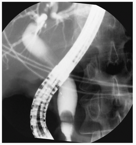
Figure 41.1 Common bile duct stone.


Gallbladder Disease


Cholelithiasis and Acute Cholecystitis

The prevalence of gallstones increases with age. In a large population-based study, the prevalence in the 50- to 65-year-old age-group was 13.7%, as compared to 1.7% in the 18- to 25-year-old age-group.16 As in younger patients, therapy for asymptomatic gallstones found in elderly patients is not indicated (Evidence Level B). Elderly patients with cholelithiasis are more likely than younger patients to present with an acute complication of gallstones, such as acute cholecystitis, gallstone pancreatitis, or common bile duct stones.17


Clinical Manifestations

Acute cholecystitis commonly presents as constant right upper quadrant abdominal pain that may radiate to the back or right shoulder. Nausea, emesis, and anorexia may be associated symptoms, and fatty food ingestion may have preceded the onset of pain. Symptoms such as fever and prolonged pain (i.e., hours) should alert the clinician to the likelihood of acute cholecystitis as opposed to a limited attack of biliary colic.

Patients with acute cholecystitis tend to have fever, tachycardia, and an overall ill appearance, as well as peritoneal signs on physical examination. A “Murphy’s sign” may also be present. The patient is asked to inspire deeply while the gallbladder fossa is palpated. As the gallbladder descends, the patient typically experiences increased discomfort. The sensitivity of the Murphy’s sign, however, may be decreased in the elderly.18


Complications

There is a high rate of complications of untreated cholecystitis. The most common complication of acute cholecystitis is gangrenous cholecystitis (up to 20% of cases), which occurs more frequently in older patients. Sepsis is typically present in patients with gallbladder gangrene, and subsequent gallbladder perforation occurs in 2% of cases.19 The additional complications of acute cholecystitis are listed in Table 41.3.


Diagnostic Testing

In the patient with symptoms of cholecystitis, laboratory work often shows a leukocytosis with an increased percentage of bands. Abnormalities in liver function tests, including hyperbilirubinemia and elevated alkaline phosphatase, should raise the suspicion for complicating factors such as cholangitis or choledocholithiasis.

The initial imaging study is usually ultrasonography, which can often confirm the diagnosis. The presence of gallbladder stones, gallbladder wall thickening or edema, and a “sonographic Murphy’s sign” all support the diagnosis of cholecystitis. A systematic review of 30 studies of ultrasonography for the evaluation of gallstones and acute cholecystitis demonstrated that the sensitivity
and specificity of ultrasound for the detection of gallstones were in the range of 84% and 99%, respectively. The sensitivity and specificity for acute cholecystitis were 88% and 80%, respectively.20


TABLE 41.3 COMPLICATIONS OF ACUTE CHOLECYSTITIS


  1. Gangrenous cholecystitis


  2. Gallbladder perforation


    1. Localized perforation: pericholecystic abscess


    2. Free perforation: generalized peritonitis (associated with high mortality)


  3. Cholecystenteric fistula: More often secondary to pressure necrosis from gallstones


  4. Gallstone ileus: Results from passage of a stone through a fistula, leading to mechanical small bowel obstruction


  5. Emphysematous cholecystitis


    1. Secondary infection of the gallbladder with a gas-forming organism such as Clostridium sp


    2. May lead to gallbladder perforation


Adapted from Zakko SF, Afdhal NH. Clinical features of acute cholecysitis. In: Rose Bd, ed. UpToDate. Waltham, MA: UpToDate; 2006.


Cholescintigraphy (also referred to as hepatic iminodiacetic acid [HIDA] scan) can be helpful if the ultrasound is inconclusive. A technetium-labeled HIDA is administered intravenously and is then selectively taken up by hepatocytes and excreted into the bile. This agent will enter the gallbladder if the cystic duct is patent and will be visualized within 30 to 60 minutes. The test is positive if the gallbladder is not visualized secondary to the cystic duct obstruction from gallbladder edema or an obstructing gallstone. The sensitivity and specificity of this nuclear medicine test are approximately 97% and 90%, respectively.20,21

An abdominal computed tomography (CT) scan can demonstrate findings associated with acute cholecystitis such as gallbladder wall edema and pericholecystic stranding. A CT scan can be helpful when complications of acute cholecystitis are suspected or when alternative diagnoses are being considered.22


Treatment

Laparoscopic cholecystectomy is the treatment of choice for symptomatic cholecystitis. In a retrospective study of patients undergoing laparoscopic cholecystectomy, elderly patients were significantly more likely than younger patients to present with acute cholecystitis, gallstone pancreatitis, and common bile duct stones. The open conversion rate in elderly patients with complicated gallstone disease was significantly higher compared to younger patients. Elderly patients with uncomplicated gallstone disease, however, appeared to be excellent candidates for laparoscopic cholecystectomy. The study concluded that early conversion or open cholecystectomy may be indicated in the elderly with acute complications of cholelithiasis.17

Because of increased complications from acute cholecystitis in elderly patients, percutaneous cholecystomy may be considered as an alternative therapy in the acute setting. In an uncontrolled trial, percutaneous cholecystomy was performed in a group of elderly patients who were at high risk for surgery due to a variety of comorbid conditions. Percutaneous cholecystomy was successful in 95% of patients, with low morbidity and overall zero mortality.23


Gallbladder Carcinoma

As in gallstone disease, gallbladder carcinoma, although uncommon overall, occurs more frequently in the elderly. In a study of 4,500 autopsies in Japan, the incidence of gallbladder carcinoma was 2.1%. From 50% to 88% of patients with gallbladder carcinoma have gallstones.24,25


Choledocholithiasis and Cholangitis

Acute cholangitis is a potentially life-threatening infection of the biliary tree. Choledocholithiasis is the most common cause of infection, which is characterized by fever, jaundice, and abdominal pain.26,27 Confusion and hypotension can occur in patients with suppurative cholangitis. Hypotension may be the only manifestation in elderly patients.28


Diagnostic Testing and Treatment

Ultrasound is generally recommended as the first imaging study in patients suspected of having cholangitis. Endoscopic retrograde cholangiopancreatography (ERCP) is utilized both to establish the diagnosis and to provide therapeutic intervention with sphincterotomy, stone removal, and/or stent insertion. Magnetic resonance cholangiopancreatography is emerging as an excellent tool in the evaluation of choledocholithiasis, particularly in patients whose symptoms of cholangitis are not severe and when the risks of ERCP are high.28,29 Magnetic resonance cholangiopancreatography can also be considered in patients with suspected choledocholithiasis in the absence of cholangitis.


As in younger patients, the first line of therapy for cholangitis includes intravenous broad spectrum antibiotics to cover gram-negative organisms, for the relief of biliary obstruction and providing biliary drainage. Supportive measures such as the administration of intravenous fluids and close monitoring are also important. Biliary drainage can be accomplished mainly by ERCP. Percutaneous drainage or surgical decompression are occasionally used. In an uncontrolled study of the management of acute cholangitis in patients older than 80, the morbidity and mortality rates of methods for drainage were compared. Endoscopic drainage was associated with the lowest rates of morbidity and mortality (17% and 6%), followed by percutaneous drainage (36% and 9%), and surgical drainage, which was associated with rates of 88% and 25% (Evidence Level C). It should be noted, however, that morbidity and mortality rates for all interventions were significantly higher in older patients than in younger patients.27

For patients with a gallbladder, cholecystectomy usually follows endoscopic treatment for choledocholithiasis. For elderly patients, especially those at high risk for surgery, endoscopic sphincterotomy alone may be the treatment of choice. In a study of the long-term outcome of endoscopic treatment for choledocholithiasis, this approach was found to be relatively favorable. No significant risk factors for cholecystitis as a late complication were identified.30


ESOPHAGEAL DYSPHAGIA


Age-Related Physiologic Changes of the Esophagus

Although the esophagus generally ages well under normal conditions, changes can be documented. Age-related changes in the oropharynx and esophagus are listed in Table 41.4. Presbyesophagus is a term that formerly described age-related changes in the esophageal body. It is now felt, however, that these changes in motility are most likely secondary to medical conditions found in the elderly. These medical conditions include neurologic disorders, diabetes mellitus, and the effects of medications.31


TABLE 41.4 AGE-RELATED PHYSIOLOGIC CHANGES OF THE OROPHARYNX AND ESOPHAGUS


  1. Decreased upper esophageal sphincter pressure


  2. Delays in upper esophageal sphincter relaxation after deglutition


  3. Decreased amplitude of esophageal body contractions


  4. Nonperistaltic contractions


  5. Diminished visceral perception


  6. Hiatal hernia


From Greenwald DA. Aging, the gastrointestinal tract, and the risk of acid-related disease. Am J Med. 2004;117(5A):8S-13S.


Dysphagia

Dysphagia is the sensation of impaired passage of either solids or liquids from the mouth to the stomach. Dysphagia is a common symptom that can affect patients of all ages, but it may be more common in older patients. In one study, the prevalence of dysphagia appeared to increase with age, and at least 10% of people older than age 50 complained of some dysphagia.32,33 Dysphagia is an alarm symptom and requires prompt evaluation to determine the etiology and to provide therapy. Dysphagia in elderly patients should not be attributed to normal aging without further prompt evaluation.34,35

Dysphagia can be classified into two types: Oropharyngeal and esophageal (transfer and transport dysphagia). Oropharyngeal dysphagia arises from diseases of the upper esophagus and pharynx or from upper esophageal sphincter dysfunction. Esophageal dysphagia arises within the body of the esophagus or the lower esophageal sphincter, and it is most commonly due to a mechanical cause or motility disturbance.34 The focus of this section will be on esophageal or transport dysphagia.


Careful history is important in the evaluation of elderly patients with dysphagia. Dysphagia should be distinguished from odynophagia, which refers to pain with swallowing in contrast to difficulty swallowing. A variety of mechanical and neuromuscular disorders are associated with esophageal dysphagia. There are three main factors that can help determine the source of dysphagia:


  • The type of food that produces symptoms (i.e., solids, liquids, or both)


  • The temporal progression of symptoms (intermittent vs. progressive)


  • Associated symptoms or findings, such as heartburn.

Dysphagia to both solids and liquids at the onset of the symptoms is suggestive of but not diagnostic of a motility disorder. Dysphagia for solids that progresses to include liquids is more likely to reflect a mechanical obstruction.36 Symptoms such as heartburn may suggest the presence of a complication of gastroesophageal reflux disease (GERD), such as a benign or a malignant stricture. There are numerous conditions that present with esophageal dysphagia (see Table 41.5). The more common causes will be reviewed here.


TABLE 41.5 DIFFERENTIAL DIAGNOSIS OF GASTROINTESTINAL COMPLAINTS


Causes of Constipation (564.00)


Causes of Fecal Incontinence (787.91)


Conditions Associated with Esophageal Dysphagia


Slow transit constipation (564.01)
Defecatory disorders (536.9)
Normal transit constipation (564.00)


Infectious (009.2)
Inflammatory bowel disease (569.9)
Radiation enteritis (558.1)
Short-gut syndrome (579.3)
Carcinoid syndrome (259.2)
Celiac disease (579.0)


Achalasia (530.0)
Hypermotility or spastic motility disorders (536.8/564.9)
Diabetes mellitus (250.00)
Systemic sclerosis (710.0)


Mechanical obstruction
Colon cancer (239.0)
Rectocele (male 564.49) (female 618.04)
Sigmoidocele
Stricture (569.2)
Extrinsic compression


Neurologic conditions
Multiple sclerosis (340)
Strokes (434.91)
Diabetes (250.00)
Central nervous system disorders (e.g., dementia, strokes, tumors, myelomeningoceles, spinal cord lesions)


Mechanical causes (luminal)
Peptic stricture (537.9)
Malignancy (i.e., adenocarcinoma)
Radiation-induced injury
Medication-induced esophagitis/stricture


Metabolic and endocrine
Diabetes mellitus (250.00)
Hypothyroidism (244.9)
Hyperthyroidism (242.90)
Hypokalemia (276.8)
Pregnancy (V22.2)
Pheochromocytoma (194.0)
Panhypopituitarism (253.2)
Porphyria (277.1)
Heavy metal poisoning (e.g., lead, mercury, arsenic intoxication)


Overflow incontinence
Fecal impaction (560.39)
Neoplasm (239.9)


GERD (530.81)


Medications
Calcium channel blockers (e.g., verapamil)
μ-Opioid agonists (loperamide, morphine, fentanyl)
Anticholinergics (e.g., antispasmodics, antipsychotics, tricyclic antidepressants, antiparkinsonian drugs)
Anticonvulsants (e.g., phenobarbital, carbamazepine, phenytoin)
Antacids (e.g., aluminum- or calcium-containing antacids)
5-HT3 antagonists (e.g., alosetron)
Iron supplements
Nonsteroidal anti-inflammatory agents (e.g., ibuprofen)
Diuretics (e.g., furosemide)
Chemotherapeutics (e.g., vinca derivatives)


Pelvic floor denervation
Obstetrical injury of the pudendal nerve (959.14)
Chronic straining leading to descending-perineum syndrome
Rectal prolapse (569.1)


Extrinsic compression
Enlarged aorta (dysphagia aortica) (447.8)
Enlarged left atrium (429.3)
Aberrant subclavian artery (747.60)
Mediastinal mass (786.6)


Neuropathies and myopathies
Systemic sclerosis (710.0)
Amyloidosis (277.3)
Dermatomyositis (710.3)
Multiple sclerosis (340)
Parkinson disease (332.0)
Spinal cord injury (952.9)
Autonomic neuropathy (337.9)
Chagas disease (086.2)
Intestinal pseudo-obstruction (564.89)
Cerebrovascular accidents (434.91)
Shy-Drager syndrome (333.0)


Anal sphincter injury (959.19)
Obstetrical injury (665.9)
Anorectal surgery (959.19)
Accidental injury (959.9)


GERD, gastroesophageal reflux disease.

Only gold members can continue reading. Log In or Register to continue

Stay updated, free articles. Join our Telegram channel

Jul 21, 2016 | Posted by in GERIATRICS | Comments Off on Common Gastrointestinal Disorders in the Elderly

Full access? Get Clinical Tree

Get Clinical Tree app for offline access