Cold antibody types account for about 25% of autoimmune hemolytic anemias. Primary chronic cold agglutinin disease (CAD) is characterized by a clonal lymphoproliferative disorder. Secondary cold agglutinin syndrome (CAS) complicates specific infections and malignancies. Hemolysis in CAD and CAS is mediated by the classical complement pathway and is predominantly extravascular. Not all patients require treatment. Successful CAD therapy targets the pathogenic B-cell clone. Complement modulation seems promising in both CAD and CAS. Further development and documentation are necessary before clinical use. We review options for possible complement-directed therapy.
Key points
- •
Primary chronic cold agglutinin disease (CAD) is a clonal lymphoproliferative disorder and a distinct clinicopathologic entity.
- •
Secondary cold agglutinin syndrome (CAS) occasionally complicates specific infections or aggressive lymphomas.
- •
In both CAD and CAS, hemolysis is entirely complement dependent.
- •
Hemolysis is predominantly extravascular, mediated by the classical complement pathway.
- •
Targeting the pathogenic B-lymphocyte clone has resulted in successful therapy for CAD. Complement modulation is promising in specific situations, but has to be further developed and documented before clinical use.
Introduction
Cold antibody types account for approximately 25% of autoimmune hemolytic anemias (AIHA) and are classified as shown in Box 1 . Most cold-reactive autoantibodies are cold agglutinins (CA). CA are antibodies that bind to erythrocyte surface antigens at low temperatures, causing agglutination and complement-mediated hemolysis. We review the etiology, pathogenesis, clinical features, and therapy of CA-mediated AIHA, highlighting the role of complement involvement. Paroxysmal cold hemoglobinuria is not addressed, because it is described elsewhere in this issue and the involved autoantibodies are not agglutinins.
Warm antibody type
Primary
Secondary
Cold antibody type
Primary chronic cold agglutinin disease
Secondary cold agglutinin syndrome
Associated with malignant disease
Acute, infection associated
Paroxysmal cold hemoglobinuria
Mixed cold and warm antibody type
Data from Refs.
Cold agglutinins
Cold hemagglutination was first described in 1903. CA are determined semiquantitatively by their titer, based on their ability to agglutinate erythrocytes at 4°C. A proportion of the adult population has demonstrable CA in serum without any evidence of hemolysis or disease; a frequency of positive screening tests at 0.3% has been reported in a cohort of patients with nonrelated disorders. These normally occurring CA are polyclonal and are found in low titers, usually below 64 and rarely exceeding 256. In 172 consecutive individuals with monoclonal immunoglobulin (Ig)M in serum, on the other hand, significant CA activity was found in 8.5% with titers between 512 and 65,500, and all individuals with detectable CA had hemolysis. Thus, monoclonal CA are generally far more pathogenic than polyclonal CA.
The thermal amplitude is defined as the highest temperature at which the CA reacts with the antigen. In general, the pathogenicity of CA depends more on the thermal amplitude than on the titer. The normally occurring CA have low thermal amplitudes. If the thermal amplitude exceeds 28°C or 30°C, erythrocytes agglutinate in the circulation in acral parts of the body, even at mild ambient temperatures and, often, complement fixation and complement-mediated hemolysis ensues. CA should not be confused with cryoglobulins. In rare cases, however, the cryoprotein can have both CA and cryoglobulin properties.
Most CAs are directed against the Ii blood group system. The I and i antigens are carbohydrate macromolecules and the density of these antigens on the erythrocyte surface are inversely proportional. Neonatal red blood cells almost exclusively express the i antigen, whereas the I antigen predominates in individuals of 18 months of age and older. Therefore, CA with anti-I specificity are more pathogenic in children as well as adults than those specific for the i antigen. Occasionally, CA show specificity against the erythrocyte surface protein antigen designated Pr and such CA can be highly pathogenic. Other specificities have been reported, but are probably very rare. More than 90% of pathogenic CA are of the IgM class and these IgM macromolecules can be pentameric or hexameric. Hexameric IgM is more pathogenic than pentameric IgM.
The terms cold agglutinin disease (CAD) and cold agglutinin syndrome (CAS) have been used in the literature in a rather random way. We should distinguish, however, between these concepts. CAD is a well-defined clinicopathologic entity, as shown herein and should be called a disease, not syndrome. The term CAS is appropriate for the secondary CA-mediated syndrome occasionally complicating specific infections or malignancies.
Chronic cold agglutinin disease
Epidemiology
Primary CAD has been reported to account for about 15% of AIHA. In Scandinavia, the prevalence has been estimated to 16 per million inhabitants and the incidence rate to 1 per million per year, which is probably a slight underestimation. There seems to be a slight female preponderance with a male-to-female ratio of approximately 0.6:1. In the same population-based, descriptive study, the median age of the patients was 76 years (range, 51–96) and the median age at onset of clinical symptoms 67 years (30–92).
Clonality and Histopathology
The first monoclonal protein ever described was a CA from a patient with CAD ; early studies showed that, in most patients, the CA was monoclonal IgM with kappa light chain restriction. In a more recent study of 86 patients, the CA was found to be monoclonal IgM-kappa in more than 90% of patients. Monoclonal IgG, IgA, or lambda light chain restriction were rare findings. In 6% of the patients, monoclonal Ig could not be detected despite otherwise typical primary CAD. This is probably a matter of sensitivity. Furthermore, 90% of patients in whom flow cytometry of bone marrow aspirate had been performed, had a clonal expansion of kappa-positive B cells. The CA in CAD are almost always specific for the I antigen and show restriction to the IGHV4-34 gene segment. These findings have led to the conclusion that patients with CAD must have a clonal B-cell lymphoproliferative disorder, which has not been elucidated fully until recent years.
Two large, retrospective studies of consecutive patients found signs of a bone marrow clonal lymphoproliferation in most patients. Undoubtedly, this majority represents the same group of patients that has traditionally been diagnosed with “primary” or “idiopathic” CAD. Within each series, however, the individual hematologic and histologic diagnoses showed a striking heterogeneity. In 1 series, lymphoplasmacytic lymphoma was the most frequent finding, whereas marginal zone lymphoma, unclassified clonal lymphoproliferation, and reactive lymphocytosis were also reported frequently.
The explanation for this perceived heterogeneity was probably revealed by a recent study in which bone marrow biopsy samples and aspirates from 54 patients with CAD were reexamined systematically by a group of lymphoma pathologists using a standardized panel of morphologic, immunohistochemical, flow cytometric, and molecular methods. The bone marrow findings in these patients were consistent with a surprisingly homogeneous disorder termed ‘primary CA-associated lymphoproliferative disease’ by the authors and distinct from lymphoplasmacytic lymphoma, marginal zone lymphoma, and other previously recognized lymphoma entities ( Fig. 1 ). The MYD88 L265P somatic mutation, typical for lymphoplasmacytic lymphoma, could not be detected in any of 15 samples from patients with CAD tested for this mutation, even though a sensitive, polymerase chain reaction-based method was used.
Complement-Mediated Hemolysis
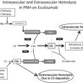
Cooling of blood during passage through acral parts of the circulation allows CA to bind to erythrocytes and cause agglutination ( Fig. 2 ). Being a strong complement activator, antigen-bound IgM CA on the cell surface binds complement protein 1 (C1) and thereby initiates the classical complement pathway. C1 esterase activates C4 and C2, generating C3 convertase, which results in the cleavage of C3 to C3a and C3b. Upon returning to central parts of the body with a temperature of 37°C, IgM CA detaches from the cell surface, allowing agglutinated cells to separate, while C3b remains bound. A proportion of the C3b-coated erythrocytes is sequestered by macrophages of the reticuloendothelial system, mainly Kupffer cells in the liver. On the surface of the surviving red blood cells, C3b is cleaved, leaving high numbers of C3d molecules on the cell surface. These mechanisms explain why the monospecific direct antiglobulin test (DAT) is strongly positive for C3d in patients with CA-mediated hemolysis and, in the majority, negative for IgM and IgG.
Complement activation may proceed beyond the C3b formation step, resulting in C5 activation, formation of the membrane attack complex (MAC), and intravascular hemolysis. Owing to surface-bound regulatory proteins such as CD55 and CD59, however, the complement activation is usually not sufficient to produce clinically significant activation of the terminal complement pathway. The major mechanism of hemolysis in stable disease, therefore, is the extravascular destruction of C3b-coated erythrocytes. Obviously, however, C5-mediated intravascular hemolysis does occur in severe acute exacerbations and in some profoundly hemolytic patients, as evidenced by the observation of hemoglobinuria in 15% of the patients, the rather frequent finding of hemosiderinuria (M. J. Stone 2014, personal communication) and the beneficial effect of C5 inhibition in at least occasional patients.
Clinical Features
By definition, all patients with CAD have hemolysis, but occasional patients are not anemic because the hemolysis is fully compensated. Most patients, however, have manifest hemolytic anemia. The anemia can be more severe than often stated in textbooks and review articles. Of 16 patients described in an early publication, 5 had hemoglobin (Hgb) levels below 7.0 g/dL and 1 had levels below 5.0 g/dL. Hgb levels ranged from 4.5 g/dL to normal in a more recent, population-based, descriptive study of 86 Norwegian patients. The median Hgb level was 8.9 g/dL and the lower tertile was 8.0 g/dL. Hemoglobinuria has been reported in at least 15% of the patients. About 50% of the patients are considered transfusion dependent for shorter or longer periods during the course of the disease. In many patients, therefore, CAD is not an indolent disease in terms of clinical symptoms and quality of life.
Approximately 90% of the patients experience cold-induced acrocyanosis and/or Raynaud phenomena. The circulatory symptoms can range from slight to disabling. In cool climates, characteristic seasonal variations in the severity of hemolytic anemia have been well-documented. In at least 70% of patients, exacerbation of hemolytic anemia is also triggered by febrile infections or major trauma. The explanation for this paradoxical exacerbation is that, during steady-state CAD, most patients are complement depleted with low levels of C3 and, in particular, C4. During acute phase reactions, C3 and C4 are replete and complement-induced hemolysis increases.
The median overall survival of patients with CAD has been estimated to be 12.5 years, similar to that of a general age- and sex-matched population. Transformation of the lymphoproliferative bone marrow disorder to aggressive lymphoma is rare, probably with a cumulative rate of 3% to 4%. The clinical course is unpredictable; patients can experience either worsening or improvement with time, quite stable disease or, occasionally, a shift in the respective clinical manifestations.
Diagnosis
The diagnosis of CAD should be based on history and clinical findings, assessment of hemolysis, the DAT pattern, and the CA titer. An additional electrophoretic, histopathologic, and flow cytometric workup should always be done, but demonstration of clonality may sometimes be difficult and is not absolutely required for diagnosis in the routine clinical setting. Table 1 shows the diagnostic criteria. Correct handling of samples as indicated in Table 1 is essential for reliable assessment.
| Level | Criteria | Procedures and Comments |
|---|---|---|
| Required for diagnosis | Chronic hemolysis | |
| Polyspecific DAT positive | ||
| Monospecific DAT strongly positive for C3d | DAT is usually negative for IgG, but occasionally weakly positive | |
| CA titer ≥64 at 4°C | Blood specimen must be kept at 37°C–38°C from sampling until serum is removed from the clot | |
| No overt malignant disease | Clinical assessment; radiology as required | |
| Confirmatory but not required for diagnosis | Monoclonal IgMκ in serum (or, rarely, IgG, IgA, or λ phenotype) | Serum must be obtained as for CA titer Immunofixation should be done even if no band is visible on electrophoresis |
| Cellular κ/λ ratio >3.5 (or, rarely, <0.9) in B-lymphocyte population | Flow cytometry in bone marrow aspirate | |
| CA-associated lymphoproliferative bone marrow disorder by histology | Trephine biopsy |
Nonpharmacologic Management
Given that drug therapy has been largely ineffective until the last 10 to 15 years, counseling has been considered the mainstay of management. Owing to the high thermal amplitude of the CA, however, the physiologic cooling of the blood in the peripheral vessels is usually sufficient to cause hemolysis and circulatory symptoms even at mild ambient temperatures.
Most authors agree that patients should avoid cold exposure, particularly of the head, face, and extremities. Those living in cool climates often, even before the diagnosis has been established, tell the physician that they use warm clothing and, in many cases, stay indoors during winter. Many patients experience improvement of Hgb levels and circulatory symptoms when temporarily relocating to a warmer climate during the cold season, but severely symptomatic CAD does exist even in the subtropics. Any liquids infused should be prewarmed, and surgery under hypothermic conditions should be avoided or specific precautions undertaken.
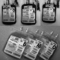
Erythrocyte transfusions can be given safely, provided that appropriate precautions are undertaken. In contrast with the compatibility problems encountered in warm antibody AIHA, it is usually easy to find compatible donor erythrocytes, and screening tests for irregular blood group antibodies are most often negative. Antibody screening and, if required, compatibility tests should be performed at 37°C. The patient and, in particular, the extremity chosen for infusion should be kept warm, and the use of an in-line blood warmer is recommended. Failure to adhere to required precautions has resulted in dismal or, very rarely, even fatal outcomes. Because complement proteins can exacerbate hemolysis, transfusion of blood products with a high plasma content should probably be avoided.
Based on theoretical considerations and clinical experience, plasmapheresis is regarded an efficient “first-aid” in acute situations or before surgery requiring hypothermia, because almost all IgM is located intravascularly. Such remissions, however, are very short lived and concomitant specific therapy should usually be initiated. Complement inhibitor-based alternative approaches to this situation are discussed elsewhere in this article. Given that the extravascular hemolysis predominantly takes place in the liver, splenectomy should not be used for the treatment of CAD. Three splenectomized patients were registered in our population-based descriptive study; none of them responded. Improvement after splenectomy has been reported occasionally among the rare patients with CAD mediated by an IgG CA instead of IgM.
Unspecific Immunosuppression and Supportive Drug Therapy
In textbooks and review articles, it is often postulated that typical patients with CAD are just slightly anemic and do not require pharmacologic therapy. Based on the Hgb levels and clinical features described herein, this holds true for a minority only. In Norway as well as the United States, drug therapy had been attempted in 70% to 80% of unselected patients studied in 2 relatively large, retrospective series.
In contrast with warm antibody AIHA, corticosteroids are of little or no value in CAD. Monotherapy with alkylating agents has shown some beneficial effect on laboratory parameters and clinical improvement has been observed. The clinical response rates, however, are probably in the same low order of magnitude as for corticosteroids. In 2 small series of patients treated with interferon-α or low-dose cladribine, these drugs were not shown to be useful, although some conflicting data do exist for interferon-α. Only a few patients treated with azathioprine have been reported; none of them responded.
Exacerbations precipitated by febrile illnesses should warrant immediate treatment of any bacterial infection. Supportive therapy with erythropoietin or its analogs seems to be used widely in North America, but less often in Scandinavia and Western Europe. No studies have been published to support or discourage its use. Although poorly documented, folic acid supplements have often been recommended.
Therapies Directed at the Pathogenic B-Cell Clone
The relative success in therapy for CAD during the last 10 to 12 years has been achieved by targeting the pathogenic B-cell clone.
Rituximab monotherapy
Monotherapy with rituximab 375 mg/m 2 weekly for 4 weeks was studied in 2 prospective, uncontrolled trials of 37 and 20 treatment courses. The response criteria used in the Norwegian study are shown in Table 2 ; similar strict definitions were used in the Danish study. The overall response rate was 54% and 45% in the 2 trials. With the exception of 1 complete response (CR) observed in the Norwegian trial, all remissions were partial responses (PR). Ten patients were treated for relapse after previously having received rituximab therapy and 6 of them responded to a second course. In our study, the responders achieved a median increase in Hgb levels of 4.0 g/dL. We found a median time to response of 1.5 months (range, 0.5–4.0) and median response duration of 11 months (range, 2–42).


