SUMMARY
Evaluation of a hemostatic disorder is commonly initiated when (1) a patient or referring physician suspects a bleeding tendency, (2) a bleeding tendency is discovered in one or more family members, (3) an abnormal coagulation assay result is obtained from an individual as part of a routine examination, (4) an abnormal assay result is obtained from a patient during preparation for surgery, or (5) a patient has unexplained diffuse bleeding during or after surgery or following trauma. Evaluation of a possible hemostatic disorder in each of these scenarios is a stepwise process that requires knowledge of the various classes of hemostatic disorders commonly found under the particular circumstances. The patient’s history, the results of physical examination, and an initial set of hemostatic tests usually enable a tentative diagnosis. However, more specific tests are commonly necessary to make a definitive diagnosis. This chapter reviews the necessary steps.
Hemostatic disorders can conveniently be classified as either hereditary or acquired (Table 6–1). Alternatively, hemostatic disorders can be classified according to the mechanism of the defect. Of the acquired disorders, the thrombocytopenias are the most frequently encountered entities. Thrombocytopenias can result from reduced production of platelets, excessive destruction caused by antibodies or other consumptive processes, or pooling of platelets in the spleen, as in hypersplenism (Chap. 7); however, if hypersplenism is the sole cause of a hemostatic disorder, it is rarely severe enough to cause pathologic bleeding.
| Major Types | Disorders | Examples |
|---|---|---|
| Acquired | Thrombocytopenias | Autoimmune and alloimmune, drug-induced, hypersplenism, hypoplastic (primary, myelosuppressive therapy, myelophthisic marrow infiltration), disseminated intravascular coagulation (DIC), thrombotic thrombocytopenic purpura, hemolytic uremic syndrome (Chaps. 7, 19, and 22) |
| Liver diseases | Cirrhosis, acute hepatic failure, liver transplantation (Chap. 18), thrombopoietin deficiency | |
| Renal failure | ||
| Vitamin K deficiency | Malabsorption syndrome, hemorrhagic disease of the newborn, prolonged antibiotic therapy, malnutrition, prolonged biliary obstruction | |
| Hematologic disorders | Acute leukemias (particularly promyelocytic), myelodysplasias, monoclonal gammopathies, essential thrombocythemia | |
| Acquired antibodies against coagulation factors | Neutralizing antibodies against factors V, VIII, and XIII, accelerated clearance of antibody-factor complexes, e.g., acquired von Willebrand disease, hypoprothrombinemia associated with antiphospholipid antibodies (Chaps. 16, 17, and 21) | |
| DIC | Acute (sepsis, malignancies, trauma, obstetric complications) and chronic (malignancies, giant hemangiomas, retained products of conception) (Chap. 19) | |
| Drugs | Antiplatelet agents, anticoagulants, antithrombins, and thrombolytic, hepatotoxic, and nephrotoxic agents (Chaps. 23–25) | |
| Vascular | Nonpalpable purpura (“senile,” solar, and factitious purpura), use of corticosteroids, vitamin C deficiency, child abuse, thromboembolic, purpura fulminans; palpable purpura (Henoch-Schönlein, vasculitis, dysproteinemias; Chap. 12), amyloidosis | |
| Inherited | Deficiencies of coagulation factors | Hemophilia A (factor VIII deficiency), hemophilia B (factor IX deficiency), deficiencies of fibrinogen factors II, V, VII, X, XI, and XIII, and von Willebrand disease (Chaps. 13–16) |
| Platelet disorders | Glanzmann thrombasthenia, Bernard-Soulier syndrome, platelet granule disorders (Chap. 10) | |
| Fibrinolytic disorders | α2-Antiplasmin deficiency, plasminogen activator inhibitor-1 deficiency (Chap. 25) | |
| Vascular | Hemorrhagic telangiectasias (Chap. 12) | |
| Connective tissue disorders | Ehlers-Danlos syndrome (Chap. 12) |
Acronyms and Abbreviations:
aPTT, activated partial thromboplastin time; DIC, disseminated intravascular coagulation; ELISA, enzyme-linked immunosorbent assay; PT, prothrombin time; RCF, ristocetin cofactor.
The bleeding history is a crucial element in the evaluation of a patient with a hemorrhagic disorder. The bleeding history helps define the subsequent diagnostic approach and the likelihood of future bleeding. Eliciting and interpreting all of the relevant information requires a systematic and methodical approach. The following points are worth considering:
Patients vary in their responses to hemorrhagic symptoms. Some patients ignore significant symptoms, whereas other patients are highly sensitive to even minor symptoms. When asked in standardized questionnaires, many normal, healthy people indicate they have excessive bleeding or bruising.1,2 Therefore, some experts believe the question “Do you bruise easily?” is virtually worthless. Women are more likely to respond that they have excessive bleeding or bruising than are men.
Patients with severe hemorrhagic disorders invariably have very abnormal bleeding histories, for example, severe hemophilia A or hemophilia B, type 3 (homozygous) von Willebrand disease, and Glanzmann thrombasthenia. Importantly, these patients may experience spontaneous bleeding episodes.
The diagnostic value of any specific symptom varies in the different disorders. Therefore, recognizing typical patterns of bleeding is important (Table 6–2). Unprovoked hemarthroses and muscle hemorrhages suggest one of the hemophilias, whereas mucocutaneous bleeding (epistaxis, gingival bleeding, menorrhagia) is more characteristic of patients with qualitative platelet disorders, thrombocytopenia, or von Willebrand disease.
Assessing the extent of hemorrhage against the background of any trauma or provocation that may have elicited the hemorrhage is important. If a patient has never had a significant hemostatic challenge, such as tooth extraction, surgery, trauma, or childbirth, the lack of a significant bleeding history is much less valuable in excluding a mild hemorrhagic disorder. For example, a significant percentage of patients with mild von Willebrand disease or mild forms of hemophilia may have negative bleeding histories,1 even though they may be at considerable risk for excessive bleeding after surgery or other interventions. Thus, these diagnoses must be considered even in elderly patients if their first severe hemostatic challenge occurs at that age.
Obtaining objective confirmation of the subjective information conveyed in the bleeding history is valuable. Objective data include (1) previous hospital or physician visits for bleeding symptoms, (2) results of previous laboratory evaluations, (3) previous transfusions of blood products for bleeding episodes, and (4) a history of anemia and/or previous treatment with iron.
Although self-administered questionnaires may provide useful background information, they cannot substitute for a dialogue between the physician and the patient. Thus, history taking in general, but especially in the often subtle histories related to hemostatic disorders, is an intellectually active process involving data collection, hypothesis development, new question formulation, additional data gathering, and new hypothesis development. However, this iterative procedure has its limitations even when it is carefully pursued.3,4
A medication history is a crucial component of the bleeding history, with particular attention to nonprescription drugs, such as aspirin and nonsteroidal antiinflammatory agents, which may affect bleeding symptoms. A medication history is especially important in patients with thrombocytopenia, because drug-induced thrombocytopenia is common (Chap. 10 and see Table 6–1). Medication also may affect hemostasis through deleterious effects on the liver or kidney functions. The increased use of herbal and alternative medicines poses particular problems, because patients may not readily share information about what they are taking, and the dose they are taking of any particular active ingredient may be difficult to determine. Ginkgo biloba and ginseng are the most commonly used herbals that can cause platelet dysfunction and induce bleeding.5 Other dietary supplements can display similar effects.5,6
A nutrition history should be obtained to assess the likelihood of (1) vitamin K deficiency, especially if the patient also is taking broad-spectrum antibiotics; (2) vitamin C deficiency, especially if the patient has skin bleeding consistent with scurvy (perifollicular purpura); and (3) general malnutrition and/or malabsorption.
Several tissues have an increased local fibrinolytic activity. Such tissues include the urinary tract, endometrium, and mucous membranes of the nose and oral cavity. These sites are particularly likely to have prolonged oozing of blood after trauma in patients with hemostatic abnormalities. Excessive bleeding following tooth extraction is one of the most common manifestations. Bleeding resulting from defects in fibrin crosslinking (factor XIII deficiency), or fibrinolytic defects may often manifest as delayed bleeding after trauma.
Bleeding isolated to a single organ or system (e.g., hematuria, hematemesis, melena, hemoptysis, or recurrent nosebleeds) is less likely to result from a hemostatic abnormality than from a local cause such as neoplasm, ulcer, or angiodysplasia. Thus, careful anatomic evaluation of the involved organ or system should be performed.
Bleeding may result from blood vessel disorders such as hereditary hemorrhagic telangiectasias, Cushing disease, scurvy, or Ehlers-Danlos syndrome. Many primary dermatologic disorders also have a purpuric or hemorrhagic component and must also be considered in the differential diagnosis (Chap. 12).
A family history is particularly important when hereditary disorders are considered. Patients usually will not spontaneously offer a history of consanguinity, so specific inquiry should be made about this possibility. A diagram of the patient’s genealogic tree, extending back at least two generations, should be included to document consideration of genetic disorders. A sex-linked pattern of inheritance is consistent with hemophilia A or B (Chap. 13). An autosomal dominant pattern is characteristic of most forms of von Willebrand disease (Chap. 16). An autosomal recessive pattern is typical for all other coagulation factor deficiencies (Chap. 14), inherited platelet disorders (Chap. 10), and the rare, severe (homozygous), type 3 von Willebrand disease. Population genetic information may be helpful; for example, the higher prevalence of factor XI deficiency in Ashkenazi Jews (Chap. 14).
The history should include information on diseases and organs that may affect hemostasis, such as cirrhosis, renal insufficiency, myeloproliferative neoplasms (e.g., essential thrombocythemia), acute leukemia, myelodysplasia, systemic lupus erythematosus, and Gaucher disease.
| Clinical Manifestations | Hemostatic Disorders |
|---|---|
| Mucocutaneous bleeding | Thrombocytopenias, platelet dysfunction, von Willebrand disease |
| Cephalohematomas in newborns, hemarthroses, hematuria, and intramuscular, intracerebral, and retroperitoneal hemorrhages | Severe hemophilias A and B, severe deficiencies of factor VII, X, or XIII, severe type 3 von Willebrand disease, afibrinogenemia |
| Injury-related bleeding and mild spontaneous bleeding | Mild and moderate hemophilias A and B, severe factor XI deficiency, moderate deficiencies of fibrinogen and factors II, V, VII, or X, combined factors V and VIII deficiency, α2-antiplasmin deficiency |
| Bleeding from stump of umbilical cord and habitual abortions | Afibrinogenemia, hypofibrinogenemia, dysfibrinogenemia, factor XIII deficiency |
| Impaired wound healing | Factor XIII deficiency |
| Facial purpura in newborns | Glanzmann thrombasthenia, severe thrombocytopenia |
| Recurrent severe epistaxis and chronic iron deficiency anemia | Hereditary hemorrhagic telangiectasias |
Individual hemorrhagic symptoms often require detailed analysis before the significance of the symptoms and the resulting diagnosis or therapy can be determined. Some of the more common symptoms are discussed below, and Table 6–2 summarizes clinical manifestations that are typical for specific hemostatic disorders.
Epistaxis is one of the most common signs of platelet disorders and von Willebrand disease. It also is the most common symptom of hereditary hemorrhagic telangiectasia. In the latter condition, epistaxis almost always becomes more severe with advancing age. Epistaxis is not uncommon in normal children, but it usually resolves before puberty. Dry air heating systems can provoke epistaxis even in otherwise normal individuals. Bleeding confined to a single nostril more likely results from a local vascular problem than a systemic coagulopathy.
Gingival hemorrhage is very common in patients with both qualitative and quantitative platelet abnormalities and von Willebrand disease. Occasional gum bleeding occurs in normal individuals, especially if they use a hard bristle tooth brush and dental hygiene procedures. Thus, establishing whether the bleeding is excessive may be difficult. Frequent gingival hemorrhage can occur in individuals with normal hemostasis if they have gingivitis.
Oral mucous membrane bleeding in the form of blood blisters is a common manifestation of severe thrombocytopenia. Such bleeding usually has a predilection for sites where teeth can traumatize the inner surface of the cheek.
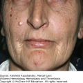
Skin hemorrhage in the form of petechiae and ecchymoses is a common manifestation of hemostatic disorders. However, skin hemorrhage also is common among individuals without hemostatic disorders. Excessive bruising is more common in women than men. Moreover, women frequently note that the severity of their bruising varies with the phase of their menstrual cycle, although the most severe phase of the cycle may differ in different women. Features that help establish the severity of skin hemorrhage include the size of the bruises, the frequency of bruising, whether the bruises occur spontaneously or only with trauma, and the appearance of bruises on regions of the body that usually are not traumatized, such as the trunk and back. The color of the bruise may yield information. Red bruises on the extensor surfaces of the arms and hands indicate loss of supporting tissues, as occurs in Cushing syndrome, glucocorticoid therapy, senile purpura, and damage from chronic sun exposure. Jet-black bruises may be caused by warfarin-induced skin necrosis and similar disorders. Easy bruising can also occur in patients with Ehlers-Danlos syndrome manifested by distensible skin or extraordinary ligament laxness and in patients with hyperflexibility of the thumb.7
Tooth extractions are common hemostatic challenges and may be helpful in defining the risk of bleeding. Molar extractions are greater hemostatic challenges than extractions of other teeth. Objective data regarding excessive bleeding based on the need for blood products or the need to pack or suture the extraction site are valuable.
Excessive bleeding in response to razor nicks is common in patients with platelet disorders or von Willebrand disease.
Hemoptysis almost never is the presenting symptom of a bleeding disorder and is rare even in patients with serious bleeding disorders. However, blood-tinged sputum in association with upper respiratory tract infections may be more common in patients with hemostatic disorders.
Hematemesis, like hemoptysis, almost never is the presenting symptom of a hemostatic disorder. However, a hemostatic disorder may lead to hematemesis because of an anatomic abnormality in the upper gastrointestinal tract, and bleeding may be more severe than expected. Some hemostatic disorders more likely result in hematemesis because of a combination of effects, such as liver disease with deficient synthesis of coagulation proteins and with esophageal varices and aspirin ingestion with gastritis.
Hematuria is rarely the presenting symptom of a hemostatic disorder except for the hemophilias. However, hemostatic disorders can exacerbate hematuria caused by other disorders, including simple urinary tract infections.
Rectal bleeding in individuals with normal hemostasis most often results from hemorrhoids. However, von Willebrand disease and platelet disorders may contribute to repeated episodes of rectal bleeding when associated with a number of different underlying causes, including diverticula, hemorrhoids, or angiodysplasia. Melena is also only rarely the presenting symptom of a hemorrhagic disorder. However, repeated episodes of melena may occur in patients with hemorrhagic disorders.
Menorrhagia is common in women with platelet disorders and von Willebrand disease. In general, menstrual bleeding is considered excessive if the patient indicates she has heavy flow for more than 3 days or total flow for more than 7 days. However, an objective distinction between menorrhagia (loss of more than 80 mL blood per period) and normal blood loss can only be made by a visual assessment technique using pictorial charts of towels or tampons.8
Postpartum hemorrhage. Childbirth poses a considerable hemostatic challenge. Consequently, patients with bleeding disorders commonly manifest excessive bleeding during or after labor necessitating blood transfusion. An exception may be mild and moderate von Willebrand disease due to the vast increase in von Willebrand factor during pregnancy.
Habitual spontaneous abortions raise the possibility that the patient has a quantitative or qualitative abnormality of fibrinogen (Chap. 15), factor XIII deficiency (Chap. 14), or the antiphospholipid syndrome (Chap. 21). There is also an association between infertility and spontaneous abortion in patients with inherited thrombophilia (Chap. 20).
Hemarthroses are the hallmark abnormality in the hemophiliac; they are rare in other disorders except in severe factor VII deficiency and type 3 von Willebrand disease (Chaps. 14 and 16). Because discoloration of the skin overlying the joint with hemarthroses does not occur, patients may not recognize that their symptoms (pain, swelling, and limitation of motion) are caused by bleeding into their joints.
Excessive hemorrhage associated with surgical procedures is common in patients with hemorrhagic disorders. Procedures involving tissues with increased local fibrinolytic activity, such as the urinary tract, nose, tonsils and oral cavity, are particularly prone to bleed.
Excessive bleeding following circumcision is common in males with severe hemostatic disorders such as hemophilia A, hemophilia B, or Glanzmann thrombasthenia and often is the patient’s first symptom.
Bleeding from the umbilical stump is characteristic of factor XIII deficiency (Chap. 14) and afibrinogenemia (Chap. 15).
Related posts:

Stay updated, free articles. Join our Telegram channel

Full access? Get Clinical Tree


