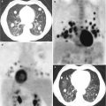
Fig. 5.1
In this picture, it is possible to see CTCs with different morphology detected with CellSearch platform. CTCs are immune-magnetically enriched with a specific antibody for epithelial cell adhesion molecule (EpCAM) coupled with ferrofluid. In a second step, the enriched cells were stained with a nucleic acid dye, DAPI (in purple), and a monoclonal antibody directed against cytokeratins (CK) 8, 18, and 19 (in green)
Currently there are many methods in order to isolate and detect CTCs, follow you find an overview of strategies used to capture CTCs and specific examples from every kind (see Table 5.1).
Table 5.1
Methods for CTC detection
Assay | Enrichment | Detection | Key features |
|---|---|---|---|
CellSearch® | EpCAM mAb coupled ferrofluid | Immunofluorescence: CTC is positive for CKs 8, 18, and 19 and nucleus positive for DAPI negative for CD45 | Semiautomated system with FDA approval for metastatic breast, colon, and prostate cancer. CTC can be enumerated and visualized |
Adna Test | Antibody cocktail (MUC1, EpCAM) coupled microbeads | Molecular biology: RT-PCR positive for at least one of the following markers: MUC1, Her2, EpCAM | This system does not quantify the tumor cell load; false-positive results are due to unspecific amplification |
MACS | EpCAM mAb coupled beads | Microscope visualization: morphology, high surface area to volume | Possibility to positive/negative enrichment |
MagSweeper | EpCAM mAb coupled ferrofluid | Microscope visualization: morphology | High purity can process WB, 9 ml/h throughput |
Ariol system | CK antibodies and EpCAM antibodies coupled to microbeads | Positive markers: CKs | Possibility to detect of EpCAM + and EpCAM- |
CTC-Chip | Microsoft array: EpCAM coupled microspots | Immunofluorescence: CTC is positive for CKs 8, 18, and 19 and nucleus positive for DAPI negative for CD45 | Microspots are optimized for cell-antibody contact, 1–2 ml/h, high detection rate even in M0 patients |
Ephesia | Self-assembly of magnetic beads in columns | Immunofluorescence or immunocytochemistry: CTC is positive for CKs 8, 18, and 19 and nucleus positive for DAPI negative for CD45 | Flexibility with capture antibody |
Isoflux | EpCAM-coated magnetic beads combined with microfluidic processing | Immunocytochemistry for cytokeratin, CD45, and Hoechst | Automated, continuous flow |
CTC iChip® | Magnetic bead capture combined with microfluidic inertial focusing | Immunocytochemistry or RT-PCR | Positive/negative enrichment, remove nucleated cells from whole blood by size-based deflection by using a specially designed array of posts performed in CTC-iChip1, inertial focusing to line up cells to prepare for precise magnetic separation and magnetophoresis for sensitive separation of bead-labeled WBCs and unlabeled CTCs |
GILUPI cell collector | Functionalized EpCAM-coated medical wire | Immunocytochemistry for EpCAM, cytokeratin, and DAPI | In vivo collection |
Ficoll-Paque® | Density | Immunocytochemistry | Inexpensive, easy to use |
OncoQuick | Density/size | Immunocytochemistry/RT-PCR | Density gradient centrifugation with OncoQuick results in higher relative tumor cell enrichment than Ficoll density gradient centrifugation |
ISET® | Filtration based on cell size | Immunocytochemistry/FISH | Epithelial and mesenchymal tumor cells can be isolated |
ScreenCell® | Filtration based on cell size | Immunocytochemistry/FISH | Epithelial and mesenchymal tumor cells can be isolated |
VyCAP | Filtration based on cell size | Filtration based on cell size | Epithelial and mesenchymal tumor cells can be isolated |
Dean flow fractionation | Size-based selection using centrifugal force | Immunocytochemistry for cytokeratin, EpCAM, CD45, and Hoechst | Non-epithelial cells can be isolated |
Dielectrophoretic field-flow fractionation | Membrane capacitance | Immunocytochemistry | CTCs selected are viable |
DEPArray™ | Enables movement of cells within chip by electric field changes | Fluorescence imaging | Requires pre-enrichment step/isolation of purified single cells for downstream analysis |
ApoStream® | Dielectrophoretic technology in a microfluidic flow chamber | Fluorescence imaging | Isolation of purified single cells for downstream analysis |
EPISPOT assay | CD45 depletion and short-term culture in plates coated in antibody against MUC-1, PSA, or cytokeratin-19 | Immunofluorescence secondary antibodies to MUC-1, PSA, or cytokeratin-19 | Detection of only viable CTCs |
Vita-AssayTM or Collagen Adhesion Matrix (CAM) technology | Density gradient centrifugation and cells applied to CAM for short-term culture | Immunocytochemistry for cell-surface markers | Detection of only viable CTCs with the invasive phenotype |
Methods that use immunoaffinity purification strategy have proven to be an efficient way to capture CTCs and for this is the most widely used. They typically use anti-EpCAM antibodies but also other antibodies that recognized tumor-associated antigen, acting as capturing elements for CTCs from human whole blood. The main example is the CellSearch platform, but there are also CTC-chip, an array of 78,000 microspots coated with anti-EpCAM antibodies, Adna Test and Mag-Sweeper Isoflux that use a cocktail of antibodies specific to kind of cancer, and the GILUPI CellCollector® that is the first in vivo CTC isolation product worldwide which is CE approved. This device resembles a venous blood withdrawal. The GILUPI CellCollector® is placed directly into the bloodstream of a patient via an indwelling catheter (size 20 G, pink), remains in the arm vein for 30 min, and thus enables the capture of a large number of CTCs in vivo [37].
It is also known that tumor cells are a heterogeneous population, and EpCAM is not constantly expressed on them. Furthermore, it has been noted that circulating tumor microemboli (CTM) or CTCs with epithelial mesenchymal transition (EMT) which are attracting attention these years show no or weak expression of EpCAM, and therefore they are not detectable by the method above. For this reason, methods to isolate CTCs based on their physical properties, including density, size, deformability, and electrical properties have been developed.
Some groups use density gradient centrifugation methods for separating CTCs in mononuclear fraction based on cell density as centrifugation with Ficoll-Paque solution or OncoQuick (combine a porous filter for size-based separation in conjunction with gradient centrifugation). The isolation is in general followed by an RT-PCR specific for CK. The most promising method is leukapheresis in which white blood cells are separated from a sample of blood. In this way, a large volume of patient’s blood could be analyzed for CTCs; the result is an improvement in the number of CTCs isolated and in sensitivity for downstream analysis and characterization.
Microfiltration and microfluidics are also employed: with microfiltration CTCs are retained on the basis of size, assuming that CTCs are larger than leukocytes. The two main techniques are ISET [38] that uses a polycarbonate filter with 8 μm diameter circular pores for CTC enrichment and ScreenCell that uses circular track-etched filters; the pores’ range is 7.5–6.5 μm. This methods’ advantage is that CTCs can be isolated as living cells without fixation. Nowadays, inexpensive and convenient devices are available, but they are disadvantageous in that the blood samples have to be isolated in a short time after drawing. Recently De Wit and colleagues [39] were able to isolate CTCs onto a silicon membrane with 5 μm diameter circular pores. Using microfluidic tool to retain CTCs, the size and deformability of these cells can be explored.
The dielectrophoresis (DEP) exploits the electrical properties of CTCs, to discriminate them from leukocytes by applying a nonuniform electric field. Gupta and colleagues developed ApoStream instrument for file flow fractionation [40], and Manaresi and colleague [41] developed DEPArray, based on a microfluidic cartridge that contains an array of individually controllable electrodes, each with embedded sensors. This circuitry enables the creation of dielectrophoretic (DEP) cages around cells. After imaging, individual cells of interest are gently moved to specific locations on the cartridge, e.g. for cell-cell interaction studies or into the holding chamber for isolation and recovery.
Related posts:
 Markers of Prostate Cancer: The Role of Circulating Tumor Markers in the Management of Bone Metastases
 Bone Metastases from Prostate Cancer: Radiotherapy
 Biology and Pathophysiology of Bone Metastasis in Prostate Cancer
 Bone Homing and Metastasis
 Imaging of Glycolysis with 18F-FDG PET
 Surgery: Treatment of Oligometastatic Disease
Stay updated, free articles. Join our Telegram channel

Full access? Get Clinical Tree


