Figure 21.1 Bilateral facial erysipelas. Note discrete, raised edge of erythema.
Because erysipelas can produce lymphatic obstruction, it tends to recur in areas of earlier infection. Such recurrences are the most common complication, occurring in approximately 30% of cases. Other complications, including progression to cellulitis or sepsis, are uncommon and are usually restricted to patients with significant comorbid conditions.
Microbiology
Most cases of erysipelas are caused by β-hemolytic group A streptococci (GAS), including Streptococcus pyogenes. Less often, groups C, D, and G streptococci are the causative organisms in adults. Group B streptococcus is often the cause of erysipelas in newborns and postpartum women. Bullae formation may indicate primary or secondary infection with Staphylococcus aureus, including strains of methicillin-resistant S. aureus (MRSA). Infrequent causative agents include Pneumococcus species, Klebsiella pneumoniae, Yersinia enterocolitica, and nontypeable Haemophilus influenzae.
Diagnosis and differential diagnosis
Characteristic skin disease usually suggests the diagnosis. Swabs for Gram stain and culture from a suspected portal of entry may be helpful but are usually not necessary. Skin biopsy for tissue culture and the injection-reaspiration method of tissue fluid collection both yield poor results and have little diagnostic value. Blood cultures are positive in only 5% of patients. Routine lab tests are usually unrevealing, but some patients will have leukocytosis.
Differential diagnosis, especially for patients with facial erysipelas, includes allergic or photosensitive contact dermatitis, but fever, pain, and leukocytosis do not occur. Autoimmune diseases, including systemic lupus erythematosus and dermatomyositis, often exhibit facial rash and fever, but they evolve slowly, have other clinical findings, and are typically bilateral, less sharply demarcated, and lack intense erythema. Other diagnostic considerations include erythema infectiosum, Sweet’s syndrome, angioedema, and erysipeloid.
Cellulitis
Clinical manifestations
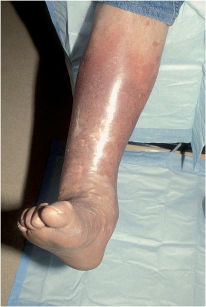
Cellulitis is an infectious inflammation of the deep dermis with variable extension into the subcutaneous tissues. Clinically, cellulitis typically presents as an ill-defined, erythematous firm plaque occurring in conjunction with the four cardinal signs of inflammation: rubor, calor, dolor, and tumor (Figure 21.2). Affected patients often have associated fever, chills, and malaise. Severe disease and signs of clinical toxicity mandate a search for systemic infection.

Figure 21.2 Right leg cellulitis. Note irregular margin.
In healthy patients, antecedent trauma often produces a defect in the skin barrier leading to cellulitis, in contrast to a bloodborne route in immunocompromised patients. If the leg is affected, interdigital tinea pedis is often the portal of entry. Other predisposing factors include peripheral vascular disease, alcoholism, intravenous drug abuse, malignancy, diabetes, or a concomitant skin disease such as stasis dermatitis or ulceration of any type.
Fever, lymphangitis, regional lymphadenopathy, focal abscess formation, and bullae may accompany cellulitis. In addition, necrosis of the skin and subcutaneous tissues may be a complication. Sepsis is a more common complication in children or immunocompromised adults. If the causative organism is GAS, cellulitis may lead to acute glomerulonephritis and a streptococcal toxic shock-like syndrome.
Microbiology
Streptococcus pyogenes and S. aureus, including MRSA, are the most frequent causes of cellulitis. Infection with MRSA typically presents with purulence, either primary or secondary. Other specific causes of cellulitis should be considered in particular clinical situations or because of certain patient exposures; these are listed in Table 21.1.
| Type/scenario | Likely organism |
|---|---|
| Postsurgical cellulitis | Staphylococcus aureus |
| Perianal cellulitis | Group A streptococcus |
| Preseptal cellulitis | S. aureus, S. pyogenes |
| Orbital cellulitis | S. aureus, Streptococcus pneumoniae |
| Facial cellulitis | Haemophilus influenzaea |
| Neonatal cellulitis | Group B streptococcus |
| Crepitant cellulitis | Clostridium species |
| Salt water exposure | Vibrio vulnificus |
| Freshwater exposure | Aeromonas hydrophilia |
| Hot-tub exposure | Pseudomonas aeruginosa |
| Soil exposure | Clostridium species |
| Dog/cat bite | Pasteurella multocida, Capnocytophaga canimorsus |
| Human bite | Eikenella corrodens |
| Immunocompromised | Often mixed infection with gram-negative organisms, fungi |
| Nosocomial | Pseudomonas |
| Handling of raw poultry, meat | Erysipelothrix rhusiopathiae |
| Handling of raw fish | Streptococcus iniae, Erysipelothrix rhusiopathiae |
| Intravenous drug use | S. aureus |
a Less common since advent of vaccine
Diagnosis and differential diagnosis
Stay updated, free articles. Join our Telegram channel

Full access? Get Clinical Tree


