Case 91
Presentation
A 47-year-old menopausal woman presents to your office with dyspnea, progressive abdominal distention and discomfort, anorexia, fatigue, lower extremity swelling, and weight loss over 8 weeks. Past medical history is significant for her having had a total abdominal hysterectomy (for fibroids) and bilateral salpingo-oophorectomy several years ago. She is on no medications, has never smoked, and drinks alcohol on rare occasions. Her family history is significant for a maternal aunt with breast cancer diagnosed at age 60, and a father who died from non-small cell lung cancer at age 72. Examination is significant for cachexia and ascites, with no palpable adenopathy or masses. Breast, pelvic, and rectal examinations are unrevealing.
Complete blood cell count, comprehensive metabolic profile, and urinalysis are all normal, except for the following values: platelets 748 K/uL (normal range for institution, 150 to 375) and alkaline phosphatase 144 U/L (normal range, 38 to 126).
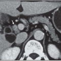
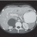
Chest x-ray reveals minimal to moderate bilateral pleural effusions, and several subcentimeter-sized nodules in the right lower and left upper lobes. Abdominal ultrasonography shows massive ascites, no organomegaly, and multiple hepatic lesions. Subsequent computed tomography (CT) scans of the chest, abdomen, and pelvis show pulmonary nodules as previously described, moderate bilateral pleural effusions, and no mediastinal or hilar adenopathy. The CT scans also show multiple hepatic lesions (ranging in size from 1 to 3 cm), omental thickening and nodularity, retroperitoneal adenopathy, ascites, and sclerotic lesions in the thoracic and lumbar spine on bone windows. Kidneys, adrenal glands, spleen, stomach, pancreas, and small and large bowel all appear normal.
Differential Diagnosis
In this clinical scenario, cancer should be considered as the primary diagnosis until proven otherwise. In a young, previously healthy woman with evidence of likely widespread metastases involving lung, liver, omentum, nodes, peritoneum, and bone, potential primary sites include ovarian, peritoneal, gastrointestinal (colorectal, pancreatic, gastric), renal, breast, lung, lymphoma, melanoma, and germ cell tumors, among others.
Recommendation
Open biopsy of omental, nodal, or hepatic lesion(s) with complete morphologic and immunohistochemical analysis including evaluation for estrogen and progesterone hormone receptors. Additionally, this patient should have a mammography and positron emission tomography (PET) scanning, if possible, to help rule out potential primary sites. As well, upper and lower endoscopy should be considered if feasible and if it does not delay other evaluation and treatment. Head imaging (CT or magnetic resonance imaging [MRI]) should be performed to rule out brain involvement and the need for prompt surgical and/or radiation intervention. Additional laboratory evaluation should include serum human chorionic gonadotropin (HCG) and alpha-fetoprotein (AFP) to help exclude a primary germ-cell tumor. Additional tumor markers are unlikely to be helpful in diagnosing and confirming a primary site, but may be useful as baseline values in following disease response with eventual treatment. These markers include CA 125, carcinoembryonic antigen (CEA), CA 19-9, CA 15-3 or CA 27, 29, and lactate dehydrogenase (LDH). In this patient, it may also prove useful to review the operative and pathology reports from her hysterectomy
and oophorectomy from several years ago to rule out any suspicious or premalignant findings.
and oophorectomy from several years ago to rule out any suspicious or premalignant findings.
Case Continued
A biopsy of an omental mass is performed by laparotomy without complication. No additional information is obtained at surgery to help confirm a primary site. Additional examination yields no new suspicious findings. She reports having recently had “normal” gynecologic evaluations, mammography, esophagogastroduodenoscopy, and colonoscopy in the last 2 years.
Pathology is consistent with a poorly differentiated adenocarcinoma. Complete immunohistochemical analysis is nonspecific for a primary tumor site. Immunohistochemistry is positive for cytokeratin-7, and TTF-1 and negative for CD-20, S-100, CD-117 (or C-KIT), Mart-1, hormone receptor, and neuroendocrine markers.
HCG and AFP are within normal limits. Abnormal tumor markers otherwise include LDH of 670 U/L (reference range, 313 to 618); CEA of 68 ng/mL (reference value <2.5 for a nonsmoker); CA -125 of 687 U/mL (reference value <35); and CA 19-9 of 5,395 U/mL (reference value <37).
PET scanning shows hypermetabolic activity correlating with the lesions seen on CT in the lungs, liver, peritoneum, and bones, including uptake in the right midfemur, right humerus, and lower cervical spine. Head imaging (CT with contrast) reveals no evidence of metastases.
Diagnosis and Recommendation
The diagnosis is carcinoma of unknown primary site. Despite a thorough evaluation, no specific finding(s) definitively establish a primary diagnosis. Therapeutic paracentesis and bilateral thoracenteses should be offered for symptomatic relief (of abdominal discomfort, dyspnea). Systemic chemotherapy, ideally as part of a clinical trial, should be offered.


