Case 75
Presentation
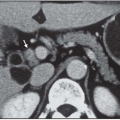
A 45-year-old woman with a history of node-negative breast cancer undergoes a computed tomography (CT) scan as part of her routine follow-up. The CT scan was supposed to be of the chest, but the upper abdomen was imaged and a 6-cm tumor was seen in the right adrenal gland. Her physician ordered a magnetic resonance imaging (MRI) scan, which further delineated a 7-cm tumor in the right adrenal gland. She is referred to you for management of this incidentaloma. She is not cushingoid, nor does she have any evidence of virilization. Her pulse equals 100 beats per minute and blood pressure is 145/95 mm Hg. She is not taking any blood pressure medications. She does not have headaches or spells of nervousness.
Differential Diagnosis
The differential diagnosis includes benign and malignant adrenal medullary and cortical tumors as well as metastatic breast cancer to the adrenal gland.
The workup of an incidentaloma is designed to address two issues that may require surgical intervention. First is the presence of a hormonally functional adrenal tumor. Second is whether or not the adrenal tumor is malignant. Hormonally functional tumors include those with excessive secretion of aldosterone, cortisol, male or female hormones, and catecholamines. Aldosteronoma is excluded by measuring serum levels of potassium (K+), as long as the patient is not taking any blood pressure medications. If the serum K+ is >3.5 mEq/L, aldosteronoma is excluded. This patient had a serum K+ of 4 mEq/L. Hypercortisolism can be identified by its signs and symptoms. Moreover, 24-hour urine levels of hydrocortisone or free cortisol should be measured before and following low-dose dexamethasone. Patients with Cushing syndrome will not suppress levels following low-dose dexamethasone, while normal individuals will. This patient did not have elevated urinary levels of free cortisol and reduced levels after dexamethasone. Finally, pheochromocytoma should be excluded by measuring a 24-hour urine concentration of vanillylmandelic acid (VMA), metanephrine, normetanephrine, and total catecholamines. A newer study designed to rule out pheochromocytoma is measurement of blood levels of plasma-free metanephrine and normetanephrine which have an accuracy of over 95%. This patient had elevated levels of plasma-free normetanephrine and elevated urinary levels of VMA and total catecholamines consistent with the diagnosis of pheochromocytoma.
The issue of malignancy is determined primarily by the size of the adrenal tumor. A critical criterion for the diagnosis of adrenal cortical cancer is tumor weight >100 g, which equals a diameter of 6 cm. Tumors that are >4 cm in diameter should be removed based on the possibility that they may be malignant. Smaller tumors that are nonfunctional should be re-imaged by CT or MRI in 6 months; if they have increased in size, they should be removed. For this patient with a history of breast cancer, if the biochemical studies exclude pheochromocytoma and there is no other site of disease, the tumor could be aspirated to diagnose breast cancer metastases to the adrenal. Resection of metastatic cancer to the adrenal may be indicated if there is a long disease-free interval and it is the sole site of tumor. Beware of needle aspiration of unexpected pheochromocytoma because sudden death has been reported in this context.
Stay updated, free articles. Join our Telegram channel

Full access? Get Clinical Tree


