Case 74
Presentation
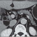
A 41-year-old woman is admitted to your hospital with hypertension and severe headache. Workup shows an increased level of noradrenaline in urine, 15 times greater than the reference level. Computed tomography (CT) scan reveals a large tumor close to the right adrenal expanding into the hilus of the liver.
Differential Diagnosis
This patient has a large tumor in close proximity to the liver and the right adrenal. The tumor produces noradrenaline and dopamine. It is evidently a paraganglioma; whether it is adrenal or extra-adrenal may be difficult to discern, but magnetic resonance imaging (MRI) is often helpful. It is hard to know preoperatively if this tumor is malignant, but the location and size strongly suggest a malignant tumor.
Recommendations
Patients with catecholamine-producing tumors are usually advised to have preoperative treatment with alpha-receptor blocking agents to reduce the vascular sensitivity to circulating catecholamines. Phenoxybenzamine 30 to 200 mg daily has been widely used. Doxazosin 4 to 12 mg can also be recommended. Other agents also have been used. So far, there are no
studies done to compare the efficiency of various types of drugs used for preoperative treatment.
studies done to compare the efficiency of various types of drugs used for preoperative treatment.
Case Continued
For alpha-receptor blockade, you chose to use phenoxybenzamine. This patient is treated with phenoxybenzamine 160 mg daily, which results in an orthostatic reaction and a stuffy nose. This is a high dose expected to cause hypotension when the tumor has been removed.
▪ Surgical Approach
The patient is explored through a thoracoabdominal incision. During the operation, periods of hypertension are treated with adenosine infusion, and similarly, periods of hypotension are treated with noradrenaline infusion. The venous drainage is carefully isolated and ligated in continuity and divided. Next, the arterial blood supply is controlled. Following this, the tumor is resected en bloc.
Case Continued
The tumor is 10 cm in diameter, highly vascularized, and difficult to resect. The perioperative blood loss was 6 liters. Postoperatively, the patient spends 48 hours in intensive care and is discharged from the hospital on postoperative day 15. The resected specimen is sent to pathology.
▪ Perioperative Image
Perioperative Report
Stay updated, free articles. Join our Telegram channel

Full access? Get Clinical Tree


