Case 60
Presentation
A 64-year-old man presents to your office complaining of a 7-month history of a lump in the left breast, with a bloody discharge from the left nipple that occurs with mild pressure noted over the last 2 months. He has no significant past medical history. He is a nonsmoker and is not receiving hormone therapy for any disease. He has no family history of breast carcinoma. On physical examination, a retroareolar hard, irregular mass fixed to the thoracic wall is evident. No skin retraction can be detected. You observe a bloody discharge from the left nipple with mild pressure. No enlarged axillary nodes are palpable.
Differential Diagnosis
The differential diagnosis includes several conditions of breast mass; the most frequent is benign breast hypertrophy during adolescence. This condition develops around puberty or some years later than male breast carcinoma (mean age of male breast cancer, 64 years), and usually presents as a discoid mass under the areola. The young age of the patient and the well-delimited, mobile lump help to make the correct diagnosis.
Gynecomastia, which affects men in the second and third decades of life, is a diffuse enlargement of the breast gland of soft consistency, with characteristics similar to the small female breast. Male breast hypertrophy, which may be idiopathic or related to liver malfunction, can present as a round, mobile, sometimes painful breast mass under the areola.
Male breast carcinoma is so unusual under 30 years of age that there is no indication for biopsy in a clinically benign breast mass in this subgroup of men.
In patients older than 50 years, breast carcinoma should be differentiated from hypertrophy related to different clinical conditions or to medications, which interact with hormonal functions. Benign hypertrophy is usually an elastic, sharp, and regular mass under the areola.
Recommendation
Mammogram, breast ultrasound, and magnetic resonance imaging (MRI) for further characterization are recommended. Because the patient is older than 50 years, and the breast lump has suspicious clinical features, fine-needle aspiration biopsy (FNAB) is recommended to establish a tissue diagnosis.
▪ Mammogram
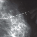
Mammography Report
A well-defined density (arrow) is seen in the retroareolar region of the left breast.
▪ Ultrasound and Doppler Analysis
Ultrasonography and Doppler Analysis Report
Ultrasound reveals a 1.9-cm complex lesion in the left breast with irregular margins. Enhanced vascular flow is noted on Doppler analysis.
▪ MRI
MRI Report
MRI demonstrates a 19 × 18 × 18-mm cystic mass with high T1 and T2 signals containing an irregular, 11- to 12-mm, enhancing intracystic solid component and septation. Kinetic assessment demonstrates rapid initial rise with washout of the solid intracystic component. The appearance is highly suspicious for a cystic malignancy.


