Case 58
Presentation
A 60-year-old woman presented with an itching right nipple, with some crust, redness, and occasional bloody discharge. This abnormality has developed gradually over the past 5 months. An ointment containing corticosteroids, prescribed by the family practitioner about 2 months before, did not improve the symptoms. The family practitioner referred the patient to your breast clinic for further evaluation.
History reveals no specific breast complaints or symptoms, previous surgery, or mammographic screening. There is no family history of breast cancer. The patient has had psoriasis for many years, but is otherwise healthy.
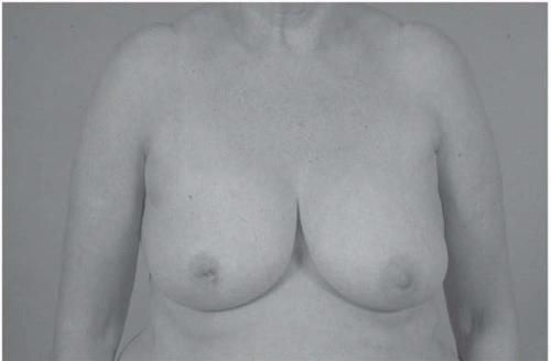
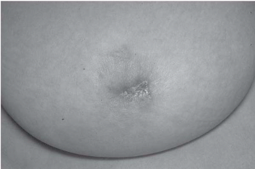
▪ Clinical Photographs
Physical Examination Report
On physical examination, the right nipple shows a crusted, eczematous, elevated redness over an area of about 2 cm. The areola feels somewhat indurated without any lumps or firm tissues in the retroareolar region. The remaining breast tissue is unremarkable without suspicious enlarged axillary lymph nodes.
Differential Diagnosis
Paget disease of the nipple is suspected. Nipple adenoma, papillomatosis, invasive ductal carcinoma, squamous cell carcinoma, eczema of the nipple, and chronic infection or fistulae by duct ectasia cannot be excluded on clinical grounds.
Discussion
Paget disease of the nipple is an uncommon manifestation of breast carcinoma, representing approximately 1% to 3% of all cases. Patients present clinically with eczematous changes of the nipple occasionally associated with itching, ulceration, and bleeding. Histologically, Paget disease is characterized by intraepidermal spread of large round or ovoid tumor cells with abundant pale cytoplasms and large pleomorphic and hyperchromatic nuclei with prominent
nucleoli. Regarding the exact origin of these Paget cells, currently most authors postulate the epidermotropic theory, which assumes that Paget cells are ductal carcinoma cells that have migrated from the underlying mammary ducts to the epidermis of the nipple. This theory is supported by the presence of underlying ductal carcinoma in situ (DCIS) or invasive breast carcinoma in nearly all patients (97%).
nucleoli. Regarding the exact origin of these Paget cells, currently most authors postulate the epidermotropic theory, which assumes that Paget cells are ductal carcinoma cells that have migrated from the underlying mammary ducts to the epidermis of the nipple. This theory is supported by the presence of underlying ductal carcinoma in situ (DCIS) or invasive breast carcinoma in nearly all patients (97%).
Paget disease can only be diagnosed with a punch (2 to 3 mm) biopsy. Punch biopsy is advised when any abnormality of the nipple is encountered. Further, a two-view mammography should be performed to evaluate for any abnormality within the breast parenchyma, such as microcalcifcations that could be associated with DCIS, or invasive cancer. Spot magnification view of the retroareolar region may be helpful for improved definition of the extent of microcalcifications. Ultrasound of the breast may show signs of invasive cancer. If this is encountered, ultrasound-directed core biopsies should be taken to confirm this suspicion. The role of magnetic resonance imaging (MRI) of the breast is not completely clear, but if further extension of the disease in the retroareolar area or further in the breast is suspected, MRI may be helpful to exclude the possibility of invasive cancer.
Case Continued
The following tests are performed: two-view mammography of both breasts and retroareolar spot compression view of the right breast; ultrasound of the retroareolar area of the right breast; and a punch biopsy (3 mm) of the right nipple in the abnormal area.
Stay updated, free articles. Join our Telegram channel

Full access? Get Clinical Tree


