Case 53
Presentation
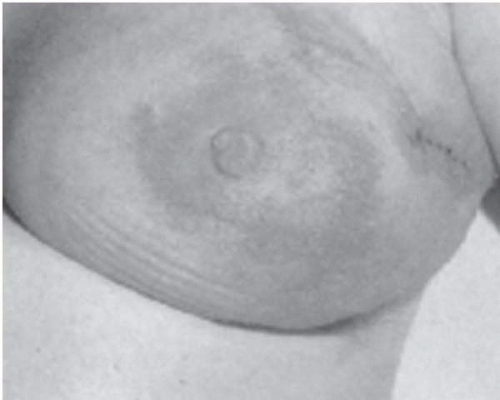
A 55-year-old woman presents with a change in the shape of her left breast and the appearance of her nipple for approximately 2 months. In the 2 weeks prior to her appointment, she noted a pink discoloration of the central portion of her breast. She denies fever, chills, weight loss, or systemic symptoms. On physical examination, the left breast is erythematous with peau d’orange of the central third. There is loss of projection of the nipple and distortion of the inferolateral contour of the breast. There are no palpable axillary, supraclavicular, or infraclavicular lymph nodes. The central portion of the left breast is firm and diffusely thickened throughout the inferior half of the gland. Examination of the left breast is normal.
Differential Diagnosis
The differential diagnosis in this case includes inflammatory breast cancer, breast infection, and fat necrosis.
Discussion
Although inflammatory breast cancer (IBC) is included in the group designated as locally advanced breast cancer (LABC), it has some distinguishing characteristics, which are important both for treatment selection and prognosis. Inflammatory breast cancer represents only 1% to 5% of newly diagnosed breast cancers. This case illustrates the classic clinical signs of inflammatory cancer: an ill-defined mass, due to diffuse infiltration of the breast tissue with tumor, and skin erythema and edema (the classic peau d’orange), caused by obstruction of the dermal lymphatics with tumor cells. The extent of disease is often underestimated mammographically, with nonspecific signs of asymmetric density and skin thickening, as in this case. These features, coupled with the absence of an obvious breast mass, often result in confusion with breast infection. Patients with IBC are often initially prescribed antibiotics for a suspected breast infection, which does not resolve with treatment. There are several other infectious processes with similar clinical signs and symptoms, which must be distinguished from IBC. Periductal mastitis can occur in a single duct or in multiple ducts, and can present with erythema and tenderness in the skin overlying the involved duct. Breast abscess may have clinical findings that are very similar to the clinical features of IBC. In general, IBC is distinguished from infection by the absence of pain and fever. If abscess is suspected, it can be diagnosed by ultrasound examination and
aspiration of purulent fluid from the mass. In patients suspected of having mastitis, a 1-week trial of antibiotics can be considered, but unless the clinical examination reverts to normal, a biopsy is indicated.
aspiration of purulent fluid from the mass. In patients suspected of having mastitis, a 1-week trial of antibiotics can be considered, but unless the clinical examination reverts to normal, a biopsy is indicated.
Trauma to the breast can produce fat necrosis or hematoma with surrounding inflammation, which occasionally is difficult to distinguish from inflammatory cancer. Patients who have had a previous breast or thoracic cancer and have received radiation therapy can have persistent erythema and skin edema lasting several years. This diagnosis should be easily made on the basis of the history. Edema of the breast can occur in patients with congestive heart failure or nephrotic syndrome who have generalized edema, but inflammatory changes and thickening of the breast parenchyma are not usually present. These conditions can usually be distinguished by a thorough history and physical examination. Definitive diagnosis may require a tissue biopsy, particularly in the case of fat necrosis.
Histologically, IBC is most commonly high-grade, poorly differentiated, estrogen-receptor (ER) and progesterone-receptor (PR) negative, infiltrating ductal carcinoma without a propensity for any particular subtype. Mutations of p53 and epidermal growth factor receptor are often present. The presence of tumor cells in the dermal lymphatics provides pathologic confirmation of the diagnosis of IBC, but the diagnosis can also be made on the basis of clinical signs of diffuse erythema and edema in the presence of a tumor in the breast parenchyma.
Recommendation
Bilateral mammogram, ultrasound scan, and full-thickness skin biopsy in the edematous/erythematous region.
Case Continued
The patient is unable to tolerate breast compression; therefore, a magnetic resonance imaging (MRI) study of the breast is obtained.
Stay updated, free articles. Join our Telegram channel

Full access? Get Clinical Tree


