Case 46
Presentation
A 66-year-old woman with no significant past medical history presents with a 3-day history of right upper quadrant pain. She is referred by her primary medical doctor. Physical examination reveals no jaundice, anemia, lymphadenopathy, or specific abdominal findings.
Ultrasonogram
Ultrasonography Report
Ultrasonography shows a sessile protruding mass (arrowheads), measuring 3.5 × 3.0 cm, in the fundus of the gallbladder, which does not involve the liver. A gallstone is present in the gallbladder lumen (not shown).
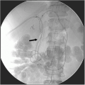
Endoscopic Retrograde Cholangiogram
Differential Diagnosis
There are a variety of tumors or tumor-like lesions arising in the gallbladder, which often present as protruded (polypoid) lesions. Epithelial tumors include carcinoma, dysplasia, and adenoma, whereas tumor-like lesions comprise hyperplasia, adenomyomatosis, polyps, and others. Among protruded lesions of the gallbladder, the cholesterol polyp is the most common. Once protruded lesions are detected on imaging, the discrimination between benign and malignant is clinically important, and it mainly depends on the size of the lesion. Most protruded lesions smaller than 1 cm in maximum diameter are benign. In contrast, many cancers appear as large polypoid lesions, and those exceeding 1.5 cm in maximum diameter are very likely to be malignant. The shape of the lesion is another useful hint when discriminating benign from malignant lesions. Carcinoma is usually sessile (with the exception of carcinoma in pedunculated adenoma), while most benign lesions are pedunculated.
In this patient, with a sessile protruded lesion far exceeding 1.5 cm in the gallbladder, one must consider gallbladder carcinoma as the primary diagnosis. The irregular (granular) contour of the lesion on cholangiography (Fig. 46.2) also supports this diagnosis.
▪ CT Scan
CT Scan Report
A 3-cm mass with contrast enhancement is depicted in the fundus of the gallbladder (not shown). It does not involve the adjacent organs, including the liver. There are neither liver metastases nor peritoneal seeding. A 2 × 1-cm soft tissue mass with heterogeneous contrast enhancement (arrow), suggestive of a positive lymph node, is located on the right side of the main portal vein.
Diagnosis and Recommendation
Gallbladder carcinoma. A computed tomography (CT) scan of the abdomen is recommended for staging of the tumor.
▪ Approach
Although gallbladder carcinoma has been considered to be a highly lethal disease, resection provides the only chance for a cure. The outcome of resection depends on the extent of the primary tumor. The Tis or T1 tumor is designated as early carcinoma and bears a favorable prognosis after resection (cumulative 5-year survival rate >90%). The outcome of radical resection of a T2 tumor is better
(cumulative 5-year survival rate >50%) than the poorer resection outcomes of T3 or more advanced tumors.
(cumulative 5-year survival rate >50%) than the poorer resection outcomes of T3 or more advanced tumors.
Stay updated, free articles. Join our Telegram channel

Full access? Get Clinical Tree


