Case 45
Presentation
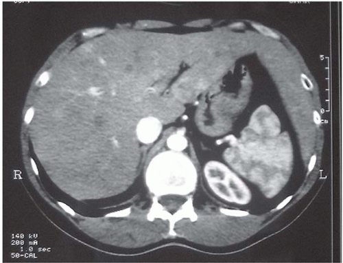
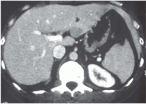
A 40-year-old man with no past medical history is admitted for long-standing pain in the left upper quadrant. He is not a smoker or a drinker. Physical examination is unremarkable; the patient was in a good condition with normal blood pressure and stools. There is no sign of portal hypertension. Abdominal ultrasound shows hepatomegaly with several hypoechoic nodules.
Differential Diagnosis
Differential diagnoses for intrahepatic tumors include hemangiomas, hepatocarcinomas, metastases of thyroid and kidney cancers, multiple nodular hyperplasias, and polyadenomas, even when there is no washout.
Case Continued
Esophagogastric endoscopy and colonoscopy are normal. Similarly, plasma levels of gastric vasoactive intestinal peptide (VIP) and calcitonin are normal. Levels of plasma serotonin and urinary 5-hydroxyindoleacetic acid (5-HIAA) are one and a half and two times normal, respectively, on two successive
assays. Levels of carcinoembryonic antigen (CEA) and cancer antigen (CA) 19-9 are normal.
assays. Levels of carcinoembryonic antigen (CEA) and cancer antigen (CA) 19-9 are normal.
At this stage, we suspect the diagnosis of liver metastases of a carcinoid tumor located in the appendicular region (suspicion of a positive lymph node in the right inferior quadrant).
A CT-scan-guided biopsy leads to the diagnosis of well-differentiated endocrine (carcinoid) tumor.
▪ MRI
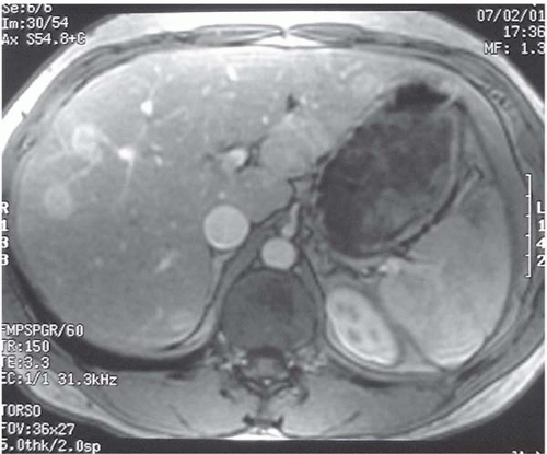
MRI Report
Magnetic resonance imaging (MRI) of the liver did not show the classic bulb sign, which would favor hemangioma, but disclosed more intrahepatic lesions in the right lobe.
▪ Octreotide Scintigram
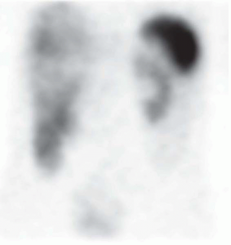
Octreotide Scintigraphy Report
There is increased uptake of radionucleotide by the terminal ileum in the location of the primary tumor, and uptake is revealed in other areas. There are several ileal and lymphatic foci in the right iliac region; multiple foci are identified in the liver.
Stay updated, free articles. Join our Telegram channel

Full access? Get Clinical Tree


