Case 40
Presentation
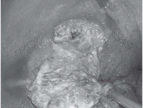
A 52-year-old man presents with perirectal pain, firm area, and bleeding that has worsened over the last 6 months. He was treated for a “boil” on the right anterior cheek 1 year ago. He had a hemorrhoidectomy 5 years ago. He has a 30-pack-year smoking history, and uses alcohol socially.
Differential Diagnosis
At the time of initial presentation with suppuration, a chance to make an early diagnosis with a biopsy was probably missed. Given the appearance, a neoplastic process is most likely a squamous or basal cell carcinoma. Extensive hidradenitis or Crohn disease are less likely.
Discussion
Anal margin cancers arise from skin lateral to the intersphincteric groove and are usually well-differentiated, keratinized variants of squamous cell carcinoma, which rarely metastasize, compared to anal canal cancers. Other lesions in this area are Bowen disease, basal cell carcinoma, melanoma, verrucous carcinoma, and Paget disease. Bowen disease and Paget disease can present as a persistent pruritic rash. Basal cell carcinoma has raised edges with central ulceration and is often mistaken for a fissure or chancre. Verrucous carcinoma may present as a slowly growing invasive cancer in long-standing large condyloma acuminata. Melanomas in the perianal area are frequently amelanotic and carry a very poor prognosis, with most patients succumbing to distant disease. The differential diagnosis includes numerous benign diseases and dermatoses like fungal infection and psoriasis. The importance of a high index of suspicion is critical to prevent the frequent delay in diagnosis. A biopsy is diagnostic.
Recommendation
Perform examination under anesthesia to evaluate the extent of disease and obtain biopsy for tissue diagnosis. Obtain computed tomography (CT) scans of abdomen and pelvis for staging purposes.
Case Continued
The patient is taken to the operating room for examination under anesthesia because pain and tenderness preclude satisfactory examination in the clinic. CT scans of the abdomen and pelvis with intravenous (IV) and rectal contrast did not show any liver lesions or pelvic or groin lymphadenopathy.
▪ Surgical Approach
Stay updated, free articles. Join our Telegram channel

Full access? Get Clinical Tree


