Case 33
Presentation
A 52-year-old healthy man complains of recurrent colicky abdominal pain following an infectious gastroenteritis 6 months ago, and reports hematochezia, mucinous diarrhea, and changes in stool habit for several weeks. His medical history includes a transurethral resection of a urinary bladder tumor 7 years ago. Current nicotine abuse has been present for many years. Family history is devoid of colorectal cancer (CRC). Abdominal and rectal examinations are unremarkable.
Differential Diagnosis
A history of changes in bowel habit associated with hematochezia in a 52-year-old patient is highly suspicious for CRC. However, hematochezia may be the dominant symptom in several other diseases, such as inflammatory bowel disease, infectious colitis, telangiectasias, diverticular disease, and hemorrhoids, which must also be considered. Changes in stool habit, especially the presence of pencil-shaped stool, may indicate advanced colonic obstruction and may be accompanied by colicky pain.
Recommendation
Colonoscopy, including biopsies.
Case Continued
Colonoscopy reveals a circular stenosis caused by a rectal mass 15 cm from the anal verge. The tumor shows firm central parts with bleeding ulcerations and peripheral areas with softer adenoma-like growth patterns. The stenosis cannot be overcome and colonoscopy is incomplete. The exact distance from the lower border of the tumor to the anal verge is determined by rigid proctoscopy and is found to be 14 cm. Histologic examination of the biopsies reveals a moderately differentiated colorectal adenocarcinoma.
Discussion
The risk for developing CRC rises exponentially with increasing age, starting at about age 45 and ending with an incidence of 400 to 500 in 100,000 at the age of 80. The average incidence in Western countries of CRC is about 50 in 100,000. More than 90% of CRC cases are thought to be sporadic CRC, and less than 10% are found in the context of hereditary syndromes such as hereditary nonpolyposis colorectal cancer (HNPCC) syndrome, familial adenomatous polyposis (FAP) syndrome, Gardner syndrome, Turcot syndrome, Peutz-Jeghers syndrome, juvenile polyposis, and inflammatory bowel disease (ulcerous colitis and Crohn disease).
Rectal cancer is the most common abdominal malignancy and accounts for 35% to 40% of all large bowel cancers. Rectal bleeding and anemia are the most common symptoms associated with rectal cancer (60%). Changes in bowel habit and stool shape are frequently reported, too, and may be present in up to 40% of patients. Patients may experience constipation in obstructive disease as well as mucous diarrhea from villous components of the tumor. Digital rectal examination is of limited value in proximal rectal cancer, as the investigator cannot reach the tumor. However, digital rectal examination allows exclusion of a tumor within the distal third of the rectum, estimation of the prostate’s size in men (important for rectal cancer surgery), and clinical examination of anal resting and squeeze pressures with regard to postoperative functional
results. Exact measurement of the distance from the tumor to the anal verge is mandatory and is best done by rigid proctosigmoidoscopy. Rectal cancer is defined as a carcinoma within 15 cm from the anal verge, even if only the lower border of the tumor is still within this margin. Therapeutic strategies strongly depend on this definition.
results. Exact measurement of the distance from the tumor to the anal verge is mandatory and is best done by rigid proctosigmoidoscopy. Rectal cancer is defined as a carcinoma within 15 cm from the anal verge, even if only the lower border of the tumor is still within this margin. Therapeutic strategies strongly depend on this definition.
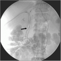
Complete colonoscopy is indicated to exclude synchronous colon cancer (3% to 5%) and to search for underlying large bowel diseases. If the endoscope cannot be passed through the stenosis, as in this case, preoperative double-contrast enema can be performed to exclude additional colonic polyps. However, the stenosis usually prevents a proper large bowel preparation, limiting the diagnostic value of the contrast enema.
Abdominal/pelvic multislice triple-contrast-enhanced computed tomography (CT) or magnetic resonance imaging (MRI) is performed to investigate locoregional tumor spread (i.e., depth of invasion of the primary tumor and presence or absence of enlarged or inhomogeneous lymph nodes) and to scan for liver metastasis as well as signs of peritoneal carcinomatosis (difficult to visualize with CT scan). Additional ultrasonography of the liver may clarify doubtful liver lesions detected on the CT scan. Workup for distant metastasis also includes chest x-ray or chest CT scan. Positron emission tomography (PET) is generally not used in a standard workup, but may be helpful before treatment of distant metastasis. The tumor markers carcinoembryonic antigen (CEA) and cancer antigen (CA) 19-9 should not be used as screening tools, but have proved valuable in monitoring for tumor recurrence during routine follow-up if the markers were elevated preoperatively.
Diagnosis and Recommendation
A proximal rectal moderately differentiated adenocarcinoma. Perform multislice triple-contrast-enhanced CT scans of the pelvis, abdomen, and chest. Determine preoperative CEA level for comparison in follow-up examinations. Perform colonoscopy of remaining colon 3 months postoperatively.
Stay updated, free articles. Join our Telegram channel

Full access? Get Clinical Tree


