Case 25
Presentation
A 62-year-old man presents to the emergency department with diffuse crampy abdominal pain, bloating, nausea, and vomiting, and obstipation for 2 days. His history is notable for vague abdominal pain for the past 2 years for which he has undergone extensive workup, including esophagogastroduodenoscopy (EGD), colonoscopy, and abdominal computed tomography (CT) scan, which were all normal. His primary care physician had empirically started him on a proton-pump inhibitor, and he had been scheduled to undergo an outpatient small bowel follow-through the following month. He also has a remote history of an uncomplicated laparoscopic cholecystectomy.
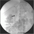
A plain abdominal radiograph of the kidneys, ureter, and bladder (KUB) reveals moderately dilated loops of small bowel with multiple air fluid levels in the left abdomen, indicating ileal versus small bowel obstruction. Abdominal CT scan shows a 4-cm mass involving the mid-ileum with evidence of proximal small bowel dilatation. Visceral angiography showed mild aortic calcification, but no stenosis of the mesenteric vessels.
Differential Diagnosis
The differential diagnosis for small bowel obstruction related to mural thickening includes mesenteric ischemia, a neoplastic process or, less likely, an infectious process. The patient’s lengthy history of vague abdominal pain is characteristic of either of the first two etiologies. Delay in diagnosis of a small bowel tumor is characteristic given the low sensitivities of imaging tests.
Recommendations
Small bowel obstruction from a mechanical cause due to findings on the CT scan of the abdomen is diagnosed. An exploratory laparotomy is necessary after initial resuscitation, placement of nasogastric tube decompression, and Foley catheter for monitoring of urine output.
Case Continued
At operation, an obstructing distal ileal mass is noted along with several firm masses in the mesentery. The small bowel and attached mesentery is resected, and a primary anastomosis is performed. The length of the bowel is inspected, and no further abnormalities are discovered. The liver is likewise palpated with no appreciable masses. The patient tolerates the procedure.
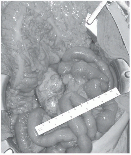
▪ Intraoperative Image
Intraoperative Report
Carcinoid tumor in small bowel visible at base of transverse mesocolon. A 25-cm length of small bowel specimen reveals a 4-cm carcinoid tumor invading the muscularis, mesenteric fat, and serosa. Three of 15 lymph nodes reveal carcinoid metastases.
Discussion
Carcinoid tumors are rare neuroendocrine tumors. In 1907, Obendorfer first described these tumors using the term “carcinoid” to denote their slower rate of growth compared to adenocarcinomas. Carcinoid tumors secrete hormones and biogenic amines, the most common of which is serotonin, but also include histamine, kallikrein, and prostaglandins. These substances are responsible for the symptoms of the carcinoid syndrome: episodic flushing, diarrhea, wheezing, and eventual right-sided valvular heart disease. The liver normally metabolizes serotonin, but once metastases occupy the liver, the hormone is released to systemic circulation and the syndrome may occur. Carcinoid syndrome is the initial presentation in 5% to 10% of carcinoids. Small bowel carcinoid tumors occur most commonly in patients in their sixth or seventh decade of life.
The small bowel is the most commonly affected location, containing 25% of all carcinoids. Carcinoid tumors comprise roughly one third of all small bowel malignancies.
Carcinoid tumors may be multicentric. Patients with small bowel carcinoid tumors can present with protean disease manifestations such as abdominal pain, nausea and vomiting, and weight loss. Patients may present with gastrointestinal bleeding, although this is less common given the deep submucosal location of most tumors and their relatively small size. Most tumors are less than 2 cm in diameter, but mass effects are responsible for most symptoms. Acutely they may cause bowel obstruction, as in this case. Additionally, they may be discovered incidentally or may present as a lead point of small bowel intussusception.
Because of their relatively small size and location, many lesions are difficult to identify with conventional imaging. Reported CT scan sensitivities for detecting primary tumors range from rare to 20%. Enteroclysis similarly has a poor rate of detection. Most small bowel carcinoids are located in the distal ileum, beyond the reach of push enteroscopy. There are little data on the use of magnetic resonance imaging (MRI) for small bowel carcinoids. The sensitivity of capsule endoscopy for detecting these lesions remains to be determined. If the tumors are actively secreting serotonin, an octreotide scan may help to localize them. However, this modality is better suited to identifying hepatic and extra-abdominal metastases. Elevation of urinary levels of 5-hydroxy-indolacetic-acid (5-HIAA), a serotonin metabolite, may be diagnostic.
Stay updated, free articles. Join our Telegram channel

Full access? Get Clinical Tree


