Case 23
Presentation
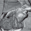
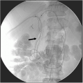
A 55-year-old male with no significant past medical history is referred with the recent diagnosis of anemia. Physical examination is normal with the exception of stools that are positive for occult hemoglobin. Colonoscopy shows only two benign tubular adenomas. An upper gastrointestinal (GI) endoscopy is performed.
Differential Diagnosis
The differential diagnosis for duodenal polypoid lesions includes benign villous adenomas or invasive adenocarcinoma.
Discussion
Villous adenomas, especially those larger than 3 cm, have a malignant potential similar to that of colonic tumors, and total excision is necessary. Up to 50% of such large tumors that are benign on endoscopic biopsy may harbor foci of invasive cancer. Symptoms usually are associated with GI blood loss, although tumors in the periampullary region may obstruct the ampulla of Vater, causing either obstructive jaundice or, rarely, acute pancreatitis. Risk factors for duodenal cancer include familial colonic polyposis syndromes (familial adenomatous polyposis, Gardner’s syndrome) and hereditary nonpolyposis colon cancer (HNPCC).
Duodenal neoplasms may present with symptoms due to GI blood loss or, if circumferential, duodenal obstruction. Lesions in the periampullary area may present with obstructive jaundice.
Recommendation
Endoscopic ultrasound to determine the presence of invasion.
▪ Endoscopic Ultrasound Image
Stay updated, free articles. Join our Telegram channel

Full access? Get Clinical Tree


