Risk factor
Examples
Notes
Comorbid conditions
Diabetes, end-stage renal disease, thyroid disease
Conditions that increase risk of wounds by affecting patient’s immune response, skin integrity or environment risks.
Drugs
Steroids, antimetabolites
Drugs that hinder proliferation of fibroblasts and collagen synthesis.
History of healed ulcer
History of Stage III or IV ulcer
Patient may still have the risk factors that predisposed to these ulcers
Impaired blood flow
Atherosclerosis, lower-extremity arterial insufficiency
Decrease blood flow to wounds for healing
Impaired or decreased mobility and functional ability
Bed bound, decreased lower extremity use, altered mental status (e.g., dementia)
Environmental risk of developing wounds due to increased pressure on skin, friction, or shear from transfer by others
Malnutrition and hydration deficit
Protein–calorie malnutrition and deficiencies of vitamins A, C, and zinc impair normal wound-healing mechanisms.
The poor blood supply seen in peripheral arterial disease, hinders wound healing by depriving the injured area of oxygen and nutrients. Diabetics are at additional risk of foot wounds due to the triad of peripheral neuropathy, microvascular disease, and suboptimal glycemic control. It is estimated that among patients with diabetes, 15 % will develop a foot wound, and 12–24 % of those will eventually require amputation [2, 14]. Even with successful wound healing, the recurrence rate of diabetic foot wounds is 66 % [14].
End stage renal disease and thyroid disease are also risk factors for wound formation. Wounds may occur when medications such as steroids and anti-metabolites hinder proliferation of fibroblasts and collagen synthesis placing a patient at risk [4].
Cognitive impairment, seen in 45–67 % of assisted living residents and 69 % of nursing facility residents, creates an array of risk factors for skin breakdown [15–17]. These include functional disabilities, poor nutritional status, and a higher incidence of skin exposure to pressure, friction, or shear. Moisture-related skin breakdown is often associated with excess perspiration; heavy wound exudates, urine and/or fecal incontinence. Diarrhea is caustic and urine contains urea and ammonia, both of which damage normal skin. With prolonged exposure, these fluids soften the outer protective layer of skin and result in skin breakdown [18]. Data also suggest an association between fecal incontinence and skin ulcer development, likely related to skin exposure to bile acid and gastrointestinal enzymes [19].
The poor nutritional status frequently seen in patients with advanced dementia is another risk factor for wound development. Dehydration, deficiencies of arginine, vitamins A, C, and zinc, as well as protein–calorie malnutrition, have been implicated in wound development and impairing wound healing [20]. In dehydration, skin integrity and wound healing are impaired due to decreased tissue perfusion. Severe protein–calorie malnutrition hinders tissue regeneration, the inflammatory reaction and immune function. Albumin and prealbumin levels are indicators of protein–calorie nutritional status (see chapter “Weight and Nutrition” for further discussion) [18]. Obesity also places patients at risk for skin breakdown under the pannus or in skin folds [21]. The warm and moist environment in skin folds promote the growth of yeast and bacteria [18], that will further increase the risk for wounds to occur.
Assessment
An initial skin evaluation should be performed immediately upon admission of a resident to the LTC facility. An ulcer can develop after only a few hours of pressure. Any discovered wounds should prompt thorough patient assessment and a subsequent treatment plan that includes a timeline for wound reassessment. This assessment involves a complete medical evaluation of the patient including careful attention to conditions that may affect both wound development and healing. A comprehensive wound history should note the onset and duration of a wound, and any previous wound care. Assessment of a person’s cognitive status, behavior, financial resources, and access to caregivers can impact a treatment plan.
Assessment of the patient’s personal environment is also critical. Frequency of repositioning, surfaces, turning schedules, transferring techniques, and durable medical equipment (such as assistive devices, trapeze, bed rails, and padding) can all impact wound development and healing [9]. Risk assessment scales may increase awareness, but have limited predictability and effectiveness in pressure ulcer prevention [22]. A meta-analysis of 33 studies demonstrated a lack of evidence for risk assessment scales in decreasing pressure ulcer incidence, but the scales did increase preventive interventions [23]. The two most commonly used tools are the Braden and Norton scales. No conclusive evidence exists showing that one is superior to the other.
The Braden scale evaluates six categories: sensory perception, moisture, activity, mobility, nutrition, friction and shear for predicting pressure ulcer development. Research has shown that patients with scores of 18 or less are at risk for the development of pressure sores [24].
The Norton Score is another commonly used tool for assessing pressure ulcer risk that evaluates five categories: physical condition, mental condition, activity, mobility and incontinence.
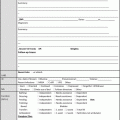
Most nursing facilities use a pressure ulcer report to document identified wounds: location, stage, measurement, and description. Pressure ulcer reports fulfill standardized documentation as mandated by both state and federal (F314) regulations in the nursing facility. The clinician should document the number, location, and size (length, width, and depth in centimeters) of wounds and assess for the presence of exudates, odor, sinus tracts, necrosis or eschar formation, tunneling/undermining, infection, healing (granulation and epithelialization), and wound margins. For a pressure ulcer, the clinician should determine the stage of the ulcer according to the National Pressure Ulcer Advisory Panel (NPUAP) Staging System (see Table 2).
Table 2
2007 NPUAP staging of pressure ulcers
Stage | Definition | Comment |
|---|---|---|
Suspected deep tissue injury (SDTI) | Pressure-related necrosis of soft tissue with intact overlying skin | Discoloration (crimson→purple), changes in temperature, texture, tenderness. May progress rapidly |
Stage I | Localized area of nonblancheable erythema. Skin is intact and sandwiched between a bony prominence and external surface. | Clinically similar to SDTI. May be harder to detect as skin pigmentation deepens |
Stage II | Partial thickness destruction of dermis characterized as either a shallow open ulcer with a crimson wound bed (without slough or bruising) or as an intact or ruptured fluid-filled blister. | Do not use to describe skin tears, tape burns, dermatitis, maceration, or excoriation |
Stage III | Full thickness tissue loss. Subcutaneous fat may be visible, but bone, tendon or muscle is not exposed. | Slough may be present. May include tunneling or undermining. Depth varies by anatomical location. |
Stage IV | Full thickness tissue loss characterized by exposed bone, tendon or muscle. Extensive destruction, necrosis, or damage to the muscle, bone, or supporting structures. | Slough or eschar may be present on some parts of the wound bed. Often include undermining and tunneling |
Unstageable | Full thickness tissue loss which cannot be staged until slough or eschar in the ulcer bed is removed | Do not remove eschar present on heels |
Risk of developing pressure ulcers is significantly high within the first 4 weeks after admission to a long-term care facility [25]. After an initial assessment, a weekly reassessment should occur for the first 4 weeks, followed by at least a quarterly assessment. Reassessments should also occur when there is a change in patient status [26]. The patient’s overall clinical condition should be reassessed whenever a pressure ulcer fails to show evidence of healing within 2–4 weeks of any intervention [25]. Every nursing facility is required to develop and implement its own comprehensive wound care plan in accordance with CMS regulations.
Some patients develop pressure ulcers 2–3 days before death. These are referred to as Kennedy terminal ulcers and are markers of the dying process. The Kennedy ulcer often develops suddenly on the sacrum as a blister or stage 2 and rapidly progress to stage 3 or 4. These ulcers can be pear-, horseshoe-, or butterfly-shaped with irregular borders. Kennedy terminal ulcers are initially red/purple, then turn to yellow, and finally turn black. The etiology is unclear, but is thought to be part of multiorgan failure. Most of the ulcers do not heal and their treatment is the same as that for pressure ulcers [27].
Types of Wounds
A wound may not necessarily fit into one of the four general categories of wounds (pressure ulcer, diabetic, arterial and venous), but may be of mixed etiology. Usually the type of wound can be distinguished by its location, combined with inspection of the wound and the patient’s clinical history. If the wound type remains uncertain, laboratory and/or radiographic studies may help clarify its type. For example, in lower extremity ulcers, an ankle-brachial index or Doppler arterial studies can help determine whether the ulcer is caused by vascular insufficiency, pressure or a combination of the two.
Pressure Ulcers
95 % develop on the lower body, 65 % over the sacrum and pelvic area, and 30 % in the lower extremities. Other common pressure ulcer sites include the coccyx, heels, ischium, iliac crest, lateral foot, lateral malleolus, and greater trochanter.
There are three mechanical forces that, when combined, create tissue damage. These are pressure, friction, and shear. A shear injury occurs beneath the skin and cannot be seen at the skin level, but a friction injury is superficial and easily visible (e.g., an abrasion or superficial laceration). Shear and friction injuries usually occur together [28].
A pressure ulcer is a localized area of damaged or necrosed tissue that develops when soft tissue is compressed between a bony prominence and an external surface for a prolonged period of time.
In 2009, NPUAP-EPUAP redefined pressure ulcer, and friction was deleted from the definition. As such it defined a pressure ulcer as a compressive tissue injury that is caused by pressure alone or by pressure combined with shearing. Friction alone is not a direct cause of pressure ulcers, but it does cause shear strain in tissue, which in turn can increase the risk of tissue breakdown [28].
Pressure ulcers can range from nonblancheable erythema of intact skin (or in dark-skinned individuals, it may have a blue or purple hue, i.e., stage I) to deep ulcers extending down to the bone (i.e., stage IV).
Diabetic Wounds
Commonly occur over the metatarsal heads.
Are due to vascular complications of diabetes mellitus including decreased healing and peripheral neuropathy.
Are typically painless; therefore the wound is usually not noticed until symptoms of infection occur such as malodor, fever or chills.
When inspecting those ulcers, providers should probe the depth of the wound with a sterile instrument to help determine if any undermining or osteomyelitis maybe present [29].
Ischemic Wounds
Typically occur in the lower extremities, but can also occur in the upper extremities.
Are due to decreased arterial blood flow seen in peripheral vascular disease. Diabetes mellitus and smoking have also been implicated.
Clinical signs of arterial insufficiency that often precede an ischemic wound include: a cold, pale or cyanotic foot, absence of digital and lower extremity hair and thin atrophic skin.
Present either as a painful wound with discrete borders (a “punched out” appearance), or wet or dry gangrene.
The base of the ulcer can be covered with a dry black or brown eschar or appear pale pink and fibrous.
Venous Wounds
Commonly seen in the lower extremities.
Are different from ischemic wounds, as these wounds are caused by peripheral edema due to venous insufficiency/stasis, medications or organ dysfunction (i.e., heart, liver, and kidney) [30].
Are less painful than ischemic wounds.
Have irregular borders and can be seen clustered together with hyperpigmented skin changes of surrounding skin.
Prevention
Paramount to wound care management is prevention. The Agency for Health Care Policy and Research (AHCPR ) published two companion practice guidelines in 1994 with recommendations for prediction, prevention and early treatment of pressure ulcers in adults [31, 32]. The companion practice guidelines were pioneering in their scope and are still widely utilized today because they remain applicable in many settings. The first step recommended for the prevention of pressure ulcers by the Institute for Healthcare Improvement (IHI) is to identify patients at risk, and then implement prevention strategies in these patients [24]. The IHI suggests “six essential elements of pressure ulcer prevention” in its guidelines.
The six steps are [24, 33]:
Conduct a pressure ulcer assessment of admission for all patients.
Reassess risk for all patients daily
Inspect skin daily
Manage moisture
Optimize nutrition and hydration
Minimize pressure
The IHI recommends that prevention measures include a comprehensive treatment plan with risk factor reduction, multidisciplinary interventions, functional adaptation, environmental modifications, and a psychosocial evaluation. Evaluating and optimizing any of the residents predisposing conditions and comorbidities can help prevent the development of wounds [9]. Inspecting skin daily during bathing or personal care, as well as scheduled turning and positioning of patients, has been shown to help prevent wounds. Provide support surfaces with special mattresses and overlays to help eliminate friction, shear, and moisture [34]. Minimize pressure over bony prominences. Seats should be padded with air, foam, or gel cushioning and avoid use of donut–shaped devices [9]. Residents at highest risk (those who completely compress a static surface, or have pressure ulcers that fail to heal), should be placed on a dynamic surface [7, 9]. A patient should never be directly placed on the greater trochanter for more than momentary positioning. Use padding (i.e., heel or “bunny” boots, egg crates, heel lifts, suspension devices, etc.) for off-loading of heels and elbows [7, 9]. Patients should have a static support surface such as a foam overlay or gel mattress placed on their standard mattress. A supine patient should be maintained at the lowest head elevation below 30° as tolerated; head elevation ≥30° provides as much pressure as being in a seated position [35]. Repositioning every four hours has been shown to be as effective as two-hour intervals in wound healing, but this repositioning or partial turning does not always remove pressure from the sacrum or heels. Care should be taken to minimize shearing or friction during repositioning. If necessary, lift devices should be used to prevent soft tissue injury. Slow gradual turns should be used in patients with hemodynamic instability [21].
Inadequate hydration and nutrition are predisposing conditions strongly associated with pressure ulcer development [36]. The caloric intake of 30–35 kcal/kg and daily protein intake of 1.2–1.5 g/kg of body weight is recommended for nutritionally compromised patients who either have or are at risk of pressure ulcers [5]. Adequate hydration is provided by 30–35 mL of fluid per kg body weight per day, or 1 mL of fluid per calorie for persons receiving enteral tube feeding [9]. Enteral nutritional support can significantly reduce the risk of developing pressure ulcers in selected patients, by up to 25 % in some studies. The benefits of nutritional support in facilitating wound healing are still debated [37–39]. Use of vitamin C supplementation in wound healing is also disputed. Two well-designed randomized controlled trials compared high dose vitamin C with either low dose vitamin C or placebo, and had contradictory results [40]. (For a more in-depth discussion on nutrition please see the chapter “Weight and Nutrition.”)
In clinical circumstances such as metastatic cancer, multiple organ failure, cachexia, severe vascular compromise and terminal illness, unavoidable wounds may develop [41]. The clinician should judiciously document the reasons why preventive interventions may not be appropriate or feasible, such as frequent repositioning that is causing discomfort or severe pain.
Unavoidable Pressure Ulcers
Pressure ulcers are considered to be quality of care indicator in LTC. There are many factors that are responsible for “unavoidable” pressure ulcers. According to the National Pressure Ulcer Advisory Panel (NPUAP), a pressure ulcer is considered to be unavoidable if it developed despite the following interventions.
1.
The patient’s clinical condition and risk for pressure ulcers were evaluated.
2.
Interventions were consistent with the patient’s needs and goals, as well as recognized standards of practice, were assessed and implemented.
A pressure ulcer is considered to be avoidable if the facility failed to implement the above interventions. Many LTC patients are either bed or chair-bound, which significantly limits mobility and places them at risk for pressure ulcers. Patients with hemodynamic instability can make turning or repositioning difficult, as they may develop bradycardia, hypotension or hypoxemia with minimal body movement. In addition, some patients are comfortable in a particular position so they move themselves back into that same position after being turned. Vasoconstrictive medications to increase low blood pressure can predispose to ischemia and increase pressure ulcer risk. When an advanced directive for health care states that artificial nutrition and hydration are not to be used, this can put the patient at risk for malnutrition and ulcer development. Refusal of care is a common issue, especially in patients who are confused and cognitively compromised, and can significantly frustrate the staff who are attempting to offload tissue pressure. Medical device–related pressure ulcers can occur, if they are applied before the development of edema [21].
Staging of Pressure Ulcers
AMDA—The Society for Post-Acute and Long Term Care Medicine follows the guidelines set forth by the National Pressure Ulcer Advisory Panel (NPUAP) that define, classify and stage pressure ulcers as summarized in Table 2. Staging is based on the extent of observable tissue damage [42]. The latest version of the these guidelines describe stages I–IV along with two adjunctive terms, “ suspected deep tissue injury ” and “unstageable” utilized to more accurately classify these wounds (see website http://www.npuap.org). Reverse staging should not be used. For example, a lesion may be referred to as a, “healing stage IV” but it cannot be described as progressing from a stage IV to a stage III. If a Stage IV pressure ulcer reopens at the same anatomical site, it is always considered as stage IV. An ulcer covered by eschar should be categorized as a Stage IV until the eschar has been debrided. Staging cannot be used for mucosal pressure ulcers [9].
Stay updated, free articles. Join our Telegram channel

Full access? Get Clinical Tree


