Carcinoid Tumors
Nathan J. O’Dorisio
Thomas M. O’Dorisio
M. Sue O’Dorisio
DEFINITION
Neuroendocrine tumors are characterized by the production, storage, and secretion of polypeptides, biogenic amines, and, in some cases, hormones. Carcinoid tumors, first termed in the early 1900s by Oberndorfer, fall under this designation. Oberndorfer noted small tumors of the gastrointestinal (GI) tract that were more indolent than adenocarcinomas and similar to the multiple tumors initially found and described by Lubarsch at autopsy of two patients in 1888. He termed these tumors “karzinoide,” or cancer-like. Carcinoid syndrome is an uncommon constellation of symptoms (<10%) of these tumors and is associated with diarrhea, flushing (usually facial), hypotension, wheezing, and/or edema. Nocturnal perspiration is also a common symptom, although not routinely listed.
ETIOLOGY
Carcinoid tumors arise from neuroendocrine cells and can be classified according to where they occur in the body. The location has classically been based on the embryonic divisions of the alimentary tract and is divided into foregut (lungs, bronchi, and stomach), midgut (small intestine, appendix, and proximal colon), and hindgut (distal colon, rectum, and genitourinary tract). Among the foregut tumors, those of the pulmonary system are thought to derive their origin from the Kulchitsky cells located within the mucosa. Within the stomach, the tumors arise from the enterochromaffin cells (ECs) and have been associated with hyperplasia secondary to hypergastrinemic states. Studies in humans taking proton pump inhibitor (PPI) have not borne out gastric hyperplasia. However, when higherpotency PPIs are used for long periods of time, gastrin levels rise as does chromogranin A; this is due to the enterochromaffin-like (ECL) cells from the stomach.
An association between gastric carcinoid and Zollinger-Ellison with MEN1 syndrome (see below) is almost 100% [1]. Within the midgut tumors, those of the small intestine are thought to derive from hyperplasia of the serotonin-producing EC intraepithelial cells. Those tumors of the appendix presumptively arise from the subepithelial cells within the submucosa and lamina propria [2]. The reason for the hyperplasia is unknown. Of the hindgut tumors, colon carcinoids also
arise from the serotonin-producing cells of the epithelium, while in the rectum, the cells contain “glicentin” (100-amino-acid-residue peptide) and glucagon-like peptides [2]. The carcinoid syndrome is related to the release of those peptides and amines produced and stored within carcinoid tumors. Most symptoms are due to overproduction of tryptophan (especially the amine serotonin) or decreased elimination of the breakdown products. This is especially true of serotonin overproduction when carcinoid tumors metastasize to the liver.
arise from the serotonin-producing cells of the epithelium, while in the rectum, the cells contain “glicentin” (100-amino-acid-residue peptide) and glucagon-like peptides [2]. The carcinoid syndrome is related to the release of those peptides and amines produced and stored within carcinoid tumors. Most symptoms are due to overproduction of tryptophan (especially the amine serotonin) or decreased elimination of the breakdown products. This is especially true of serotonin overproduction when carcinoid tumors metastasize to the liver.
EPIDEMIOLOGY
Carcinoid tumors are the most common type of neuroendocrine tumor in adults, accounting for approximately 55% of all new occurrences yearly affecting the GI tract. The overall occurrence is, however, still rare. Several studies have postulated the incidence to be 1 to 2 cases per 100,000 people [3, 4]. The rate is similar for women, men, and among races, with slight variations based on the location and type of tumor expressed. Carcinoids occur across the age spectrum, with the peak incidence occurring between 50 and 70 years of age. Among carcinoid tumors, the most common site is the appendix followed by the rectum and then the ileum. Carcinoid is the most common tumor of the appendix and can account for up to 1/3 of small bowel neoplasms. In contrast, carcinoid tumors represent less than 2% of organ-specific tumors in the pulmonary, gastric, and colonic/rectal systems. Carcinoid syndrome occurs only in a minority of patients (<10%). The presence of this syndrome varies by site of tumor, as well as size and metastatic disease.
Pathology
As previously mentioned, carcinoid tumors are classified according to the different embryonic divisions of the GI tract (fore-, mid-, and hindguts). The individual cells are further divided as “typical” and “atypical,” with typical cells classified as insular, trabecular, glandular, undifferentiated, and mixed [2, 5]. Malignant versus benign is based on cellular histology as well as tumor size at surgery and site of primary occurrence. “Typical” pulmonary carcinoids are indolent and perihilar in location, with less than 15% metastasizing. In contrast, “atypical” carcinoids of the lung have a 30% to 50% incidence of metastases and are often more aggressive. They may secrete ACTH and result in Cushing syndrome. Of the gastric carcinoids, up to 75% are type 1 and associated with chronic atrophic gastritis, 10% to 15% type 2 and associated with Zollinger-Ellison syndrome, and the remainder sporadic or type 3. Most tumors are less than 1 cm in diameter, and types 1 and 2 have a more benign course. Those tumors greater than 2 cm (and type 3 tumors) are more likely to have metastatic disease at the time of diagnosis. Small bowel carcinoid is most commonly found in the ileum and often has multiple local sites of involvement. Unlike gastric tumors, size greater or less than 2 cm is a less reliable predictor of metastasis [6]. It has been observed that ileal tumors greater than 2 cm in size at surgery will metastasize locally or regionally 100% of the time. Within the appendix, most tumors occur at the tip and, most, are not associated with symptoms. Of appendiceal tumors, 95% are less than 1 cm; however, the 2-cm size delineation appears to correlate with both metastatic disease and the carcinoid syndrome [6]. Colonic tumors are often right sided with most occurring around the cecum. These tumors tend to be larger at diagnosis (5 cm) and more commonly associated with distant metastases, although less than 5% exhibit features of carcinoid syndrome. Rectal carcinoids occur predominantly (99%) within the zone defined as 4 to 13 cm above the dentate line, though the reason for this is not known. The majority (2/3) are less than 1 cm, but the association with metastatic disease and carcinoid syndrome is again defined by an absolute size of 2 cm.
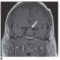
Among all carcinoid tumors, more than 80% express somatostatin receptors. Of the five somatostatin receptor subtypes currently known to exist, types 2, 3, and 5 have been demonstrated on carcinoid tumors with type 2 predominating. As previously noted, carcinoid syndrome can be attributed to altered metabolism of tryptophan and subsequent conversion to serotonin or other breakdown products. Diarrhea, flushing, nocturnal perspiration, and cardiac manifestations (especially right-sided valve disease) have all been linked with this mechanism, while histamine from some gastric tumors may be associated with atypical flushing and pruritus as well as an increased occurrence of duodenal ulcers seen in this population. The syndrome usually is directly related to the ability of the tumor to secrete serotonin into the circulation system and bypass the breakdown that occurs in the liver [7]. As well, it can exceed the liver metabolism threshold resulting in high circulating serotonin concentrations. Evidence of this can be seen in the high association with hepatic metastases and the low incidence in patients with limited or local disease. One caveat to this syndrome is ovarian carcinoids; while rare, these are more often associated with this syndrome due to their direct vascular access. It is thought that the episodic secretion of serotonin and vasoactive peptides is due to the nonautonomous nature of the neuroendocrine tumors and the presence of somatostatin subtype 2 receptors on them.
DIAGNOSIS
Due to the indolent nature (thought to be due, in part, to the binding of endogenous somatostatin to the tumor somatostatin receptors subtype 2 [sst2]) and mostly occult presentation of most carcinoid neoplasms, the diagnosis is commonly not made until later in the course of the disease. Pulmonary involvement often manifests as cough, recurrent pneumonia, or hemoptysis. Gastric tumors are commonly found on routine endoscopies for symptoms of abdominal pain or gastritis, while those in the small bowel may present with signs of obstruction or nonspecific abdominal pain; some to the point regrettably of being labeled “psychosomatic” in origin. Appendiceal lesions commonly are found during routine appendectomies for appendicitis but may present with obstruction if located in the base rather than the tip of the appendix [8]. Abdominal pain without obstruction is a common complaint in colonic tumors as well as rectal pain with associated tumors. Intestinal bleeding is an uncommon presentation for both rectal and colonic tumors and is almost nonexistent with small bowel tumors. Rectal carcinoids are often found incidentally on routine colonoscopy or sigmoidoscopy. The carcinoid syndrome is often characteristic and can be the initial presentation for the underlying tumor [9].
Related posts:

Stay updated, free articles. Join our Telegram channel

Full access? Get Clinical Tree


