Cancer of the Salivary Glands


Cancer of the Salivary Glands


Yekaterina A. Koshkareva

Robert L. Ferris


Cancers of the salivary glands are a distinctive group of neoplasms arising in the major and minor salivary glands. The distinguishing features of these cancers include their low incidence, histopathologic heterogeneity, presentation at multiple anatomic sites, and wide spectrum of clinical behaviors. The goal of this chapter is to provide a contemporary, comprehensive review of the demographics, etiology, pathology, and presentation of cancers of the salivary glands, as well as a summary of current recommendations for staging and treating these cancers.


SURGICAL ANATOMY

There are two types of salivary glands; the major salivary glands, which are readily identifiable, with anatomically distinct excretory ducts, and the minor salivary glands, which are less easily identifiable, smaller, and lack distinct ducts. The parotid, submandibular, and sublingual glands are the major salivary glands, whereas the minor salivary glands are not individually named and are found throughout the length of the upper aerodigestive tract.


Parotid Gland

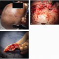
The paired parotid glands are the largest of the major salivary glands and form the lateral portions of the facial contour. The parotid gland is bounded by the cartilage of the external auditory canal posteriorly, the mandibular ramus and the masseter muscle medially, and the buccinator muscle anteriorly. The superior border is the zygoma, whereas the inferior aspect, also known as the “tail,” lies over the posterior belly of the digastric muscle medially and sternocleidomastoid (SCM) muscle laterally. The medial portion of the parotid gland abuts and sometimes extends into the parapharyngeal space (Fig. 21.1). Accessory parotid tissue may be found anterior to the body of the gland, along the course of Stensen duct, in 21% of the population.1 The gland is encased by a capsule derived from the deep cervical fascia and is further enveloped by the superficial musculoaponeurotic system (SMAS) in the face and the platysma in the neck. The parotid (Stensen) duct travels along the surface of the masseter and turns medial at its anterior border, transversing the buccal adipose tissue pad and the buccinator, and opens into the oral cavity opposite the second maxillary molar.

The arterial supply of the parotid gland comes from the two terminal branches of the external carotid artery: the superficial temporal and internal maxillary arteries. The venous outflow is collected by the respective superficial temporal and internal maxillary veins, which then join to form the retromandibular vein. The anterior division of the retromandibular vein along with the anterior facial vein forms the common facial vein. The posterior division joins the posterior auricular vein to drain into the external jugular vein.

There are as many as 20 lymph nodes within and adjacent to the capsule of the parotid gland, and these nodes are the first echelon of nodes to receive lymphatic drainage from the soft tissues of the temporal scalp, cheek, ear, and external auditory canal. Hence, the parotid lymph nodes may harbor metastatic foci from cancers that arise from these sites. Efferent parotid lymphatics drain into the lymph nodes of the superior and middle deep jugular chain.

Another important anatomic structure that must be considered in the management of parotid tumors is the facial nerve, which runs through the gland and serves as a landmark that separates the parotid into superficial and deep lobes. The main trunk of the facial nerve exits the stylomastoid foramen and enters the parenchyma of the parotid gland, where it typically divides into an upper and lower division. It then further subdivides into its five principal branches, but the arborization of the nerve can be quite varied.2 The point at which the main trunk of the facial nerve divides for the first time is referred to as the pes anserinus. The lower division contains the branches to the platysma (cervical branch) and the lower lip depressors (marginal mandibular branch). The marginal mandibular nerve is located deep to the platysma muscle and lateral to the facial vein and the capsule of the submandibular gland. The midface division (zygomaticobuccal branches) supplies innervation to the buccal, zygomatic, and lower eyelid muscle groups. The buccal branch is identified in the vicinity of the parotid (Stensen) salivary duct. The upper face division (frontal branch) travels lateral to the superficial layer of the deep temporal fascia to supply the frontalis and superior orbicularis oculi muscles.

Other nerves of importance encountered during parotid gland surgery are the greater auricular and auriculotemporal nerves. The greater auricular nerve is found on the lateral surface of SCM muscle, along the lateral border of the parotid gland. Its anterior and posterior branches provide sensation to the inferior aspect of the auricle and periauricular skin. The auriculotemporal nerve is a branch of the fifth cranial nerve (trigeminal nerve), which travels along with the superficial temporal vessels, posterior to the parotid, providing sensation to the temporal region. The auriculotemporal nerve also provides parasympathetic innervation to the parotid gland from the otic ganglion. After resection of the parotid salivary tissues, these fibers may form an aberrant innervation with sweat glands leading to perspiration in response to the gustatory stimuli, a phenomenon known as gustatory sweating (Frey syndrome).


Figure 21.1. A and B: Parotid gland anatomy. (From Snell RS, ed. Clinical Anatomy by Regions. 9th ed. Philadelphia, PA: Lippincott Williams & Wilkins; 2011.)


Submandibular Gland

The paired submandibular glands are located in the superior aspect of the anterior neck, bounded superiorly and laterally by the body of the mandible. The platysma muscle covers the gland’s lateral border, and the hyoglossus muscle lies medial to the gland. The gland resides posterior to the lateral border of the mylohyoid muscle. The submandibular (Wharton) duct originates from the multiple smaller branches originating from the medial aspect of the gland. It courses anteriorly and superiorly first between the mylohyoid and the hyoglossus muscles and then between the sublingual gland and the genioglossus muscle. The duct is situated between the lingual and hypoglossal nerves while on the surface of the hyoglossus, but at the anterior border of the muscle, the branches of the lingual nerve cross the duct to assume a more medial position. The submandibular duct travels
a total distance of ˜5 cm and empties into the anterior floor of the mouth.

The facial artery provides the main blood supply to the submandibular gland. This artery courses deep to the posterior belly of the digastric muscle and travels either along the medial aspect of the gland or through the gland parenchyma and traverses the lower border of the mandible at the antegonial notch curving over the insertion of the masseter muscle into the anterior border of the mandible. Venous drainage is supplied by the anterior facial vein.

Although there is no lymphoid tissue within the parenchyma of the submandibular gland, there are a number of pre- and postvascular lymph nodes adjacent to the facial vessels, which run over the lateral aspect of the gland, which serve as the first echelon nodes for lymphatic drainage from the lip, oral cavity, and skin of the anterior face. These lymphatics can also drain into the deep jugular chain of nodes. Enlargement of the submandibular nodes can be difficult to distinguish from a primary tumor of the submandibular gland, and a metastatic process within these nodes may also invade the adjacent gland by direct extension.

Similar to the parotid, the submandibular gland lies in anatomical proximity to the facial nerve. The marginal mandibular branch runs along the inferior border of the mandible, just deep to the platysma muscle and lateral to the fascia of the gland. Additionally, the lingual and hypoglossal nerves are located deep to the medial surface of the gland, whereas the nerve to the mylohyoid is adjacent to the superior aspect of the gland (Fig. 21.2).


Figure 21.2. A and B: Submandibular gland anatomy. (From Moore KL, Agur AMR, Dalley AF, eds. Clinically Oriented Anatomy. 7th ed. Baltimore, MD: Lippincott Williams & Wilkins; 2013.)


Sublingual Glands

The sublingual glands are also found bilaterally. They are located deep to the mucosa of the floor of the mouth and drain directly into the oral cavity through numerous small ducts. Tumors of the sublingual glands can be difficult to distinguish from those arising within the submucosal minor salivary glands of the floor of the mouth.


Minor Salivary Glands

There are over 600 minor salivary glands distributed throughout the length of the upper aerodigestive tract, and approximately half of them are located on the hard palate. Tumors can arise in these glands in such diverse sites as the oral cavity, oropharynx, larynx, pharynx, nose, nasopharynx, and paranasal sinuses. Additionally, small rests of heterotopic salivary tissue can be located within the cervical lymph nodes, mandible, thyroid gland, and the middle ear.3


ANATOMIC TUMOR CLASSIFICATION

Over 75% of salivary gland neoplasms originate in the parotid gland (mainly in the superficial lobe), with submandibular and minor glands comprising the rest. Approximately 80% of parotid gland tumors are benign. The likelihood of a submandibular gland lesion being benign is much less, around 43%. Finally, lesions arising within the minor salivary gland lesions are more likely to be malignant (up to 60%), especially if they originate outside of oral cavity (Fig. 21.3).4,5,6,7,8,9,10,11,12


HISTOLOGY AND HISTOGENESIS OF SALIVARY GLAND TUMORS

The salivary glands originate as ingrowths from the oral epithelium, which makes them of ectodermal descent. These ingrowths go on to form the ductal structure. The basic functional unit of a salivary gland is an acinus, made up of serous and mucinous cells. The acini release their secretions into an intercalated duct, which later becomes a larger striated duct, which eventually empties into an excretory duct. The myoepithelial cells surrounding the acini and intercalated ducts are capable of contraction and help with saliva transport (Fig. 21.4). The parotid gland is almost exclusively serous. The submandibular and sublingual glands are both mixed, but the serous component predominates in submandibular glands, whereas the mucinous element is dominant in sublingual glands.13


Figure 21.3. Relative ratio of benign versus malignant salivary neoplasms at various anatomic sites. (Dark blue, benign; light blue, malignant.)


Figure 21.4. Functional unit of a salivary gland. (From Rhoades RA, Bell DR, eds. Medical Physiology. 4th ed. Baltimore, MD: Lippincott Williams & Wilkins; 2012.)

There are two leading theories of the histogenesis of tumors of the salivary glands. According to the multicellular theory, each cancer arises from a particular cell type within the secretory unit.14 For example, Warthin and oncocytic tumors are thought to arise from striated ductal cells and acinic cell tumors from acinar cells.15 The bicellular reserve cell theory states that the development of any salivary gland neoplasm can be traced to the basal cells of either the excretory or the intercalated ducts, which are thought to be the reserve cells capable of differentiation. According to this model, pleuripotent intercalated duct basal cells give rise to salivary neoplasms of adenomatoid origin (pleomorphic adenoma, oncocytic tumors, acinic cell and adenoid cystic carcinomas). The epidermoid tumors (squamous cell and mucoepidermoid carcinomas) are then thought to be derived from the excretory duct reserve cells.16


HISTOLOGIC CLASSIFICATION

Salivary gland malignancies remain a diagnostic challenge for pathologists due to the rarity of these tumors as well as the wide histopathologic diversity and heterogeneity of these cancers. The modifications to the original classification scheme described by Foote and Frazell have resulted in the current World Health Organization (WHO) classification.17 The most recent WHO classification published in 2005 recognizes 13 benign and 24 malignant tumor subtypes, which are summarized in Table 21.1.18

Mucoepidermoid carcinoma, adenoid cystic carcinoma, adenocarcinoma, and malignant mixed tumor are the most common malignancies of the salivary glands, whereas acinic cell carcinoma and anaplastic carcinomas are encountered less frequently.4,5,6,7,8,9,10,11,12,19,20,21,22 Metastases from the skin of the scalp and face and squamous variants of mucoepidermoid carcinoma should be considered and ruled out prior to making the diagnosis of a primary salivary gland squamous cell carcinoma (SCC), as those are rare. In their review, Ying et al.23 found that SCC of the parotid represented metastases from an identified primary site in more than half of the patients. Cutaneous carcinomas and melanomas of the scalp and face often drain into intraparotid lymph nodes and comprise 70% to 80% of metastases to intraparotid lymph nodes. Metastases from infraclavicular primary sites are less common.24,25,26


Table 21.1 WHO Histologic Classification of Salivary Gland Neoplasms


Benign Epithelial Tumors


Malignant Epithelial Tumors


Basal cell adenoma


Canalicular adenoma


Cystadenoma


Ductal papillomas


Intraductal papilloma


Inverted ductal papilloma


Lymphadenoma


Myoepithelioma


Oncocytoma


Pleomorphic adenoma


Sebaceous adenoma


Sialadenoma papilliferum


Warthin tumor


Acinic cell carcinoma


Adenoid cystic carcinoma


Adenocarcinoma, not otherwise specified


Basal cell adenocarcinoma


Carcinoma ex pleomorphic adenoma


Carcinosarcoma


Clear cell carcinoma, not otherwise specified


Cystadenocarcinoma


Epithelial-myoepithelial carcinoma


Large cell carcinoma


Low-grade cribriform cystadenocarcinoma


Lymphoepithelial carcinoma


Metastasizing pleomorphic adenoma


Mucinous adenocarcinoma


Mucoepidermoid carcinoma


Myoepithelial carcinoma


Oncocytic carcinoma


Polymorphous low-grade adenocarcinoma


Sebaceous carcinoma


Sebaceous lymphadenocarcinoma


Salivary duct carcinoma


Sialoblastoma


Small cell carcinoma


Squamous cell carcinoma


The relative incidence rates of various malignant tumors by salivary gland subsite according to the National Cancer Institute Surveillance, Epidemiology, and End Results (SEER) Program data from 1992 to 2006 are presented in Figure 21.5. As mentioned above, perhaps erroneous SCC designation within the database may have affected the data presented in the figure. The frequency of encountering a certain neoplasm depends on the specific anatomic subsite of interest. Mucoepidermoid carcinoma is the most commonly encountered cancer in the parotid gland, whereas the submandibular gland is more likely to give rise to adenoid cystic carcinoma. Both entities occur at approximately the same rate within the minor salivary glands, followed by adenocarcinoma.


INCIDENCE AND ETIOLOGY

Approximately 7% of epithelial head and neck cancers (HNC) in the United States originate in the salivary glands.27 The annual incidence varies between 0.3 and 3 cases per 100,000 population.13 Similar numbers have been quoted in the European reports.28

Our current understanding of the pathogenesis of salivary gland malignancies is quite limited. The significance of both hereditary and environmental influences has been proposed. For example, a higher incidence of salivary gland tumors has been shown in some ethnic groups, such as Eskimos.29 Ionizing radiation increases the risk of development of salivary gland malignancies, according to the multiple descriptive studies of atomic bomb survivors in Hiroshima and individuals with a history of low-dose radiation of the head and neck.30,31,32,33,34,35 Industrial exposures to rubber and nickel have also been proposed to elevate the risk for development of salivary gland neoplasms.36 Recent genomic characterizations of salivary tumors show a unique pattern of gene alterations in certain histologic subtypes. Despite these reports, a clear understanding of the etiology of salivary tumors remains elusive.


CLINICAL PRESENTATION AND EVALUATION


History

Cancers of the major salivary glands present most commonly as an asymptomatic swelling and are often present for many months/years before a patient’s initial presentation for medical evaluation. The symptoms that arise from minor salivary gland tumors depend upon the anatomic regions involved. Thus, cancers of the paranasal sinuses can cause facial pain and/or swelling, whereas cancers of the nasal cavity can lead to obstruction of the nasal passages and/or epistaxis. Cancers of minor salivary glands of the oral cavity can lead to a mass effect, which can result in ill-fitting dentures. Cancers arising in the larynx commonly present with hoarseness and/or sore throat. Sun exposure and the presence of current or previously excised skin lesions should be ascertained when squamous cancer or melanoma is found in parotid lymph nodes.

Pain is more common in patients with malignant compared to benign salivary gland neoplasms. Between 10% and 29% of patients with cancer of the parotid gland report pain, perhaps due to perineural invasion (PNI) by the cancer.28,37,38,39,40 Pain is also reported in a few patients with benign submandibular neoplasms and up to 50% of patients with cancer of the submandibular gland.8,41,42


Physical Findings

As with any tumor, it is of paramount importance to document the exact location and size of the mass, as the details from this assessment dictate the staging, management, and treatment as well as follow-up.

As noted above, the majority of salivary tumors present as a palpable mass. Salivary lesions of the parapharyngeal
space, however, may not be palpable; hence, the lateral pharyngeal walls should be examined transorally for asymmetry. Obviously, tumor fixation to either skin or deep structures suggests malignancy. In patients with untreated cancer of the parotid, fixation to the skin was reported in 9%, whereas fixation to the deep tissues was reported in 13% to 17% from the Memorial Sloan Kettering and MD Anderson Cancer Centers.39,43


Figure 21.5. Summary of the incidence of malignant tumors of (A) parotid gland (4,265 cases), (B) submandibular glands (845 cases), and (C) sublingual glands (58 cases) according to the National Cancer Institute’s SEER database, 1992-2006. *Authors alert the audience regarding possible misclassification of squamous cell carcinomas in the database, as the majority of the cases are either intraparotid metastases or misdiagnosed high-grade mucoepidermoid carcinomas.

Facial nerve involvement manifested by weakness of the muscles innervated by one or more branches of the nerve is also among the prominent hallmarks of physical examination for malignancy. Although facial nerve paresis or paralysis is present in fewer than a quarter of cases, it is generally associated with a poor prognosis.43,44,45 Facial nerve involvement occurs most commonly in patients with adenoid cystic carcinoma, undifferentiated carcinoma, or SCC.44,46,47

The presence of cervical lymphadenopathy is another strong predictor of malignancy. Cervical lymph nodes are found to be positive at presentation in ˜13% to 25% of patients with cancers of the parotid,28,39,43,48 14% to 33% of patients with cancer of the submandibular gland,41,49,50 and 14% in patients with cancers of the minor salivary glands.12 The histopathologic subtypes of cancer of the salivary glands most likely to metastasize to regional lymph nodes are SCC, high-grade mucoepidermoid carcinoma, high-grade adenocarcinoma, and malignant mixed tumor.38,39,41


Diagnostic Imaging

As a general principle, imaging studies should only be obtained if the patient management might be altered by the result. Most minor salivary gland tumors require radiographic evaluation to better delineate the extent of disease, including the involvement of adjacent structures. A thorough history and physical examination are generally sufficient to establish the diagnosis and extent of a tumor in the major salivary glands. Imaging is usually warranted to define the extent of the primary cancer as well as the presence of any regional metastases. This is particularly true of deep lobe parotid tumors extending into the parapharyngeal space.


Several imaging modalities are available as adjuncts in evaluating a tumor of the salivary glands. In expert hands, ultrasonography is an appropriate initial step in evaluation of the major salivary gland tumors and the status of the neck. It is readily available, none invasive, and cost-effective. Ultrasonography also allows for precise localization of the tumor within the gland and readily differentiates between cystic and solid components, thereby enabling accurate fine needle aspiration or core biopsy. Heterogeneous echogenicity, ill-defined margins, and extension beyond the borders of the gland with involvement of adjacent structures are ultrasonographic signs of a malignant process.51,52,53 Computed tomography (CT) is the study of choice for the evaluation of involvement of cortical bone, whereas magnetic resonance imaging (MRI) better visualizes soft tissue details of the minor salivary gland lesions, deep parotid tumors, and perineural spread of tumor. Irregular borders, extraglandular extension, and hypointensity on T2-weighed images are suggestive of malignancy.51,53,54 Additionally, in the case of sinonasal minor salivary gland tumors, MRI can assist in differentiating between a tumor and opacification of an obstructed sinus by fluid. Combined 18 F-fluorodeoxyglucose-positron emission tomography (FDG-PET)/CT has assumed an important role in head and neck oncology. However, its applications remain limited in salivary gland neoplasms.55 Although it has been shown to be superior to CT alone in delineating the tumor extent, nodal involvement, and distant metastases,55,56,57 it is not particularly useful in distinguishing between malignant and benign neoplasms. This is due to the fact that some benign salivary gland lesions, such as pleomorphic adenomas and Warthin tumors, are capable of high glucose uptake.58


Fine Needle Aspiration Cytology

Unlike the minor salivary glands, open incisional biopsy is not recommended for parotid and submandibular glands due to the potential risk of tumor seeding and injury to the facial nerve or its branches. Fine needle aspiration cytology (FNAC) provides an insight into the preliminary diagnosis of a salivary gland lesion, which could be useful in the formulation of a management plan. For example, a poor surgical candidate may be observed once a benign result is obtained on FNAC. Additionally, a person with a diagnosis of lymphoma or benign lymphoepithelial disease of HIV on FNAC may avoid a diagnostic parotidectomy. Up to one-third of patients can be spared an unnecessary operation and risk to the facial nerve.59

In a recent meta-analysis of FNAC in salivary gland tumors by Schmidt et al., the sensitivity and specificity for diagnosis of neoplasia were 71% and 100%, respectively, and the sensitivity and specificity for diagnosis of malignancy were 76% and 97%, respectively. For differentiation between a benign and malignant salivary lesion, the positive and negative predictive values were 90% and 94%, respectively.60 In a systematic review by Colella et al., the concordance rates between cytologic and surgical pathology diagnoses were 95.61% for benign and 79.95% for malignant salivary tumors. The concordance rates are lower for particular histologic types of salivary tumors: 63% for mucoepidermoid carcinomas and 70% for adenoid cystic carcinomas.61 In summary, FNAC is a safe, fast, well-tolerated, and reliable procedure for distinguishing between benign and malignant lesions, though it is not as useful in differentiating between the various malignancies. The challenges encountered with FNAC include lack of histologic architecture within the sample and necessity of a cytopathologist with significant experience in salivary gland disease. Small-volume care center providers should maintain a low threshold for pathology consultation within large academic institutions.

Core needle biopsy (CNB) technique was introduced to address some of the shortcomings of FNAC. Histologic architecture is preserved within the sample provided by the larger needle used (14 to 21 gauge). Also, larger tissue sample size eliminates the need for a cytopathologist to assess the specimen adequacy. Lastly, CNB specimens are formalin fixed and paraffin embedded, which facilitates the immunohistochemical staining. According to the recent meta-analysis by Schmidt et al., CNB sensitivity was 92% and specificity was 100%, with a 1.2% sample inadequacy rate. When compared to FNAC, CNB had a higher diagnostic accuracy. Hematoma was reported as the main complication, occurring in 1.7% patients, none of whom required aspiration or drainage. Theoretically, because of the larger diameter needle, CNB is more painful and there is a greater risk of facial nerve damage. No cases of permanent CN VII weakness have been reported thus far.62 Regardless of the biopsy technique used, clinical judgment should be used in analyzing the results if the cytologic aspiration diagnosis is inconsistent with the clinical presentation.


STAGING

Oncologic staging is critically important in the assessment of disease extent, prediction of prognosis, formulation of a treatment plan, and follow-up evaluations. It is an integral tool in communication between the treating surgeon and the patient and within the clinical and research medical communities. The current AJCC staging system used for major salivary gland cancers is presented in Table 21.2.63 As of now, there is no separate standardized staging system for minor salivary cancers. A study from Memorial Hospital demonstrated that the staging system used for squamous carcinoma of the upper aerodigestive tract can be used in evaluation of the minor salivary gland malignancies arising in the same anatomic regions and has a similar prognostic value.64


PATHOLOGY


Mucoepidermoid Carcinoma

Mucoepidermoid carcinoma is the most common cancer of salivary gland origin. The key morphologic feature is multicystic arrangement of mucous and epidermoid cells. Based on the proportion of cystic component, neural invasion, degree of anaplasia, mitoses, and presence of necrosis, mucoepidermoid carcinomas are classified into low-grade, intermediategrade, or high-grade categories.64 Low-grade cancers manifest well-defined glandular architecture. They very rarely metastasize and have an excellent overall prognosis. In high-grade mucoepidermoid carcinomas, the glandular structure is less defined or may be absent altogether. These tumors have a propensity for early and aggressive spread, with a rate of regional metastasis up to 70%.65 As the name implies, the intermediategrade lesions contain moderate cystic components and are less clinically aggressive than their high-grade counterparts. The patients with low-grade disease have an almost negligible recurrence rate.66 The natural history of mucoepidermoid
carcinoma is less protracted than that of other salivary malignancies; hence, a 5-year disease-free interval suggests cure.


Table 21.2 AJCC Staging System for Salivary Gland Cancer (7th Edition)


Primary Tumor (T)


TX


Primary tumor cannot be assessed


T0


No evidence of a primary tumor


T1


Tumor ≤2 cm in greatest dimension without extraparenchymal extension


T2


Tumor >2 cm but ≤4 cm in greatest dimension without extraparenchymal extension but <4 cm


T3


Tumor >4 cm and/or tumor having extraparenchymal extension


T4a


Moderately advanced disease. Tumor invades the skin, mandible, ear canal, and/or facial nerve


T4b


Very advanced disease. Tumor invades the skull base and/or pterygoid plates and/or encases carotid artery


Regional Lymph Nodes (LN) (N)


NX


Regional LNs cannot be assessed


N0


No regional LN involvement


N1


Metastasis in single LN <3 cm


N2a


Metastasis in single ipsilateral LN >3 cm but <6 cm


N2b


Metastasis in multiple ipsilateral LNs >3 cm but <6 cm


N2c


Metastasis in bilateral or contralateral LNs, none >6 cm


N3


Metastasis in LN >6 cm


Distant Metastasis (M)


MX


Distant metastasis cannot be assessed


M0


No distant metastasis


M1


Distant metastasis


Stage Grouping


I


T1/N0/M0


II


T2/N0/M0


III


T3/N0/M0


T1-3/N1/M0


IVA


T4a/N0-1/M0


T1-4a/N2/M0


IVB


T4b/any N/M0


Any T/N3/M0


IVC


Any T/any N/M1


Used with the permission of the American Joint Committee on Cancer (AJCC), Chicago, Illinois. The original source for this material is the AJCC Cancer Staging Manual, Seventh Edition (2010) published by Springer Science and Business Media LLC, www.springer.com.


Adenoid Cystic Carcinoma

Adenoid cystic carcinoma is the most common cancer of the submandibular gland, and it is as common as mucoepidermoid carcinoma in the minor salivary glands. It constitutes ˜10% of all epithelial salivary tumors. It most commonly presents as a slow-growing mass and can be associated with pain, due to its propensity for neural invasion and lack of a capsule barrier.67 Patients with adenoid cystic carcinoma must be under careful clinical surveillance indefinitely, due to its protracted natural history, with disease-related deaths having been reported even after 20 years of disease-free survival.68,69 Despite excellent local control, adenoid cystic carcinomas result in poor longterm outcomes due to distal recurrence. The lung is the primary site for distant metastases; however, patients can often live for many years with asymptomatic pulmonary metastases that remain stable or progress slowly.70

Adenoid cystic carcinoma can be arranged in solid, tubular, and cribriform histologic patterns. Predominantly, tubular and cribriform lesions tend to be less aggressive than tumors with >30% solid component.71,72 The solid histologic pattern prognosticates development of distant metastases and poor long-term survival.71 The difference in survival based on histopathologic grading disappears at 10 years after the treatment, suggesting that the histology affects the disease-free survival, with minimum impact on the overall outcome.73,74,75


Adenocarcinoma, Not Otherwise Specified

Adenocarcinoma, not otherwise specified (NOS), is notable for its ductal differentiation.76 Again, histologic grading affects prognosis: high-grade lesions tend to have worse outcomes. Also, the survival decreases significantly from 5 to 10 years
after treatment, which warrants a long-term surveillance in these patients.77,78


Salivary Duct Carcinoma

Salivary duct carcinomas (SDC) are rare tumors with aggressive clinical behavior and a poor prognosis. These cancers manifest as a rapidly growing mass and occur in patients over 50 years of age, with a male to female ratio of 4:1. The majority of cases (>80%) originate in the parotid gland.79 The rate of cervical lymphatic involvement at presentation is over 50%. Most patients present with advanced stage disease: one-third of them suffer recurrence, 46% develop distant metastases, and 65% die of the disease, usually within 4 years of diagnosis.80


Carcinoma Ex Pleomorphic Adenoma

As the name implies, carcinoma ex pleomorphic adenoma originates in the setting of a pleomorphic adenoma. The malignant component usually features a poorly differentiated adenocarcinoma or an undifferentiated carcinoma. The proportions of benign and malignant components vary. This cancer is classified into noninvasive, minimally invasive, or invasive.81 Noninvasive variants display indolent behavior similar to that of pleomorphic adenomas, whereas the invasive variants are more aggressive, with recurrence of 23%, regional metastatic rates between 49% and 56%, and distal metastatic rate up to 40%.82,83


Acinic Cell Carcinoma

Acinic cell carcinoma affects all age groups and is almost never seen outside of the parotid gland. Due to its indolent course, it is considered a low-grade cancer. However, high-grade papillary-cystic variants exist. Despite its overall good prognosis, the potential of local recurrence or distant metastases exists, especially in advanced stage or improperly treated tumors.84,85 In the review of prognostic factors by Neskey et al.,86 older age and larger tumor size were found to correlate with poor overall survival. Also, distant metastases were prognostic of disease-specific death.


Squamous Cell Carcinoma

Primary salivary SCC of the salivary glands is rare. Time and care should be taken to distinguish it from the more common high-grade mucoepidermoid carcinoma or metastatic SCC from a cutaneous malignancy of the face, ear, or scalp. In the review by Ying et al.,23 over 60% of patients with SCC of the parotid had a known primary cutaneous malignancy (most commonly, auricle). Only a quarter of the patients were found to have the primary salivary SCC. Both groups had high rates of cervical node involvement and poor disease outcome.


Lymphoma

Up to 85% of salivary gland lymphomas are of the non-Hodgkin type. Lymphomas of the salivary glands arise either from intraglandular lymph nodes (nodal) or from the nondiscrete lymphoid tissue within the gland parenchyma (extranodal). Although extranodal lymphomas affect salivary glands in only ˜5% of cases, the overwhelming majority of these occur in the parotid gland. Lymphoma of the salivary glands either can be the only manifestation of the disease in primary cases or can also be a part of a disseminated lymphomatous process. Primary lymphomas of the salivary glands are usually associated with Sjögren syndrome.87 Those associated with Sjögren syndrome have a significantly worse prognosis, compared to lymphomas arising in normal salivary glands. The risk of the development of the lymphoma in patients with Sjögren syndrome is 44-fold higher than that in the general population.88


SECONDARY (METASTATIC) CANCERS

The majority of metastases to the salivary glands involve the parotid gland and are due to intraparotid lymph node spread from the cutaneous malignancies of the face, ear, and scalp. Approximately 40% of these are SCCs and 40% are melanomas. The majority of metastatic parotid SCCs appear within the first year of treatment. Therefore, every patient with a parotid mass with cutaneous histology should undergo careful examination of the skin of the head and neck.

Involvement of the salivary glands by the distant infraclavicular metastatic cancers is extremely rare. When it does occur, the parotid gland is most commonly affected, and the most common sites giving rise to these metastases are the lung, kidney, and breast.89

Only gold members can continue reading. Log In or Register to continue

Stay updated, free articles. Join our Telegram channel

Dec 18, 2016 | Posted by in ONCOLOGY | Comments Off on Cancer of the Salivary Glands

Full access? Get Clinical Tree

Get Clinical Tree app for offline access