Cancer of the Neck


Cancer of the Neck


Jesus E. Medina


Cervical lymph node metastases in patients with squamous cell carcinoma of the upper aerodigestive tract have important prognostic significance; when they are present, survival decreases by ˜50%.1 Detection of lymph node metastases at an early stage, preferably when they are microscopic, is paramount because the prognosis worsens as the extent of the metastases in the neck nodes (N stage) increases2 (Fig. 18.1). Depending on the location and stage of the primary tumor and the cervical lymph nodes, management of the neck may require surgery, radiation therapy, or both, and in some instances, it may also require chemotherapy. I begin this chapter presenting basic concepts such as surgical anatomy, staging, and nomenclature, and then, will outline the principles of management of the clinically negative (N0) and positive (N1/N3) neck.


ANATOMY

A neck dissection is the mainstay of the surgical treatment of many patients with cancer of the head and neck. A thorough knowledge of the anatomy of the neck is necessary to perform a neck dissection well. Such a discussion of the anatomy of the neck is not within the scope of this chapter. However, it is appropriate to highlight the pertinent surgical anatomy of certain structures that are particularly relevant, in the course of performing neck dissections.


Marginal Mandibular Branch of the Facial Nerve

The inferior division of the facial nerve divides into cervical and marginal branches, a short but variable distance behind the anterior border of the lower portion of the parotid gland (Fig. 18.2). The “lower” cervical branch runs downward and forward and, after exiting the parotid, is located between the anterior border of the parotid and the submandibular gland; it “terminates” in the platysma muscle, a variable distance below the submandibular gland. The marginal branch subdivides further, at about the level of the angle of the mandible, into an “upper” cervical branch and the marginal branch. The upper cervical branch overlies the posterior and then the inferior portions of the submandibular gland, as it curves upward to enter the platysma about 1.5 to 2 cm below the inferior border of the mandible. It is usually not possible to preserve the cervical branches of the facial nerve when performing a neck dissection that includes the submandibular triangle (level I). However, it is often possible to preserve these branches when performing a submandibular gland excision.


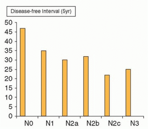
Figure 18.1. Impact of the extent of neck metastases (N stage) on prognosis. Disease-free interval according to TNM staging. (Modified from Kowalski LP, Bagietto R Lara JR, et al. Prognostic significance of the distribution of neck node metastasis from oral carcinoma. Head Neck. 2000;22:207-214.)

The marginal branch runs forward, in a slightly oblique, upward direction; it is located deep to the superficial cervical fascia, between the inferior border of the mandible and the superior border of the submandibular gland. When performing a neck dissection, this nerve is usually identified about 1 cm in front of and inferior to the angle of the mandible by incising the superficial layer of the deep cervical fascia that envelops the submandibular gland, immediately above the gland, in a direction parallel to the direction of the nerve. The incised fascia then is gently pushed superiorly, exposing the nerve that lies deep to it but superficial to the adventitia of the anterior facial vein. The submandibular retrovascular lymph nodes are usually in close proximity and medial to the nerve and must be carefully dissected away from it. As this is done, the facial vessels are exposed and can be divided.

Identifying the marginal mandibular branch is essential in performing an adequate excision of the lymph nodes in the submandibular triangle. The practice of ligating the anterior facial vein low in the submandibular triangle and retracting it superiorly to “protect the marginal branch” can also result in elevation of the prevascular and retrovascular lymph nodes, thus precluding their appropriate removal. When indicated, it is preferable to identify the nerve and thoroughly remove these lymph nodes.


Spinal Accessory Nerve

The spinal accessory nerve (SAN) is located medial to the digastric and stylohyoid muscles and lateral or immediately posterior to the internal jugular vein (IJV) (Fig. 18.3). Occasionally, the superior portion of the nerve is posterior-medial to the vein. The nerve then runs obliquely inferior and backward to reach the medial surface of the sternocleidomastoid muscle (SCM) near the junction of its superior and middle thirds (two to three fingerbreadths below the mastoiditis). Although the nerve can continue its downward course entirely medial to the muscle (18%), more commonly, it traverses and appears in the posterior border (82%).3 Here, the nerve is always located above the point where the greater auricular nerve turns around the posterior border of
the SCM, also known as Erb point (Fig. 18.4).4 The mean distance between the Erb point and the SAN is 10.7 mm, SD ± 6.3. It then runs through the posterior triangle of the neck and crosses the anterior border of the trapezius muscle. The mean distance between this point and the clavicle is 51.3 mm, SD ± 17.4 Two anatomic characteristics of this portion of the nerve are relevant to avoid injuring it in the course of a neck dissection. First, the SAN is located rather superficially as it courses through the middle and low posterior triangle of the neck, and it can be easily injured while elevating the skin flaps in the posterior neck. Second, the nerve does not enter the trapezius muscle at the anterior border of it but courses along the deep surface of the muscle in close relationship with the transverse cervical vessels. Therefore, isolating the nerve to the level of the anterior border of the trapezius does not ensure its preservation during surgical dissection below this point, particularly in a bloody operative field.


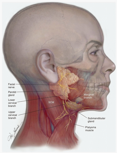
Figure 18.2. Anatomy of the inferior division of the facial nerve: 1. Marginal mandibular branch; 2. Upper cervical branch; 3. Lower cervical branch.


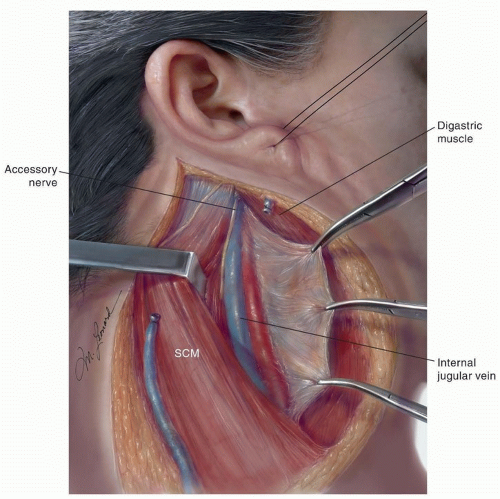
Figure 18.3. The SAN in its most common position in the superior aspect of the right neck: lateral and slightly posterior to the IJV.


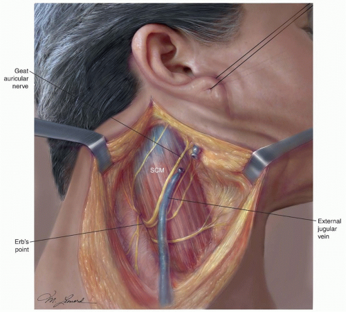
Figure 18.4. The spinal accessory in the posterior triangle of the neck. 1. Erb point; 2. The SAN is usually located about 1 cm above Erb point.


A common sequela in patients who undergo neck dissection is related to the removal of the SAN or to dysfunction of this nerve subsequent to its dissection during surgery. The resulting paralysis or paresis of the trapezius muscle, one of the most important shoulder abductors, causes destabilization of the scapula with progressive flaring of it at the vertebral border, drooping, and lateral and anterior rotation. The loss of the trapezius function decreases the patient’s ability to abduct the shoulder above 90 degrees at the shoulder. Paralysis of the trapezius muscle causes a clinical syndrome characterized by weakness and deformity of the shoulder girdle, usually accompanied by pain.5 The shoulder pain appears to be secondary to increased supportive demands on and strain of the levator scapulae and rhomboid muscles. Furthermore, adhesive capsulitis of the glenohumeral capsule may result in a “frozen shoulder” leading to a chronic disability and impaired quality of life.6

Preservation of the SAN to avoid this cumbersome sequela has been one of the reasons for the development of the modified radical and selective neck dissections (SNDs) that are commonly used today. Interestingly, varying degrees of shoulder dysfunction can occur, even when the SAN is preserved during a neck dissection. Therefore, it is important for the surgeon to understand the anatomy of the SAN and the importance of early diagnosis and rehabilitation when dysfunction occurs.


Nerve to the Levator Scapulae Muscle

The levator scapulae is a triangular muscle located deep in the lateral aspect of the neck, anterior and medial to the splenius capitis muscle. It extends from the transverse process of the atlas and the next three cervical vertebrae to the superior angle and the spine of the scapula. The action of the levator scapulae is to raise the medial angle of the scapula and incline the neck to the corresponding side with rotation of the neck in the same direction. With the trapezius muscle, the levator scapula makes a shoulder shrug possible. Because one of the functions of the levator is to draw the scapula and the shoulder upward and medially, inadvertent or unnecessary resection of the nerves to it during a neck dissection, particularly a radical neck dissection (RND), may accentuate the resulting deformity and functional disability of the shoulder.


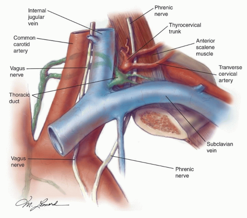
Figure 18.5. Anatomic relations of the thoracic duct. 1. Vagus nerve; 2. Common carotid artery; 3. Internal jugular vein; 4. Phrenic nerve; 5. Thoracic duct draining into the subclavian vein; 6. Transverse cervical artery.

The nerves to the levator scapulae, which vary in number from 1 to 3, branch off the 4th and 5th cervical nerves and travel posteriorly and inferiorly. They cross the anterior border of the levator scapulae and remain on the surface of the muscle for a short distance. The nerves to the levator scapulae are deep to the fascia of this muscle; thus, in the course of any neck dissection, but especially in an RND or an modified radical neck dissection (MRND), it is crucial to keep the plane of dissection superficial to the fascia of the levator in order to preserve these nerves. The dorsal scapular nerve is inconsistent in its anatomic relations in the posterior triangle of the neck and contributes to the innervation of the levator scapulae in a minority of cases.7


Thoracic Duct

Inadvertent puncture or transection of the thoracic duct at the base of the neck can result in a chylous fistula, a relatively infrequent but potentially cumbersome complication of a neck dissection. Knowledge of the usual location and course of the thoracic duct is paramount whenever a dissection involves the supraclavicular, lower jugular (level IV) region. It is, perhaps, even more important when the surgeon is called on to search for and repair a chyle fistula during or after a neck dissection.

The thoracic duct is located medial to and behind the common carotid artery and the vagus nerve (Fig. 18.5). From here, it arches upward, forward, and laterally, passing behind the IJV and in front of the anterior scalene muscle and the phrenic
nerve. It then opens into the IJV, the subclavian vein, or near the angle formed by the junction of these two vessels. The duct is anterior and medial to the thyrocervical trunk and the transverse cervical artery. To prevent a chyle leak, the surgeon also must remember that the thoracic duct may be multiple in its superior aspect and that at the base of the neck it usually receives a jugular, a subclavian, and other minor lymphatic trunks, which must be ligated or clipped individually.


Lymph Nodes of the Neck

The lymph node regions of the neck are shown in Figure 18.6. The six levels currently used encompass the complete topographic anatomy of the neck. The concept of sublevels has been introduced into the classification because certain zones have been identified within the six levels, which may have clinical significance.

Level I is divided into two sublevels. Sublevel IA (submental) includes the lymph nodes within the inferiorly based triangle bound by the anterior belly of the digastric muscles and the hyoid bone. Sublevel IB (submandibular) includes the lymph nodes within the boundaries of the anterior belly of the digastric muscle, the stylohyoid muscle, and the inferior border of the body of the mandible.


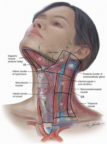
Figure 18.6. The lymph node regions/“levels” of the neck.

Level II (upper deep jugular) includes the lymph nodes located around the upper third of the IJV and adjacent SAN extending from the level of the skull base to the level of the inferior border of the hyoid bone. The anterior/medial boundary is the stylohyoid muscle (the radiologic correlate is the vertical plane defined by the posterior surface of the submandibular gland), and the posterior (lateral) boundary is the posterior border of the SCM. Two sublevels are recognized in level II: sublevel IIA, nodes located anterior/medial to the vertical plane defined by the SAN, and sublevel IIB, nodes located posterior/lateral to the vertical plane defined by the SAN.

Level III (mid deep jugular) includes the lymph nodes located around the middle third of the IJV extending from the inferior border of the hyoid bone to the inferior border of the cricoid cartilage. The anterior/medial boundary is the lateral border of the sternohyoid muscle, and the posterior/lateral boundary is the posterior border of the SCM.


Level IV (lower deep jugular) encompasses the lymph nodes located around the lower third of the IJV extending from the inferior border of the cricoid cartilage to the clavicle.

The anatomic boundary that separates the medial border of levels III and IV from the lateral border of level VI has traditionally been the lateral border of the sternohyoid muscle, a landmark that cannot be easily discerned on imaging studies. Therefore, the medial aspect of the common carotid artery has been suggested as an alternate boundary to separate these levels in an axial plane in imaging studies.8

Level V (posterior triangle) comprised predominantly of the lymph nodes located along the inferior half of the SAN and the transverse cervical artery. The supraclavicular nodes are also included in the posterior triangle group. The superior boundary is the apex formed by convergence of the sternocleidomastoid and trapezius muscles, the inferior boundary is the clavicle, the anterior boundary is the posterior border of the SCM, and the posterior boundary is the anterior border of the trapezius muscle. A horizontal plane marking the inferior border of the anterior cricoid arch separates two sublevels. Sublevel VA, above this plane, includes the spinal accessory nodes. Sublevel VB, below this plane, includes the nodes that follow the transverse cervical vessels and the supraclavicular nodes with the exception of Virchow node, which is located in level IV.

Level VI (anterior compartment) lymph nodes in this compartment include the pre- and paratracheal nodes, precricoid (Delphian) node, and the perithyroidal nodes including the lymph nodes along the recurrent laryngeal nerves. The superior boundary is the hyoid bone, the inferior boundary is the suprasternal notch, and the lateral boundaries are the common carotid arteries.

Level VII refers to the extension of the paratracheal nodes below the suprasternal notch (the dividing line between levels VI and VII) to the level of the innominate artery.


Other Lymph Node Groups

Lymph nodes involving regions not located within these levels should be referred to by the name of their specific nodal group; examples of these are the superior mediastinal, the retropharyngeal, the periparotid, the buccinator, the postauricular, and the suboccipital lymph nodes.

The retropharyngeal lymph nodes (RPLNs) lie within a pad of adipose tissue located behind the posterior wall of the pharynx and anterior to the prevertebral fascia and the cervical sympathetic trunk and ganglion. This pad of adipose tissue extends from about the level of the carotid bifurcation to just inferior to the skull base. The RPLN are divided into medial and lateral groups; the medial group of nodes lies behind the pharyngeal midline at a level between the first and fourth cervical vertebrae. The lateral group of nodes, better known as the nodes of Rouviere, are contained within a sliver of adipose tissue located immediately medial to the internal carotid artery. The RPLN receive lymphatic drainage from the nasopharynx, tonsil fossa, the walls of the oropharynx and the hypopharynx, and the posterior ethmoid sinuses.


STAGING

After completing the clinical evaluation of a patient with squamous cell carcinoma of the head and neck region, the disease should be classified according to stage. The staging for the lymph nodes proposed by the American Joint Committee on Cancer in 20099 is outlined below:

NX: Regional lymph nodes cannot be assessed.

N0: No regional lymph node metastasis.

N1: Metastasis in a single ipsilateral lymph node, 3 cm or less in greatest dimension.

N2: Metastasis in a single ipsilateral lymph node, more than 3 cm but not more than 6 cm in greatest dimension; in multiple ipsilateral lymph nodes, none more than 6 cm in greatest dimension; or in bilateral or contralateral lymph nodes, none more than 6 cm in greatest dimension.

N2a: Metastasis in a single ipsilateral lymph node more than 3 cm but not more than 6 cm in greatest dimension.

N2b: Metastasis in multiple ipsilateral lymph nodes, none more than 6 cm in greatest dimension.

N2c: Metastasis in bilateral or contralateral nodes no more than 6 cm in greatest dimension.

N3: Metastasis in a lymph node more than 6 cm in greatest dimension.

Staging of the neck in patients with carcinoma of the nasopharynx is different because the distribution and the prognostic impact of regional lymph node metastasis from cancer of the nasopharynx, particularly of the undifferentiated type, are different from that of other cancers of the head and neck mucosa and justifies the use of the following scheme:

NX: Regional lymph nodes cannot be assessed.

N0: No regional lymph node metastasis.

N1: Unilateral metastasis in lymph node(s), 6 cm or less in greatest dimension, above the supraclavicular fossa.

N2: Bilateral metastasis in lymph node(s), 6 cm or less in greatest dimension, above the supraclavicular fossa.

N3: Metastasis in a lymph node(s) >6 cm and/or to supraclavicular fossa.

N3a: Greater than 6 cm in dimension.

N3b: Extension to the supraclavicular fossa.


CLASSIFICATION OF NECK DISSECTIONS

Several cervical lymph node dissections are currently used for the surgical treatment of the neck in patients with cancer of the head and neck region. To standardize the nomenclature used to refer to these operations, it is essential to adopt a common nomenclature for the groups of lymph nodes in the neck, such as the one outlined above. The current classification of neck dissections recommended by the American Academy of Otolaryngology: Head and Neck Surgery (Table 18.1) takes into account the groups of
lymph nodes of the neck that are removed and secondarily the anatomic structures that may be preserved, such as the SAN and the IJV. Analyzing neck dissections from these two points of view, there are essentially four anatomic types of neck dissections: radical, modified radical, selective, and extended. Recently, clinicians from around the world have proposed a nomenclature for neck dissection, which, if recognized internationally, would be “logical, unambiguous, precise, and easy to remember.”10


Table 18.1 Classification of Neck Dissections


2001 Classification


2010 Proposed Classification


1. Radical neck dissection


1. ND (I-V, SCM, IJV, CNXI)


2. Modified radical neck dissection


2. ND (I-V, SCM, IJV), ND (I-V, IJV, CNXI), ND (I-V)


3. Selective neck dissection (SND):


3.


SND (I-III/IV)


ND (I, II, II/IV)


SND (II-IV)


ND (II, III, IV)


SND (II-V, postauricular, suboccipital)


ND (II, III, IV,V, postauricular, suboccipital)


SND (level VI)


ND (VI)


4. Extended neck dissection


4. ND (levels removed, nodes or structures removed)


In this classification, the following three descriptors are used to label a neck dissection:


  • “ND” to represent neck dissection, which is prefaced by either “L” or “R” for side. If bilateral, both sides must be classified independently.


  • The levels and sublevels of lymph nodes removed designated by Roman numerals I through VII in ascending order. For levels that contain sublevels (I, II, and V), listing of the level without a sublevel indicates that the entire level (both A and B) was excised.


  • The nonlymphatic structures removed designated by their internationally recognized initials, that is, SCM for sternocleidomastoid muscle and IJV for internal jugular vein.

The advantage of this classification is that it conveys precisely the groups of lymph nodes included as well as the nonlymphatic structures removed in a neck dissection. This will allow a standardized reporting and meaningful comparison of outcomes.

Irrespective of the nomenclature used, it is the responsibility of the surgeon to divide or otherwise orient the neck dissection specimen identifying the different groups of lymph nodes it contains. Only then can the pathologist render a report that is useful clinically and prognostically. Such a report describes the location and number of lymph nodes examined, the number of nodes that contain cancer, and the presence or absence of extra capsular extension of tumor.


MANAGEMENT OF THE N0 NECK

The initial management of the clinically N0 neck is dictated by the treatment modality that is ideal or is selected for the management of the primary cancer. Therefore, we will discuss the management of the N0 neck when the primary cancer is treated initially with surgery and when it is treated initially with radiation alone or in combination with chemotherapy.


Primary Cancer Treated with Surgery

Most primary squamous cell carcinomas of the oral cavity are treated with surgery. Selected carcinomas of the oropharynx and larynx are increasingly treated with transoral laser excision or robot-assisted surgery. Removing the lymph nodes, that is, performing a neck dissection in every patient with these tumors, is obviously impractical, because not everyone of them would have metastases in the lymph nodes. Ideally, dissection of the lymph nodes would be limited to those patients that are most likely to have metastases. Unfortunately, detection of “subclinical,” microscopic metastases in the lymph nodes of the neck in patients without palpable adenopathy (clinically N0) remains a challenge to the clinician.


Ultrasonography, Computed Tomography, and Magnetic Resonance Imaging

These imaging modalities have a higher sensitivity and specificity than clinical examination in the detection of metastases in lymph nodes. In a prospective study of 48 patients who were to undergo neck dissection, Haberal et al. found that the sensitivity and specificity of palpation for the detection of metastases in the lymph nodes were 64% and 85%, respectively, whereas the corresponding values for US were 72% and 96% and for computed tomography (CT) 81% and 96%.11 Adams et al.12 reported similar results for US and CT; in addition, they reported a sensitivity of 80% and a specificity of 79% for magnetic resonance imaging (MRI). However, these imaging modalities failed to detect metastases, in this and other studies, in 19% to 28% of the patients staged clinically N0; thus, a “negative” US, CT, or MRI of the neck cannot be relied upon enough to withhold elective treatment of the cervical lymph nodes.

There are several reasons for the poor performance of current imaging studies in the detection of occult metastases in lymph nodes. First, size is the imaging criterion used most often to consider a cervical node suspicious for metastasis; nodes located in levels I and II larger than 1.5 cm in maximum diameter and nodes larger than 1 cm in other regions of the neck are considered suspicious.13 Although a correlation exists between the size of a lymph node and the presence of histologic metastasis, not all enlarged lymph nodes contain metastatic deposits, and nodes smaller than 1 or 1.5 cm can contain metastases (Table 18.2). In fact, 33% of all metastases from squamous cell carcinomas of the head and neck are found in lymph nodes smaller than 1 cm.14,15 Second, 10% of cancer-positive neck dissection specimens contain only metastases <3 mm in diameter, and more importantly, 25% of all clinically occult lymph node metastases are too small to be detected by any of the currently available imaging techniques.14


Table 18.2 Nodal Size and Presence of Histologic Metastases


Node Size (cm)


Histologic Status (%)


Negative


Positive


Positive with Extranodal Extension


1


67


33


14


2


38


62


26


3


19


81


49


4


12


88


71


5


0


100


76


From Mozzillo N, Chiesa F, Botti G, et al. Sentinel node biopsy in head and neck cancer. Ann Surg Oncol. 2001;8(suppl):105S, with permission.


Positron Emission Tomography

Prospective studies using 18-fluorodeoxyglucose positron emission tomography (PET) to assess lymph node metastases from squamous cell carcinomas of the oral cavity have shown a sensitivity and specificity higher than MRI, CT, and US. However, current FDG PET techniques are also limited in the detection of tumor foci smaller than 1 cm.16,17,18

Kyzas et al.19 performed a meta-analysis of 32 studies that assessed the diagnostic performance of PET scans in patients with squamous cell carcinoma of the head and neck. In patients staged clinically N0, the sensitivity of 18FDG PET scan was only 50% (95% CI = 37% to 63%), whereas the specificity was 87% (95% CI = 76% to 93%). These authors also compared the performance of PET scan with that of “conventional diagnostic methods,” that is, CT, magnetic resonance imaging, and ultrasound with fine needle aspiration by analyzing studies that had used all these diagnostic methods on the same patients. The sensitivity and specificity of PET scan was 80% and 86%, respectively, whereas for the “conventional diagnostic tests,” they were 75% and 79%.

Ng et al.20 showed that the visual correlation of 18FDG PET with contrast-enhanced CT/MRI was more accurate than 18FDG PET alone for the detection of subclinical lymph node metastases. In 134 patients with squamous cell carcinoma of the oral cavity who were staged N0 clinically, they found a sensitivity of 51.4% for 18FDG PET, which increased to 57.1% after visual correlation with CT or MRI. This increment stemmed from the correction of false-negative 18F-FDG PET results caused by necrotic nodes. Ozer et al.21 reported a sensitivity of 57% and specificity of 82% for the detection of occult metastasis by 18F-FDG PET/CT in 112 patients with clinically negative necks on physical examination, CT, and/or MRI.

Liao et al.22 recently reported a meta-analysis comparing CT, MRI, PET, and ultrasound for the detection of cervical lymph node metastasis in patients with clinically N0 necks. They found no significant differences in sensitivity and specificity among these imaging modalities, with the exception that CT had a higher specificity than ultrasound.

Thus, at the present time, the role for PET scan in the evaluation of the N0 neck is limited as it will not detect subclinical metastases in 20% to 50% of the cases.


Ultrasound-Guided Fine-Needle Aspiration Biopsy

In an attempt to overcome the lack of sensitivity of morphologic imaging criteria, US was combined with US-guided fine needle aspiration cytology (US-FNAB). This technique appeared more promising for the preoperative evaluation of the N0 neck as it enabled sampling of lymph nodes as small as 3 mm in diameter and added the advantages of cytologic evaluation.23 However, the usefulness of this technique is strongly dependent on the skill and time of the ultrasonographer and on the experience of the cytopathologist. Furthermore, the outcomes of a wait-and-see policy after negative US-FNAB have been disappointing. In a study of 92 patients with cancer of the oral cavity, staged T1 and T2, who were observed after a negative US-FNAB, metastases in neck nodes became apparent subsequently in 19 (21%).24 In a more recent study, Wensing et al. found that palpation and US with or without US-FNAB missed occult lymph node metastases in 22% of the patients with oral cavity squamous cell carcinoma.25 These figures are troubling because the incidence of lymph node metastases in patients with such cancers, who are observed without any intervention to the neck, is about 25%.


Sentinel Lymph Node Biopsy

SLNB is feasible and useful as a staging procedure in patients with early cancer of the oral cavity and in particular for patients with cancer of the oral tongue. Proponents of this technique point out that it allows accurate histopathologic staging of the neck by examining the sentinel lymph node (SLN) with serial sectioning and immunohistochemistry, and it avoids unnecessary neck dissection and its possible complications.

The SLNB is based on the principle that cancers metastasize via lymphatics to regional lymph nodes in an orderly fashion, that this process is embolic in nature, and that the lymph node that first receives lymphatic drainage from the primary site can be identified and excised for histologic analysis.26

Early studies in patients with squamous cell carcinoma of the mucosal surfaces of the head and neck investigated the methodology and feasibility of SLNB.26,27,28,29 In general, the primary cancer should be accessible to infiltration with a radioisotope-labeled colloid to perform a lymphoscintigraphy and a blue dye injection to aid in intraoperative localization of the sentinel node. These localizing methods have been shown to be complementary. In a multicenter study, Ross and associates investigated SLNB in 134 patients with squamous cell carcinoma of the oral cavity and oropharynx, staged T1/T2 N0.27 Lymphoscintigraphy was performed preoperatively; blue dye and a gamma probe were used intraoperatively to aid in the identification of sentinel nodes. Sentinel nodes were identified in 93% of the cases. The number of sentinel nodes varied, but in a previous series of 48 patients studied by Ross et al.,30 the mean number of sentinel nodes harvested was 2.4.

Subsequent studies have examined the utility of SLNB in patients with oral cavity or oropharyngeal cancers staged T1/T2 N0. The sensitivity of the procedure is 90% when the histopathology of the sentinel node is compared with that of the neck dissection specimen.31 It results in histopathologic upstaging of the clinically N0 neck in 36% of the patients when the nodes are examined with routine hematoxylin and eosin staining; serial sectioning and immunohistochemistry upstage an additional 8% of the cases.27 The detection of micrometastases can be further enhanced by using highly specific tumor markers and molecular methods.32,33

Recently, Civantos et al. reported the results of a North American Multi-Institutional Prospective Study that evaluated the utility of SLNB in T1/T2 oral SCCs.34 The study included 140 patients (68% oral tongue, 19% floor of the mouth) from 25 institutions who underwent SLNB and SND (levels I to IV). The negative predictive value (NPV) was 94% when the SLNs were examined with hematoxylin and eosin stains and 96% when they were examined with serial sectioning and immunocytochemistry.34 The NPV among experienced surgeons was 100% versus 95% for less experienced ones. The SLN was the only positive node in 51% of the cases with a positive SLN. The false-negative rate was 9.8% overall. Interestingly, however, the false-negative rate was 10% in patients with cancer of the oral tongue, but was 25% in patients with cancer of the floor of the mouth. In the experience of the University of Miami,35 the NPV of SLNB was 88.5% in patients with cancers of the FOM and 95.8% when these patients were excluded. Similarly, Ross et al. have reported that the identification of SLNB in patients with cancer of the floor of the mouth was lower (86%) than in patients with cancer in other locations (97%); the sensitivity of SLNB in cancer of the floor of the mouth was also lower (80%), compared with other tumor locations (100%).28
It appears that lymphoscintigraphy in cancers of the floor of the mouth is not as helpful in identifying the SLN; this is most likely due to the “shine-through” effect of the radioactivity at the primary, which obscures the lymph nodes in level I, the primary echelons of lymphatic drainage for the floor of the mouth and inferior Alvestar ridge. Obviously, this limits the utility of SLNB in patients with tumors in these locations.


Probability of “Subclinical” Metastases

Because clinical examination and current imaging studies cannot reliably rule out the presence of metastases in patients clinically staged N0, therapeutic decisions in these patients are, for the most part, based on the probability of lymph node metastases for a given cancer. There is general agreement that elective treatment of the cervical lymph nodes is indicated when the risk of occult metastases exceeds 15% to 20%.36 This probability varies with the site of the primary cancer, the stage of it (T stage), and other factors related to the cancer, the patient, or both.


Carcinomas of the Oral Cavity

The probability of occult metastases derived from clinical and histopathologic data is outlined in Table 18.3.41 Based on those figures, elective treatment of the neck is indicated in patients with T2, T3, and T4 cancers of the oral cavity, regardless of the subsite of origin. On the other hand, it is not necessary in patients with T1 cancer of the retromolar trigone. However, the probability of metastases is too variable to be dogmatic in cases with T1 cancers of other oral cavity subsites. Thus, there has been a search for other parameters that may be helpful in the decision making in these patients. Some investigators have proposed elaborate scoring systems based on several parameters,42 but they have not proven practical. The thickness of the primary tumor has been shown to be variably useful in several studies and may be helpful in the decision making regarding elective treatment of the neck.43,44 A meta-analysis of studies published before 2009 revealed that occult lymph node metastases are significantly more common when the thickness of the primary tumor is >4 mm. A practical advantage of using tumor thickness is that it can be evaluated with frozen section and the decision about neck dissection can be made intraoperatively.45 Furthermore, recent reports indicate that high-resolution diagnostic intraoral US can be used in the determination of tumor thickness.46 It should be pointed out, however, that in a recent study that evaluated multiple parameters potentially predictive of lymph node metastases in patients undergoing a thorough search for occult metastases by sentinel lymph node biopsy, tumor thickness failed to achieve statistical significance.47


Table 18.3 Oral Cavity Squamous Cell Carcinoma Incidence of Lymph Node Metastases by Stage


Site of Primary Tumor


Percentage of Necks with Node Metastases


T1


T2


T3


T4


Oral tongue37


18-38


30


57


76.5


Floor of the mouth


8.7-30


29


43.5


53.5


Retromolar trigone38


7-11


21


50


50-67.5


Buccal mucosa39,40


12-40


20-52


41-80


44-64


We recently conducted a systematic review of the literature dealing with tumor markers possibly associated with cervical metastasis in cancers of the oral tongue over the last 25 years.48 Sixty-five articles met the inclusion criteria; of these, 51 papers provided adequate data for analysis. A total of 76 unique markers were reported. Adequate data was found for 61 of those markers. Thirteen markers were evaluated in two or more studies. Twenty-two markers had sensitivity >75%. Five markers achieved this in two or more studies; these were MMP-9 (0.80), VEGF (0.94), E-cadherin (0.90), cyclin D1 (0.85), and CD105 (0.82) with a combined sensitivity of 0.86. A total of 13 markers had specificity over 75%. p52 (0.86) and CD105 (0.94) achieved this in more than one study (combined specificity 0.90). Four markers had both sensitivity and specificity over 75%, namely, E-cadherin, CD105, VEGF, and osteopontin (0.87 and 0.97), respectively. Twenty-eight markers had a NPV > 75%. Four of them achieved this in two or more studies with a combined NPV of 0.91 (E-cadherin, VEGF, CD105, and cyclin D1). Two markers achieved an NPV >95%, namely, VEGF and osteopontin (combined NPV of 0.97). At the present time, we are studying five of these biomarkers (VEGF, E-cadherin, cyclin D1, CD105, osteopontin) in a cohort of patients with T1 and T2 cancers of the oral tongue with the purpose of building a predictive model for lymph node metastases.


Cancer of the Larynx

For glottic cancers, the frequency of nodal metastases is <8% for T1 and T2 tumors and varies between 11% and 16% for T3 and T4 tumors.49,50,51,52 For supraglottic cancers, the reported frequency of nodal metastases is higher, ranging from 14% to 42% for T1 and T2 tumors, 35% to 55% for T3, and 65% to 75% for T4 tumors.10,53,54

McGavran et al. found that the incidence of lymph node metastases was significantly higher in patients whose tumors measured more than 2 cm in diameter, were poorly differentiated, exhibited an infiltrating rather than a pushing margin, or exhibited perineural invasion. Unfortunately, these authors did not perform a multivariate analysis. More recently, Kowalski et al. performed a study of 103 patients with carcinoma of the larynx who underwent either unilateral or bilateral comprehensive neck dissection. A logistic regression analysis demonstrated that cancer site (supraglottic origin) and poor histologic differentiation were the only predictors of lymph node metastases. When they considered only cases staged N0, the probability of occult lymph node metastases was influenced significantly only by a supraglottic origin of the primary cancer.

Bilateral cervical lymph node metastases are present in about 6% of the patients with cancer of the larynx.55 However, the frequency of bilateral metastases is higher in advanced supraglottic cancers that involve the midline and in patients with advanced unilateral/ipsilateral lymph node metastases.25,56

The possible predictive role of a number of other factors has been investigated. Mansour et al. studied 171 patients with cancers of the larynx and pharynx and found cervical metastases in 100% of tobacco users and in only 54% of nonusers (p < 0.0001). Extracapsular spread (ECS) was observed in 100% of tobacco users and 19% of nonusers (p < 0.0001). This study suggests that tobacco use is a possible risk factor for cervical metastasis and extracapsular spread in cancer of the larynx, and thus, it may be helpful information in planning therapy for patients with a clinically N0 neck.26 In another study, carcinomas of the larynx with a depth of invasion ≥3.25 mm were associated with
a rate of cervical lymph node metastasis significantly higher than cancers with a depth of invasion <3.25 mm (p < 0.05). Expression of epidermal growth factor (EGFR) is another potentially useful biologic marker. A significant correlation between expression of EGFR and the risk of lymph node metastases was observed by Maurizi et al.,57 in a study of 140 cases of carcinoma of the larynx. In a similar study, Almadori et al.58 observed that the 5-year lymph node metastasis-free survival was 66% for patients with EGFR-negative larynx cancers compared with 15% for patients with EGFR-positive tumors.


Carcinoma of the Oropharynx

The oropharynx contains abundant lymphoid tissue (Waldeyer ring) and has a prominent network of lymphatics, which communicate freely across the midline. This explains the propensity of cancer of this region to metastasize to the regional lymph nodes, as well as the relatively high frequency of bilateral lymph node metastases (Table 18.4). The lymphatic drainage of the oropharynx occurs predominantly toward the upper and midjugular lymph nodes (levels II and III). The retropharyngeal nodes are a less common but important echelon in the lymphatic drainage of the oropharynx.

When cancer of the oropharynx is treated with surgery (open or transoral), based on the distribution of bilateral lymph node metastases shown in Table 18.5, a bilateral SND (levels II to IV) is indicated except for T1 and T2 cancers of the tonsil and well-lateralized T1 cancers of the base of the tongue and soft palate.

Dec 18, 2016 | Posted by in ONCOLOGY | Comments Off on Cancer of the Neck

Full access? Get Clinical Tree

Get Clinical Tree app for offline access