Cancer of the Nasal Cavity and the Paranasal Sinuses


Cancer of the Nasal Cavity and the Paranasal Sinuses


Ehab Y. N. Hanna

Shirley Su

Michael E. Kupferman

Shaan M. Raza

Franco DeMonte


INTRODUCTION

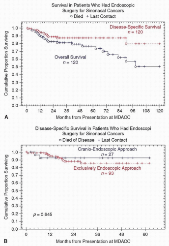
Over the last two decades, significant advances have been made in both the diagnosis and management of cancer of the nasal cavity and paranasal sinuses. The most significant advances in diagnosis are office endoscopy and high-resolution imaging. These diagnostic tools have allowed more accurate delineation of the extent of sinonasal tumors and, hence, improved treatment planning. Significant advances in treatment include progress made in cranial base surgery allowing for safe excision of tumors involving the cranial base. In addition, the development of microvascular free tissue transfer has made effective reconstruction of more extensive surgical defects possible. Advances have also been made in both planning and delivery of radiotherapy such as intensity-modulated radiation therapy (IMRT) and proton therapy. Both modalities allow optimal radiation dosimetry to the tumor while sparing normal surrounding tissue. Various new combinations of effective cytotoxic chemotherapeutic and targeted biologic agents are also being increasingly incorporated in the overall management of patients with sinonasal cancer.

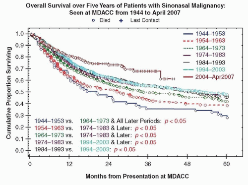
Recent advances made in diagnosis and treatment of patients with sinonasal cancer have clearly impacted our ability to control the disease and improve survival. Over the last 40 years, survival rates have improved from 25% to 40% in the 1960s to 65% to 75% in the last decade (Fig. 10.1). Despite these improvements, a significant number of patients die of their disease. The rarity of these tumors and the similarity of their presenting symptoms to more common benign conditions, coupled with the propensity for early spread and involvement of surrounding critical structures, are reflected in the fact that most patients still present with advanced stage disease. This has clearly hampered attempts to further improve prognosis. In this chapter, we present the current trends in diagnosis, classification, staging, and treatment of patients with cancers of the nasal cavity and paranasal sinuses. We also discuss some of the strategies to improve the outcome of these patients.


Figure 10.1. Improvement in 5-year OS of Patients with Sinonasal Malignancy. Data of 2,698 patients with sinonasal cancer seen at MD Anderson Cancer Center (MDACC) from 1944 to 2007.


ANATOMY


Nasal Cavity

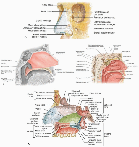
The nasal cavity is bounded by the bony pyriform aperture and the external framework of the nose (Fig. 10.2A). The nasal cavity opens anteriorly through the skin-lined nasal vestibule into the nares and communicates posteriorly through the choanae with the nasopharynx (Fig. 10.2B). The nasal cavity is divided in the midline by the nasal septum, which includes both cartilaginous and bony components (Fig. 10.2C). The cartilage of the septum is somewhat quadrilateral in form and is thicker at its margins than at its center. Its anterior margin is connected with the nasal bones and is continuous with the anterior margins of the lateral cartilages; below, it is connected to the medial crura of the greater alar cartilages by fibrous tissue (Fig. 10.2A). Its posterior margin is connected with the
perpendicular plate of the ethmoid, its inferior margin with the vomer and the palatine process of the maxilla.


Figure 10.2. A: Anatomy of the external nose. (From Chung KW, Chung HM, Halliday NL, eds. Gross Anatomy. 8th ed. Philadelphia, PA: Wolters Kluwer; 2015.) B: Anatomy of the nasal septum. (From Pansky B, Gest TR, eds. Lippincott’s Concise Illustrated Anatomy: Head and Neck (vol. 3). Philadelphia, PA: Wolters Kluwer Health; 2014.) C: Skeletal framework of the nasal septum. (From Chung KW, Chung HM, Halliday NL, eds. Gross Anatomy. 8th ed. Philadelphia, PA: Wolters Kluwer; 2015.)

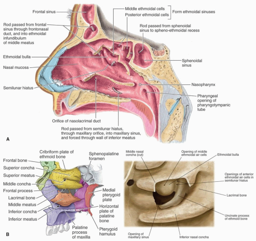
On the lateral nasal wall are the superior, middle, and inferior nasal turbinates, and below and lateral to each turbinate (concha) is the corresponding nasal passage or meatus (Fig. 10.3A and B). Above the superior turbinate is a narrow recess, the sphenoethmoidal recess, into which the sphenoid sinus opens. The superior meatus is a short oblique passage extending about halfway along the upper border of the middle turbinate; the posterior ethmoid cells open into the front part of this meatus. The middle meatus is below and lateral to the middle turbinate. The anatomy of the middle meatus is fully displayed by removing the middle turbinate (Fig. 10.3A and B). The bulla ethmoidalis is the most prominent anterior ethmoid air cell. The hiatus semilunaris is a curved cleft lying below and in front of the bulla ethmoidalis. It is bounded inferiorly by the sharp concave margin of the uncinate process of the ethmoid bone and leads
into a curved channel, the infundibulum, bounded above by the bulla ethmoidalis and below by the lateral surface of the uncinate process of the ethmoid. The anterior ethmoid air cells open into the front part of the infundibulum. The frontal sinus drains through the nasofrontal duct, which in ˜50% of subjects will also drain into the infundibulum; but when the anterior end of the uncinate process fuses with the front part of the bulla, this continuity is interrupted and the frontonasal duct then opens directly into the anterior end of the middle meatus. Below the bulla ethmoidalis, and partly hidden by the inferior end of the uncinate process, is the ostium of the maxillary sinus. An accessory ostium from the maxillary sinus is frequently present below the posterior end of the middle nasal concha. The inferior meatus is below and lateral to the inferior nasal turbinate. The nasolacrimal duct opens into the inferior meatus under cover of the anterior part of the inferior turbinate.


Figure 10.3. A: Anatomy of the lateral nasal wall. Removal of the middle turbinate demonstrates the anatomy of the middle meatus. (From Moore KL, Agur AMR, Dalley AF, eds. Clinically Oriented Anatomy. 7th ed. Philadelphia, PA: Wolters Kluwer Health; 2013.) B: Skeletal framework of the lateral nasal wall. Removal of the middle turbinate demonstrates the anatomy of the middle meatus. (Left image from Moore KL, Agur AMR, Dalley AF, eds. Clinically Oriented Anatomy. 7th ed. Philadelphia, PA: Wolters Kluwer Health; 2013. Right image from Pansky B, Gest TR, eds. Lippincott’s Concise Illustrated Anatomy: Head and Neck (vol. 3). Philadelphia, PA: Wolters Kluwer Health; 2014.)

The roof of the nasal cavity is narrow from side to side and slopes downward (at about a 30-degree angle) from front to back. The cribriform plate, which transmits the filaments of the olfactory nerve, forms the roof of the nasal cavity medial to the superior attachment of the middle turbinate. Lateral to the middle turbinate, the fovea ethmoidalis forms the roof of the ethmoid sinuses. Careful assessment of the anatomy of the
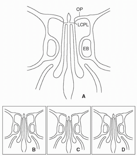
nasal roof, especially the relationship of the cribriform plate to the fovea ethmoidalis, is critical in avoiding a cerebrospinal fluid (CSF) leak during surgery in this region. The cribriform plate is usually at a slightly lower horizontal plane than the fovea ethmoidalis forming a shallow olfactory groove. This configuration is known as Keros type I (Fig. 10.4). However, the cribriform plate may be moderately or significantly lower than the fovea ethmoidalis resulting in a medium (Keros type II) or deep (Keros type III) olfactory groove. The topography of the roof may also be asymmetrical (Fig. 10.4).
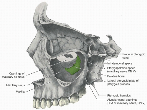


Figure 10.4. Anatomy of the ethmoid roof and lateral lamella of the cribriform plate. A: Keros type I. B: Keros type II. C: Keros type III. D: Asymmetrical ethmoid roof. Note that the right lateral lamella of the cribriform plate is very thin and long and is obliquely oriented including much of the right ethmoid roof. OP, orbital plate of frontal bone; LCPL, lateral cribriform plate lamella; EB, ethmoid bulla.

The floor of the nasal cavity is concave from side to side and almost horizontal anteroposteriorly. The palatine process of the maxilla forms the anterior three-fourths, and the horizontal process of the palatine bone forms the posterior fourth of the nasal floor (Fig. 10.2C).

The majority of the nasal cavity is lined by pseudostratified ciliated columnar epithelium, which contains mucous and serous glands (respiratory epithelium). Specialized olfactory epithelium lines the most superior portion of the nasal cavity and has direct connections with the olfactory tracts through openings in the cribriform plate.

The arteries of the nasal cavities are the anterior and posterior ethmoidal branches of the ophthalmic artery, which supply the ethmoid and frontal sinuses and roof of the nose. The sphenopalatine artery supplies the mucous membrane covering the lateral nasal wall. The septal branch of the superior labial artery supplies the anterior inferior septum. The veins form a close cavernous plexus beneath the mucous membrane. This plexus is especially well marked over the lower part of the septum and over the middle and inferior turbinates. Venous drainage follows a pattern similar to arterial supply. The lymphatic drainage from the anterior part of the nasal cavity, similar to that of the external nose, is to the submandibular group of lymph nodes (level I). Lymphatics from the posterior two-thirds of the nasal cavities and from the paranasal sinuses drain to the upper jugular (level II) and retropharyngeal lymph nodes.

The sensory nerves of the nasal cavity transmit either somatoautonomic or olfactory sensation. Somatoautonomic nerves include the nasociliary branch of the ophthalmic, which supplies the anterior septum and lateral wall. The anterior alveolar nerve, branch of the maxillary (V2), supplies the inferior meatus and inferior turbinate. The nasopalatine nerve supplies the middle of the septum. The anterior palatine nerve supplies the lower nasal branches to the middle and inferior turbinates. The nerve of the pterygoid canal (vidian) and the nasal branches from the sphenopalatine ganglion supply the upper and posterior septum and superior turbinate. The olfactory nerve fibers arise from the bipolar olfactory cells and unite in fasciculi, which form a plexus beneath the mucous membrane and then ascend passing into the skull through the foramina in the cribriform plate. Intracranially, olfactory nerve fibers enter the under surface of the olfactory bulb, in which they ramify and form synapses with the dendrites of the mitral cells of the olfactory tract.


Maxillary Sinus

The maxillary sinus (antrum of Highmore), the largest of the accessory sinuses of the nose, is a pyramidal cavity in the body of the maxilla (Figs. 10.5 and 10.6). Its base is formed by the lateral wall of the nasal cavity, and its apex extends into the zygomatic process. Its roof or orbital wall is frequently ridged by the infraorbital canal, whereas its floor is formed by the alveolar process of the maxilla and is usually 1 to 10 mm below the level of the floor of the nose. Projecting into the floor are several conical elevations corresponding with the roots of the first and second molar teeth, and in some cases, the floor is perforated by one or more of these roots. The natural ostium of the maxillary sinus is partially covered by the uncinate process and communicates with the lower part of the hiatus semilunaris of the lateral nasal wall (Figs. 10.3 and 10.5). An accessory ostium is frequently seen in, or immediately behind, the hiatus. The maxillary sinus appears as a shallow groove on the medial surface of the bone about the 4th month of fetal life but does not reach its full size until after the second dentition.


Ethmoid Sinus

The ethmoidal air cells consist of numerous thin-walled cavities situated in the ethmoidal labyrinth and bounded by the frontal, maxillary, lacrimal, sphenoid, and palatine bones. They lie in the upper part of the nasal cavity between the orbits (Fig. 10.6). The ethmoid sinuses are separated from the orbital cavity by a thin bony plate, the lamina papyracea. On either side, they are arranged in three groups, anterior, middle, and posterior. The anterior and middle groups open into the middle meatus of the nose, the former by way of the infundibulum, and the latter on or above the bulla ethmoidalis (Fig. 10.3). The posterior cells open into the superior meatus under cover of the superior nasal concha. Sometimes one or more ethmoid air cells extend over the orbital cavity (supraorbital ethmoid cells) or the optic nerve (Onodi cell). The ethmoidal cells begin to develop during fetal life.


Figure 10.5. Anatomy of the maxillary sinus (lateral wall removed). (From Scheid RC, Weiss G, eds. Woelfel’s Dental Anatomy. 8th ed. Philadelphia, PA: Wolters Kluwer Health; 2012.)


Figure 10.6. Anatomy of the ethmoid and maxillary sinuses (Coronal section). (From Moore KL, Agur AMR, Dalley AF, eds. Clinically Oriented Anatomy. 7th ed. Philadelphia, PA: Wolters Kluwer Health; 2013.)


Frontal Sinus

The paired frontal sinuses appear to be outgrowths from the most anterior ethmoidal air cells. They are situated behind the superciliary arches, are rarely symmetrical, and the septum between them frequently deviates to one or the other side of the middle line. Absent at birth, the frontal sinuses are generally fairly well developed between the 7th and 8th years but only reach their full size after puberty. The frontal sinus is lined with respiratory epithelium and drains into the anterior part of the corresponding middle meatus of the nose through the frontonasal duct, which traverses the anterior part of the labyrinth of the ethmoid. The soft tissues of the forehead are located anteriorly, the orbits are located inferiorly, and the anterior cranial fossa is located posteriorly (Fig. 10.3). Blood and neural supply is from the supraorbital and supratrochlear neurovascular bundles.


Sphenoid Sinus

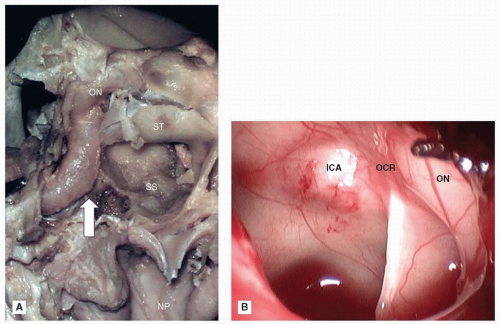
The sphenoid sinus begins at the most posterior and superior portion of the nasal cavity (Fig. 10.3). This midline structure, which is contained within the body of the sphenoid bone, is irregular and often has an eccentrically located intersinus septum. When exceptionally large, the sphenoid sinus may extend into the roots of the pterygoid processes or great wings and may pneumatize the basilar part of the occipital bone. The sphenoid sinus ostium is located on the anterior wall of the sinus and communicates directly with the sphenoethmoidal recess above and medial to the superior turbinate (Fig. 10.3). The sphenoid sinuses are present as minute cavities at birth, but their main development takes place after puberty. The posterior superior wall of the sphenoid sinus displays the forward convexity caused by the floor of the sella turcica, which contains the pituitary gland. The optic nerve and the internal carotid artery are closely related to the superior lateral wall of the sphenoid sinus, and their bony canals may be dehiscent within the sinus cavity (Fig. 10.7). Vascular and neural supplies come from the sphenopalatine and posterior ethmoidal arteries and the branches of the sphenopalatine ganglion, respectively.


Figure 10.7. A: Cadaver dissection of the sphenoid sinus (SS). The sinus is located in the midline superior to the nasopharynx (NP). The sella turcica (ST) forms a convexity in the posterior superior wall. The internal carotid artery (arrow) courses through the lateral wall of the sinus and is related superiorly to the optic nerve (ON). B: Endoscopic view of the left sphenoid sinus. Note the internal carotid artery (ICA) and optic nerve (ON) impressions on the lateral and superior walls. A bony septum within the sinus inserts into the opticocarotid recess (OCR).


Infratemporal Fossa

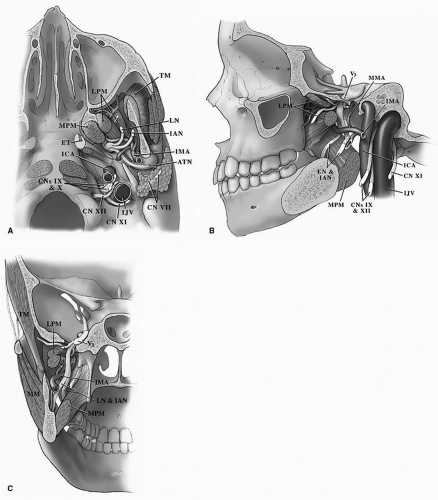
The infratemporal fossa is an irregularly shaped space, situated below and medial to the zygomatic arch. It is bounded anteriorly by the posterior surface of the maxilla; superiorly by the greater wing of the sphenoid and by the under surface of the squamous portion of the temporal bone; medially by the lateral pterygoid plate; and laterally by the ramus of the mandible. It contains the inferior aspect of the temporalis muscle and the medial and lateral pterygoid muscles (Fig. 10.8). It also contains branches of the internal maxillary vessels including the middle meningeal artery and the mandibular (V3) nerves including the lingual, inferior alveolar, and auriculotemporal nerves. The foramen ovale and foramen spinosum open on its roof and the alveolar canals on its anterior wall. The inferior orbital and pterygomaxillary fissures communicate with and may act as routes of spread of cancer to the infratemporal fossa. The infratemporal fossa also contains the upper carotid sheath including the internal carotid artery, internal jugular vein, and the last four cranial nerves (Fig. 10.8).


Figure 10.8. Anatomy of the infratemporal fossa: (A) inferior view, (B) lateral view, and (C) anterior view. TM, temporalis muscle; MM, masseter muscle; MPM, medial pterygoid muscle; LPM, lateral pterygoid muscle; IMA, internal maxillary artery; MMA, middle meningeal artery; ICA, internal carotid artery; IJV, internal jugular vein; LN, lingual nerve; IAN, inferior alveolar nerve; ATN, auriculotemporal nerve; V3, third division of the trigeminal nerve; ET, eustachian tube; CN, cranial nerve (VII, IX, X, XI, and XII).


Pterygopalatine Fossa

The pterygopalatine fossa (PPF) is a small, triangular space situated behind the maxillary sinus, in front of the pterygoid plates, and beneath the apex of the orbit. This fossa communicates with the orbit by the inferior orbital fissure, with the nasal cavity by the sphenopalatine foramen, and with the infratemporal fossa by the pterygomaxillary fissure (Fig. 10.5). Five foramina open into it. Of these, three are on the posterior wall, which are the foramen rotundum, the pterygoid canal, and the pharyngeal canal, in this order downward and medial. On the medial wall is the sphenopalatine foramen, and below is the superior orifice of the pterygopalatine canal (Fig. 10.5). The fossa contains the maxillary nerve, the sphenopalatine ganglion, and the terminal part of the internal maxillary artery.


The fissures and foramina of the PPF serve as “highways” for spread of cancer from the sinonasal region to the orbit, infratemporal fossa, and cranial base.


Anterior Cranial Fossa

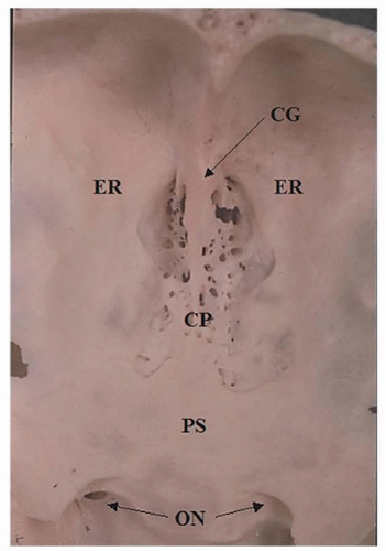
The floor of the anterior fossa is formed by the orbital plates of the frontal bone, the cribriform plate of the ethmoid, and the lesser wings and front part of the body of the sphenoid. In the midline, it presents, from anterior to posterior, the frontal crest for the attachment of the falx cerebri; the foramen cecum, which usually transmits a small vein from the nasal cavity to the superior sagittal sinus (SSS); and the crista galli, the free margin of which affords attachment to the falx cerebri (Fig. 10.9). On either side of the crista galli is the olfactory groove formed by the cribriform plate, which supports the olfactory bulb and presents foramina for the transmission of the olfactory nerves. Lateral to either olfactory groove are the internal openings of the anterior and posterior ethmoidal foramina; the anterior, situated about the middle of the lateral margin of the olfactory groove transmits the anterior ethmoidal vessels and the nasociliary nerve; the nerve runs in a groove along the lateral edge of the cribriform plate; and the posterior ethmoidal foramen opens at the back part of this margin under cover of the projecting lamina of the sphenoid and transmits the posterior ethmoidal vessels and nerve. More laterally, the cranial floor forms the orbital roof and supports the frontal lobes of the cerebrum. Farther back in the middle is the planum sphenoidale, forming the roof of the sphenoid sinus, and the anterior margin of the chiasmatic groove, running laterally on either side to the upper margin of the optic foramen (Fig. 10.9).


Orbit

The orbits are two quadrilateral pyramidal cavities, their bases being directed forward and lateral, and their apices backward and medial, so that their long axes diverge at a 45-degree angle and if continued backward would meet over the body of the sphenoid. The orbit is anatomically defined by seven bones (Fig. 10.10): frontal, zygomatic, maxillary, lacrimal, ethmoid, sphenoid, and palatine, and by the orbital
septum, which originates at the arcus marginalis, fusing with the levator aponeurosis above and the capsulopalpebral fascia below. It is bounded by the ethmoid and sphenoid sinuses at its medial aspect, the frontal sinus superomedially, the cranial vault superiorly and posteriorly, the temporal fossa laterally, and the maxillary sinus inferiorly. Each orbital cavity has a roof, a floor, a medial and a lateral wall, a base, and an apex.
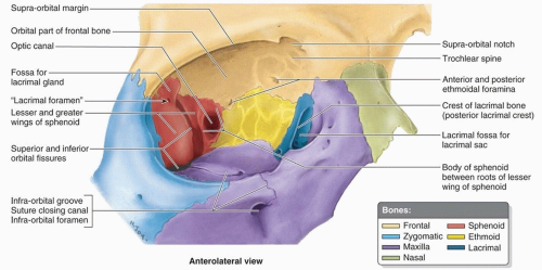


Figure 10.9. The floor of the anterior cranial fossa. The cribriform plate (CP) is characterized by the presence of foramina for the olfactory nerves on each side of the crista galli (CG), which is seen in the midline. Lateral to the CP is the ethmoidal roof (ER) and even more lateral the roof of the orbit. Posterior to the cribriform plate is the planum sphenoidale (PS). The optic nerves (ON) form the optic chiasm behind the planum sphenoidale.


Figure 10.10. Anatomy of the right orbit. (From Moore KL, Agur AMR, Dalley AF, eds. Clinically Oriented Anatomy. 7th ed. Philadelphia, PA: Wolters Kluwer Health; 2013.)

The roof is formed anteriorly by the orbital plate of the frontal bone and posteriorly by the lesser wing of the sphenoid. It presents medially the trochlear fovea for the attachment of the cartilaginous pulley of the superior oblique muscle and laterally the lacrimal fossa for the lacrimal gland.

The floor is formed mainly by the orbital surface of the maxilla, anteriorly and laterally by the orbital process of the zygomatic bone, and posterior and medially, to a small extent, by the orbital process of the palatine bone. At its medial angle is the superior opening of the nasolacrimal canal, immediately to the lateral side of which is a depression for the origin of the inferior oblique muscle. Running anteriorly near the middle of the floor is the infraorbital canal, ending anterior to the maxilla in the infraorbital foramen and transmitting the infraorbital nerve and vessels.

The medial wall is formed anteriorly to posteriorly by the frontal process of the maxilla, the lacrimal bone, the lamina papyracea of the ethmoid, and a small part of the body of the sphenoid anterior to the optic foramen. Anteroinferiorly, the lacrimal sac is situated between the anterior and posterior lacrimal crests at the junction between the medial wall and the floor. The lacrimal part of the orbicularis oculi arises from the posterior lacrimal crest. At the junction of the medial wall and the roof, the frontoethmoidal suture presents the anterior and posterior ethmoidal foramina, the former transmitting the nasociliary nerve and anterior ethmoidal vessels and the latter the posterior ethmoidal nerve and vessels. These foramina indicate the level of the cranial base within the orbit.

The lateral wall is formed by the orbital process of the zygomatic and the orbital surface of the greater wing of the sphenoid. On the orbital process of the zygomatic bone are the orbital tubercle (Whitnall) and the orifices of one or two canals, which transmit the branches of the zygomatic nerve. Between the roof and the lateral wall, near the apex of the orbit, is the superior orbital fissure (SOF). Through this fissure, the oculomotor, the trochlear, the ophthalmic division of the trigeminal (V1), and the abducens nerves enter the orbital cavity, also some filaments from the cavernous plexus of the sympathetic and the orbital branches of the middle meningeal artery. Passing posteriorly through the fissure are the ophthalmic vein and the recurrent branch from the lacrimal artery to the dura mater. The lateral wall and the floor are separated posteriorly by the inferior orbital fissure, which transmits the maxillary nerve (V2) and its zygomatic branch, the infraorbital vessels, and the ascending branches from the sphenopalatine ganglion.


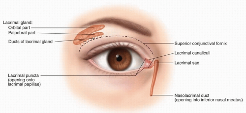
Figure 10.11. Anatomy of the lacrimal system. (From Pansky B, Gest TR, eds. Lippincott’s Concise Illustrated Anatomy: Head and Neck. Vol. 3. Philadelphia, PA: Wolters Kluwer Health; 2014.)

The base of the orbit (orbital rim), quadrilateral in shape, is formed superiorly by the supraorbital arch of the frontal bone, in which is the supraorbital notch or foramen for the passage of the supraorbital vessels and nerve; inferiorly by the zygomatic bone and maxilla, united by the zygomaticomaxillary suture; laterally by the zygomatic bone and the zygomatic process of the frontal joined by the zygomaticofrontal suture; and medially by the frontal bone and the frontal process of the maxilla united by the frontomaxillary suture.

The apex is situated in the posterior aspect of the orbit. The optic foramen is a short, cylindrical canal, through which passes the optic nerve and ophthalmic artery.

The extraocular muscles—four rectus muscles and two obliques—effect movement of the eye. The third cranial nerve innervates all but the lateral rectus and the superior oblique muscles, which are innervated by the fourth and sixth cranial nerves, respectively. The rectus muscles originate at the annulus of Zinn and insert on the globe forming a muscle cone, which is the central anatomic space in the orbit.

The lacrimal system is composed of secretory and drainage systems. Secretory glands—the glands of Moll, Krause, and Wolfring—may be found along the margin of the eyelid. The lacrimal gland with its palpebral and orbital lobes is located in the superotemporal orbit (Fig. 10.11). The lacrimal drainage system, located in the inferonasal orbit, is represented by
the puncta, canaliculi, lacrimal sac, and nasolacrimal duct. Tumor involvement of the lacrimal system may present with epiphora.

The skin of the eyelid is continuous with the palpebral and bulbar conjunctivae, which are, in turn, contiguous with the globe. Each of these epithelial surfaces represents a potential site of origin for cancer.


ETIOLOGY

The cause of sinonasal neoplasms is unknown. There is some epidemiologic evidence, however, to support an occupational risk for developing cancer of the sinonasal tract (SNT).1 Occupational exposure to inhalation of certain metal dusts or aerosols can cause loss of olfactory acuity, atrophy of the nasal mucosa, mucosal ulcers, perforated nasal septum, dysplasia of the nasal mucosa, or sinonasal cancer.2 Cancer of the nose and paranasal sinuses has been reported to be more frequent in workers exposed to nickel compounds in nickel refining, cutlery factories, and alkaline battery manufacture or to chromium in chromate production and chrome plating.3 In a report on the risk of developing sinonasal cancer in Scandinavian countries, nickel workers involved with electrolytic work for more than 15 years were found to have a 250-fold increased incidence of cancer of the sinus. In the same study, random biopsy of the middle turbinate showed evidence of dysplasia in 21% of workers. These changes were independent of their smoking history. All workers had been employed for at least 10 years, and there was an average latent period of 18 to 36 years before the development of carcinoma, most of which were squamous cell or anaplastic.4 Similarly, in a Swedish cohort of workers (n = 6,454) from seven aluminum foundries and three secondary aluminum (scrap) smelters, significantly elevated risk estimates for sinonasal cancer were observed.5

In animals, several heavy metals (e.g., Al, Cd, Co, Hg, Mn, Ni, Zn) have been shown to pass via olfactory receptor neurons from the nasal lumen through the cribriform plate to the olfactory bulb. Some metals (e.g., Mn, Ni, Zn) can even cross synapses in the olfactory bulb and migrate via secondary olfactory neurons to distant nuclei of the brain. The olfactory bulb tends to accumulate certain metals (e.g., Al, Bi, Cu, Mn, Zn) with greater avidity than other regions of the brain. The molecular mechanisms responsible for metal translocation in olfactory neurons and deposition in the olfactory bulb are unclear, but chelation by metal-binding molecules such as carnosine (beta-alanyl-L-histidine) may be involved.3

Other occupational exposures may also increase the risk of developing cancer of the SNT.6 A European case-control study revealed that exposure to leather and wood dust was associated with an excess risk of sinonasal cancer.7 Both wood and leather dusts were associated more with adenocarcinoma than SCC.6,8 Recent large-scale studies suggest that this increased risk is specifically linked to the development of intestinal-type adenocarcinoma rather than other types of sinonasal adenocarcinomas (SNACs).9,10,11 In European populations, occupation was associated with about 11% of all sinonasal cancers in women and 39% in men. A meta-analysis of 12 large case-control studies estimated that male wood workers had a summary odds ratio of sinonasal cancer of 2.6 (95% confidence interval [CI] = 2.1 to 3.3).12 The risk was greatest among men who had been employed in jobs with the highest wood dust exposure and increased with duration of exposure.13 Employment in the boot and shoe industry has been also associated with adenocarcinoma of the nasal cavity in England and Italy.14 Data from a case-control study conducted at 27 hospitals in France showed exposure to textile dust were associated with an elevated risk of SCC and adenocarcinoma of the SNT and that the risk increased with the duration and the level of exposure.15

Although epidemiologic studies have not addressed the relationship between outdoor air pollution and sinonasal malignant neoplasms, a report on the incidence of sinonasal cancer in urban polluted cities suggests such a correlation.16 Both primary and environmental (secondary) tobacco smoke appear to be also related to increase in the incidence of sinonasal cancer, particularly sinonasal squamous cell carcinoma (SNSCC).7,17,18 Recent studies have found a correlation in the development of lung cancer and SNSCC, propagating a possible causal relationship between SNSCC and smoking.19

High-risk human papillomavirus (HR-HPV) is an established cause of head and neck carcinomas arising in the oropharynx. The presence of HPV has also been reported in some carcinomas arising in the SNT, but little is known about their overall incidence or their clinicopathologic profile. In a recent study by Takahashi et al.20 of 70 patients with SNSCCs treated with surgery between 1999 and 2009 at MD Anderson Cancer Center (MDACC), high-risk HPV and its surrogate p16 were found in only 10% and 18% of tumor tissues, respectively. However, Bishop et al.21 reported higher prevalence rates of both high-risk HPV and p16 in sinonasal cancers. Of 161 sinonasal carcinomas, 34 (21%) were positive for HR-HPV DNA, including type 16 (82%), type 31/33 (12%), and type 18 (6%). Immunohistochemistry for p16 was positive in 59/161 (37%) cases, and p16 expression strongly correlated with the presence of HPV DNA: 33 of 34 (97%) HPV-positive tumors exhibited high p16 expression, whereas only 26 of 127 (20%) HPV-negative tumors were p16 positive (p < 0.0001). A trend toward improved survival was observed in the HPV-positive group (hazard ratio [HR] = 0.58, 95% CI [0.26, 1.28]). The presence of HR-HPV in 21% of sinonasal carcinomas suggests that HPV may be an important oncologic agent of carcinomas arising in the SNT. Although nonkeratinizing SCC is the most common histologic type, there is a wide morphologic spectrum of HPV-related disease that includes a variant that resembles adenoid cystic carcinoma (ACC). The distinctiveness of these HPV-related carcinomas of the SNT with respect to risk factors, clinical behavior, and response to therapy remains to be clarified.21


PATHOLOGY

The mucosal lining of the nose—the schneiderian membrane— is derived from ectoderm. This is uniquely different from the mucosa of the rest of the upper respiratory tract, which is derived from endoderm. Olfactory neuroepithelium lines the superior portion of the nasal cavity and the roof of the nose and gives rise to neuroectodermal tumors such as olfactory neuroblastoma and neuroendocrine carcinoma (NEC).22,23,24 The sinonasal epithelium also has minor salivary glands (SGs) and gives rise to SG tumors such as ACC and mucoepidermoid carcinoma (MEC).25,26,27,28 However, the most common epithelial neoplasms of the SNT are those arising from “metaplastic” squamous epithelium namely SCC and those originating from the seromucinous glands of the mucosal lining, collectively known as adenocarcinomas.29 The unique histology of this
region is reflected in the histogenesis of a complex variety of epithelial and nonepithelial tumors (Table 10.1). These tumors have a wide range of biologic behavior, and a few arise only in the SNT (e.g., inverted papilloma, olfactory neuroblastoma). Nonepithelial tumors are similar to those in other regions in the head and neck.


Table 10.1 Tumors of the Sinonasal Tract


Benign


Epithelial


Papilloma


Adenoma


Dermoid


Nonepithelial


Fibroma


Chondroma


Osteoma


Neurofibroma


Hemangioma


Lymphangioma


Nasal glioma


Intermediate


Schneiderian papilloma


Inverted


Papillary


Cylindrical


Angiofibroma


Ameloblastoma


Fibrous dysplasia


Ossifying fibroma


Giant cell tumor


Malignant


Epithelial


Squamous cell carcinoma


Differentiated (well, moderately, poorly)


Basaloid squamous


Adenosquamous


Nonsquamous cell carcinoma


Adenoid cystic carcinoma


Mucoepidermoid carcinoma


Adenocarcinoma


Neuroendocrine carcinoma


Hyalinizing clear cell carcinoma


Melanoma


Olfactory neuroblastoma


Sinonasal undifferentiated carcinoma (SNUC)


Nonepithelial


Chondrosarcoma


Osteogenic sarcoma


Chordoma


Soft tissue sarcoma


Fibrosarcoma


Malignant fibrous histiocytoma


Hemangiopericytoma


Angiosarcoma


Kaposi sarcoma


Rhabdomyosarcoma


Lymphoproliferative


Lymphoma


Polymorphic reticulosis


Plasmacytoma


Metastatic


Renal


Lung


Breast


Ovary


INCIDENCE

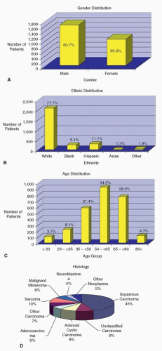
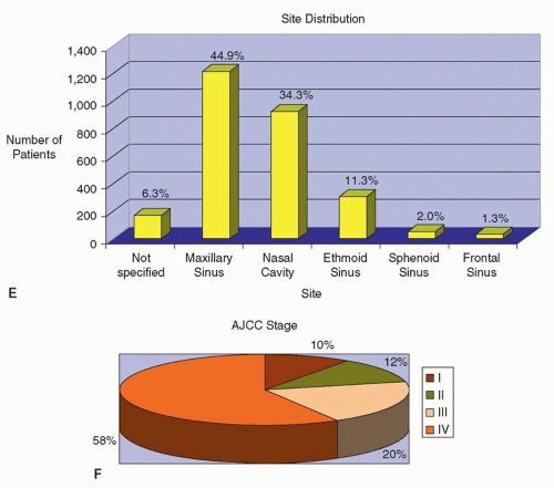
Overall, sinonasal cancer accounts for about 1% of all malignancies and ˜3% of cancer of the head and neck. There is a male predominance (Fig. 10.12A), with a strong predilection for Caucasians (Fig. 10.12B). The majority of patients are over 50 years of age at the time of diagnosis (Fig. 10.12C). The most common malignant tumor of the nasal cavity and paranasal sinuses is SCC (Fig. 10.12D). Although the maxillary antrum is the most commonly involved sinus (Fig. 10.12E), anterior skull base invasion is most frequently encountered with malignant neoplasms of the nasal cavity and ethmoid sinus. Upward extension of these neoplasms toward the cribriform plate or fovea ethmoidalis is not uncommon and heralds intracranial extension.30 Primary carcinoma of the frontal sinus is uncommon, and those arising in the sphenoid sinus are rare.31 Unfortunately, despite significant improvement in diagnostic techniques such as nasal endoscopy and highresolution imaging, most patients present with advanced stage disease (Fig. 10.12F).


SPREAD


Local Spread

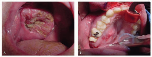
The most common route of local spread of cancer of the SNT is through direct extension. As most sinonasal cancers are relatively asymptomatic when small, it is often the manifestation of local spread that prompts patients to seek medical attention. In the maxillary sinus, direct extension may occur anteriorly into the soft tissues of the cheek, superiorly into the orbit with resultant proptosis and diplopia, inferiorly into the oral cavity, or posteriorly into the pterygomaxillary space where it may spread along the branches of the maxillary division of the trigeminal nerve (V2). Cancer of the frontal sinus is quite rare, but the most significant direct extension is posteriorly to the frontal lobes. Cancer of the ethmoid sinus often presents with medial extension to the orbit, superior extension to the cribriform plate, and posterior extension into the sphenoid sinus and nasopharynx. Cancers involving the sphenoid sinus may quickly become problematic because of proximity to the optic nerves, the cavernous sinus (CS), and the pituitary fossa (Fig. 10.7).

In addition to direct local extension, cancer of the paranasal sinuses can spread to nearby structures via the many fissures and foramina located in this region. Cancer of the maxillary sinus frequently erodes posteriorly into the PPF (Fig. 10.8). Once in the PPF, the tumor may extend laterally through the pterygomaxillary fissure into the infratemporal fossa, superiorly into the orbit via the inferior orbital fissure or into the middle cranial fossa through the foramen rotundum, posteriorly into the vidian canal with extension to the petrous portion of the temporal bone, or inferiorly into the oral cavity by way of the palatine canal or the sphenopalatine foramen.

From the frontal sinus, cancer may extend into the nasal cavity through the nasofrontal duct. Cancer of the ethmoidal sinuses may also extend into the nasal cavity through the middle meatus and the sphenoethmoidal recess, posteriorly into the nasopharynx and along the eustachian tube, or inferiorly along the nasolacrimal duct (Fig. 10.3).


Figure 10.12. A-F: Patients with sinonasal cancer seen at MD Anderson Cancer Center between 1944 and April 2007 (n = 2,698 patients).


Figure 10.12. (Continued)


Perineural Spread

The dissemination of cancer cells along nerves is a frequent pathologic finding among a variety of cancers, including head and neck, upper gastrointestinal, pancreatic, and prostate carcinomas. Tumors that have a considerable propensity to disseminate along nerves are known as neurotropic cancers. In the head and neck, the most common tumors with a predilection to invade nerves are ACCs, followed by SCCs.32,33 Tumors of the paranasal sinus that exhibit perineural invasion (PNI) may use this route to spread in a retrograde fashion to the skull base and even progress intracranially. Alternatively, they may spread in an antegrade fashion and along the involved nerve and its terminal branches. In either case, this neural spread makes surgical resection more complicated and achieving negative surgical margins less certain. Imaging particularly MRI is critical in determining the extent of neural spread of sinonasal cancers as will be discussed later in this chapter under the section on Imaging.34

Gil et al.35 reported the incidence and pattern of neural invasion (NI) in 208 patients with cancers of the paranasal sinuses and anterior skull base. Forty-one specimens (20%) had evidence of NI. Sinonasal undifferentiated, adenoid cystic, and SCC had a high propensity for NI, whereas melanoma and sarcoma rarely invaded nerves. Intraneural invasion was found in 32% of these cases, and 34% invaded more than 1 cm distal to the tumor. NI was associated with a high rate of positive margins, maxillary origin, and previous surgical treatment (p < 0.04) but not with stage, orbital invasion, or dural invasion. Patients with NI were more likely to undergo adjuvant radiotherapy (p = 0.003), which significantly improved survival in patients with minor SG carcinomas (p = 0.04).


Regional Metastases

The lymphatic drainage of the posterior nasal cavities and paranasal sinuses is primarily to the retropharyngeal and lateral pharyngeal nodes at the base of the skull and then to the upper jugular lymph nodes. Cancer of the anterior nasal cavity and those that erode through the maxilla into the soft tissues of the face spread to the submandibular and upper jugular lymph nodes.

Regional metastases from paranasal sinus cancer are relatively uncommon and have been characterized to a greater extent for maxillary sinus cancer than for other paranasal sites.36 Regional disease is evident on initial presentation in ˜10% of patients, and an additional 15% of patients will develop lymph node metastasis at some point after treatment. In patients with SCC of the maxillary sinus, the risk of having lymph node metastasis on presentation correlates with extension of the primary tumor to the nasopharynx or oral cavity. The risk of developing regional metastasis after treatment correlates with local tumor recurrence. The role of elective neck dissection or radiation has yet to be defined in patients with cancer of the maxillary sinus.37 Development of regional metastasis, however, is associated with worse prognosis.


Distant Metastases

Although distant metastasis from cancer of the paranasal sinus does occur, failure to control the disease secondary to local recurrence is far more common. For SCC of the maxillary sinus, the rate of distant metastasis is ˜10% and rarely occurs in the absence of local recurrence. Cancer of the ethmoid has a similar rate of distant metastasis, with adenocarcinoma having a slightly higher rate than squamous cell cancer (15% to 20% vs. 10%). In general, the most common sites for metastasis are the lung and bone.36,37,38


STAGING

The most widely used staging system for sinonasal cancers is the American Joint Committee on Cancer (AJCC) tumornode-metastasis (TNM) system. There is a different staging system for tumors of the maxillary sinus from that used for ethmoid sinus and nasal cavity tumors. The nodal staging system for maxillary, ethmoid, and nasal cavity tumors is the same as for other sites in the head and neck and depends on the number, size, and laterality of involved lymph nodes. There is a separate staging system for esthesioneuroblastoma (ENB). The classification from the most recent version, 7th edition,39 is shown in Table 10.2.


Table 10.2 Classification of Sinonasal Cancer According to the AJCC 7th Edition


Primary Tumor (T stage)


Maxillary sinus T1


T2


T3


T4a


T4b


Limited to the maxillary sinus mucosa with no erosion or bone destruction


Bone erosion/destruction including hard palate, middle nasal meatus, except for posterior wall of maxillary sinus and pterygoid plates


Invasion of bone of posterior wall of maxillary sinus, subcutaneous tissues, floor or medial wall or orbit, pterygoid fossa, ethmoid sinuses


Invasion of anterior orbital contents, skin of cheek, pterygoid plates, infratemporal fossa, cribriform plate, sphenoid or frontal sinuses


Invasion of orbital apex, dura, brain, middle cranial fossa, nasopharynx, clivus, or cranial nerves except for V2


Nasal cavity and ethmoid sinus


T1


T2


T3


T4a


T4b


Limited to any one subsite, with or without bony invasion


Invasion into two subsites in a single region or extending to adjacent region in the nasoethmoidal complex, with or without bony invasion


Invasion of medial wall or floor of orbit, maxillary sinus, palate or cribriform plate


Invasion into anterior orbital contents, skin of nose or cheek, minimal extension into anterior cranial fossa, pterygoid plates, sphenoid or frontal sinuses


Invasion into orbital apex, dura, brain, middle cranial fossa, nasopharynx, clivus, or cranial nerves other than V2


Olfactory esthesioneuroblastoma


T1


T2


T3


T4


Tumor isolated to nasal cavity and ethmoid sinuses


Tumor extends to sphenoid sinus or cribriform plate


Tumor extends to anterior cranial fossa or orbit, no dural invasion


Tumor invades dura or brain parenchyma


Regional Lymph Nodes (N stage)a


NO


N1


N2a


N2b


N2c


N3


No regional lymph node metastasis


Metastasis in a single ipsilateral lymph node, ≤3 cm in greatest dimension


Metastasis in a single ipsilateral lymph node, >3 cm and ≤6 cm


Metastasis in multiple ipsilateral lymph nodes, none >6 cm


Metastasis in bilateral or contralateral lymph nodes, none >6 cm


Metastasis in a lymph node >6 cm


Distant Metastatic Disease (M stage)b


M0


M1


No distant metastasis


Distant metastasis present


a Definitions apply to all subsites except for olfactory ENB, which uses a N0 vs. N1 system for positive and negative nodal metastases, respectively.

b Definitions apply to all subsites.


From Edge SB, et al. AJCC Cancer Staging Manual. 7th ed. New York, NY: Springer; 2010, Reference 39.


Used with the permission of the American Joint Committee on Cancer (AJCC), Chicago, Illinois. The original source for this material is the AJCC Cancer Staging Manual, Seventh Edition (2010) published by Springer Science and Business Media LLC, www.springer.com.


PATIENT EVALUATION

Clinical evaluation of patients with cancer of the nasal cavity, paranasal sinuses, and orbit should help to achieve three objectives: (1) establishment of the diagnosis, (2) determination of the extent of disease, and (3) development of a plan for treatment. These objectives are usually achieved through a detailed history, comprehensive clinical examination of the head and neck, imaging, and biopsy.


Imaging

Imaging of the nasal cavity, paranasal sinuses, and orbit is indicated whenever there is clinical suspicion of a neoplastic process. Imaging is also indicated for obtaining pretreatment information regarding the location, size, extent, and invasiveness of the primary tumor, as well as the presence of regional and distant metastasis. Such information is crucial in deciding on therapeutic options and for proper preoperative planning of the optimal surgical approach. Imaging also plays an important role in the posttreatment follow-up, indicating areas of residual or recurrent disease, and defining suspicious areas for biopsy.

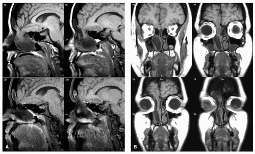
Both CT and MRI might be needed for optimum radiologic assessment of sinonasal malignancy, particularly in assessing the cranial base, orbit, and pterygopalatine and infratemporal fossae. Coronal images best delineate involvement of the orbital walls and invasion of the skull base, particularly the cribriform plate. Axial images are particularly helpful in demonstrating tumor extension through the posterior wall of the maxillary sinus into the PPF and infratemporal fossae. Sagittal images are particularly helpful in evaluating extension along the cribriform plate, planum sphenoidale, and clivus (Fig. 10.16).

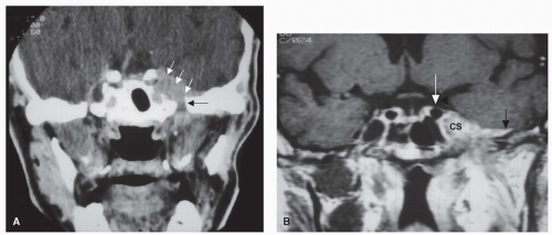
The main advantage of CT scans is in delineating the architecture of the bones, especially in “bone windows” (Fig. 10.16A and B). The addition of contrast enhancement increases tumor definition from adjacent soft tissue, especially intracranially. Bone destruction and soft tissue invasion suggest an aggressive lesion, usually a malignant neoplasm. Widening or sclerosis of the foramina of the infraorbital, vidian, mandibular, or maxillary nerves may indicate perineural spread (Fig. 10.17).

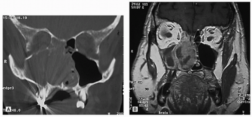
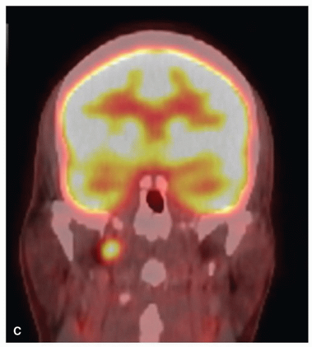
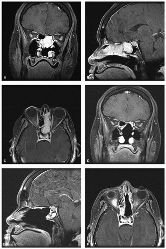
MRI with its superior soft tissue contrast and multiplanar capability is superior to CT in pretreatment evaluation of primary malignant tumors of sinonasal cavity.40 MRI is unsurpassed in delineating soft tissue detail, both intraand extracranially (Fig. 10.16). Obliteration of fat planes in the PPF, infratemporal fossa, and nasopharynx usually indicates tumor transgression along these boundaries. Dural thickening or enhancement is usually an indication of tumor involvement, and evaluation of critical structures such as the brain and carotid artery is best delineated by MRI. Similarly, enhancement or thickening of cranial nerves indicates perineural spread, which is better detected on MRI than CT34 (Fig. 10.17). Perhaps one of the most significant advantages of MRI is the ability to distinguish tumor from retained secretions secondary to obstruction of sinus drainage (Fig. 10.18). MRI is also particularly helpful in monitoring patients in the postoperative follow-up period, although this role may be supplanted by positron emission tomography (PET) scans because of its ability to distinguish between tumor recurrence and posttreatment fibrosis. PET-CT is also helpful in delineating regional and distant metastasis (Fig. 10.19).

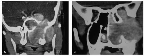
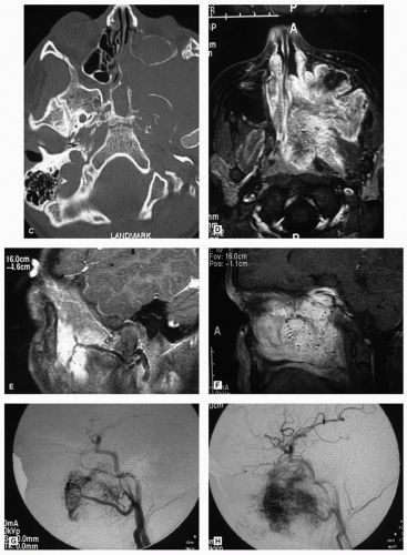
Angiography is not indicated in the routine assessment of patients with neoplasms of the nose, paranasal sinuses, and orbit. In certain selected cases, however, angiography may be necessary. These cases include vascular neoplasms of the sinonasal region, where angiography will not only delineate the tumor extent and the blood supply but also permit the use of selective embolization of the vascular supply to the tumor (Fig. 10.20). This reduces intraoperative blood loss, facilitating surgical resection.


Biopsy

The definitive diagnosis of a neoplasm of the nasal cavity and paranasal sinuses relies on expert histopathologic review of any biopsy specimens by a head and neck pathologist to confirm the exact diagnosis prior to treatment. This is critical because the treatment and prognosis of sinonasal cancer is greatly influenced by histology.41,42 This is particularly true for neuroendocrine tumors in which the misdiagnosis rate is particularly high. In a study from MDACC, patients referred with a presumed diagnosis of ENB were frequently reclassified to other types of tumors including NEC, sinonasal undifferentiated carcinoma (SNUC), melanoma, and even pituitary adenoma.43 The implications of this misdiagnosis are far reaching and required significant alteration in the initially proposed treatment plan in a significant number of patients.


Biopsy of Tumors of the Nasal Cavity

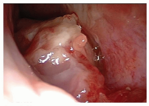
The vast majority of sinonasal neoplasms are accessible for biopsy through a strictly endonasal approach. A wide variety of rigid nasal endoscopes offer superb visualization of intranasal lesions with a high degree of optical resolution


and bright illumination (Fig. 10.13). The application of topical anesthetics and decongestants improves visualization and allows thorough examination of the nasal cavity. The site of origin of the lesion and its relation to the nasal walls (septum, floor, roof, and lateral nasal wall) should be noted. An adequate specimen should be obtained, avoiding crushing of tissue, and submitted for histopathologic examination. If the diagnosis of lymphoma is suspected, fresh tissue should be sent in saline, rather than fixed in formalin. Most endonasal biopsies can be performed in the outpatient setting with minimal discomfort to the patient. In certain cases, the diagnosis of a highly vascular neoplasm, such as angiofibroma, may be suspected on clinical grounds. Under these circumstances, it is prudent not to perform the biopsy until imaging and angiography (possibly with embolization) are performed (Fig. 10.20). Preoperative biopsy can then be performed in the operating room under controlled conditions to confirm the diagnosis before surgical resection. If a nasal mass is suspected to have an intracranial communication such as an encephalocele, meningocele, or nasal glioma, this should be confirmed with imaging to avoid inadvertent CSF leak and subsequent meningitis (Fig. 10.21).
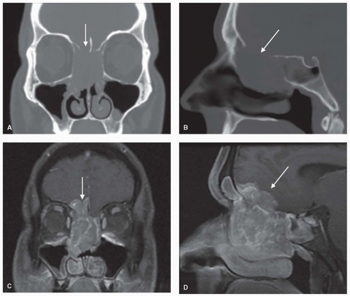


Figure 10.16. Ethmoid carcinoma. Coronal and sagittal images showing bony destruction on CT scan (A, B) and intracranial invasion (arrows) on T1-MRI with contrast (C, D).


Figure 10.17. Perineural spread of ACC along the third division of the trigeminal nerve (V3). A: A coronal CT with IV contrast showing widening of the left foramen ovale (black arrow), compared to the one on the right. There is also enhancement and thickening along the left Meckel cave (white arrows). B: A coronal T1-weighted MRI with gadolinium showing marked thickening and enhancement of V3, trigeminal ganglion, and the lateral cavernous sinus (CS). The tumor abuts the cavernous carotid artery (white arrow). There is enhancement of the dura along the floor of the middle cranial fossa (black arrow). This “dural tail” is usually a sign of involvement of the dura with tumor.


Figure 10.18. Sinonasal melanoma. A: Coronal CT scan demonstrating opacification of the right nasal cavity as well as the maxillary and ethmoid sinuses. There appears to be destruction of the lateral nasal wall and the nasal septum. The lesion is abutting the orbital floor and the cribriform plate, but it is unclear whether or not these structures are involved. B: Coronal T1-weighted MRI with gadolinium of the same patient revealing that the lesion is limited to the nasal cavity and ethmoid sinuses and that the changes in the maxillary sinuses are due to retained secretions secondary to obstruction of the ostium, rather than soft tissue involvement. It also demonstrates that the lesion does not invade the orbit or the cranial base. The presence of low signal areas within the lesion gives it a heterogeneous appearance, which is characteristic of sinonasal melanoma.


Figure 10.19. PET-CT of the head and neck. These images are from the same patient whose CT and MRI are depicted in Figure 10.16. The fused PET-CT images show ethmoid carcinoma (A) with metastasis to the retropharyngeal lymph nodes (B, C), which were not detected on CT or MRI.


Figure 10.19. (Continued)


Figure 10.20. Juvenile nasopharyngeal angiofibroma. Coronal (A, B) and axial (C) CT with contrast and axial T1 axial MRI with contrast (D) showing the mass in the nasal cavity, maxillary sinus, nasopharynx, sphenoid sinus, pterygoid plates, and pterygopalatine and infratemporal fossa. The mass involves the floor of the middle cranial fossa and extends intracranially to the cavernous sinus. T1 sagittal MRI (E, F) show flow voids of increased extensive vascular supply coming from the internal maxillary artery (E) and internal carotid artery (F). Early-phase angiogram showing the blood supply from the internal maxillary artery (G) and late phase showing significant tumor vascular blush and contribution from the internal carotid artery (H).


Biopsy of Tumors of the Paranasal Sinuses

In the unusual case where a paranasal sinus neoplasm is confined to the sinus cavity and does not present itself intranasally, a biopsy should be obtained by direct access to the involved sinus. Tumors of the maxillary sinus can still be accessed through an endoscopic approach by creating a wide antrostomy in the region of the natural ostium. Otherwise, the maxillary sinus is accessible through a sublabial incision in the canine fossa via an anterior antrostomy. The ethmoidal sinus can be approached endonasally through endoscopic ethmoidectomy. Alternatively, an external ethmoidectomy approach provides a direct access via a Lynch incision.

The sphenoid sinus is easily approached endoscopically in the vast majority of cases. The use of C-arm fluoroscopy or more recently computer-assisted three-dimensional (3-D) intraoperative imaging is sometimes used in cases with difficult access or unusual anatomy. Isolated tumors of the frontal sinus are rare. A trephination through the floor of the sinus is utilized for biopsy of lesions within its cavity.


Figure 10.20. (Continued)


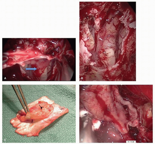
Figure 10.21. Meningoencephalocele. Sagittal (A) and coronal (B) T1 MRI with gadolinium showing a defect in the anterior cranial base at the right fovea ethmoidalis and cribriform plate. There is herniation of a meningoencephalocele, which presented as a nasal mass. Inadvertent biopsy of such lesions may lead to CSF leaks and should be avoided.


Dec 18, 2016 | Posted by in ONCOLOGY | Comments Off on Cancer of the Nasal Cavity and the Paranasal Sinuses

Full access? Get Clinical Tree
Get Clinical Tree app for offline access