Cancer of the Head and Neck in the Pediatric Population


Cancer of the Head and Neck in the Pediatric Population


Catherine G. Lam

Atmaram Pai Panandiker

Jon P. Ver Halen

Noah D. Sabin

Sandeep Samant


INTRODUCTION


Epidemiology

It is estimated that 15,780 new cases of cancer and 1,960 cancer-related deaths occurred among children and adolescents below age 20 in the United States in 2014.1 Tumors of the head and neck region account for 5% to 10% of all childhood malignancies.2,3 Similar to other childhood cancers, the incidence of head and neck malignancies in the pediatric age group has demonstrated apparent increases in the United States in recent decades.2 Given the potential for pediatric head and neck tumors or their treatment to adversely affect critical structures, functional outcomes, and quality of life, coupled with the rarity and diversity of many such tumors, multidisciplinary team management with expertise specializing in pediatric cancer is obligatory.

In an analysis using the National Cancer Institute’s population-based tumor registry, Surveillance, Epidemiology, and End Results (SEER), 3,050 children younger than age 19 years with head and neck tumors were identified from 1973 through 1996. Dominant histologic categorizations included lymphoma (27%), neural tumors (including neuroblastoma and retinoblastoma, 23%), thyroid cancers (21%), and soft tissue sarcomas (12%). The most common individual diagnoses were papillary thyroid carcinoma (n = 537), Hodgkin lymphoma (HL) (n = 515), retinoblastoma (n = 497), non-Hodgkin lymphoma (NHL) (n = 301), and rhabdomyosarcoma (n = 239). Unlike cancers of the head and neck among adults, squamous cell carcinomas in pediatric patients are rare, accounting for fewer than 2% of cases.2 Another recent analysis, looking only at sarcomas of the head and neck in the SEER database from 1973 through to 2010, identified 1,244 pediatric cases (including up to age 19 years) and 11,481 adult cases, with 10-year survival of 71% in pediatric patients compared to 61% in adult patients.4 In this analysis of head and neck sarcomas, the most commonly reported histologies were rhabdomyosarcoma (48%), malignant fibrous histiocytoma (MFH, 11%), osteosarcoma (8%), and Ewing sarcoma (6%).
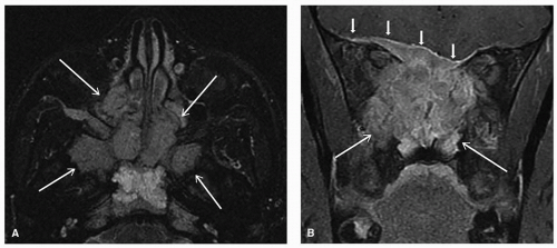
4

The age of presentation can provide important initial guidance for conditions likely to be malignant arising in the head and neck. From the SEER database, retinoblastoma, neuroblastoma, germ cell tumors, and rhabdomyosarcoma are among the most common conditions among neonates and infants.2 Among children below age 10 years, one can see various conditions, including NHL and HL. Among children and adolescents above age 10 years, dominant malignancies of the head and neck include thyroid cancer, Hodgkin and non-Hodgkin lymphoma, and melanoma.2 Certain tumors are rarely seen outside of their typical age of presentation, including malignant germ cell tumors involving the head and neck, which are mainly seen only in neonates or infants,5 and retinoblastomas, which are rarely seen in children older than 5 years.

Of note, clinical reviews of malignancies of the head and neck do not consistently include important differential diagnoses such as retinoblastoma in the pediatric age group, and leukemia presenting with chloromas. In addition to primary cancers arising in the head and neck, conditions such as malignant germ cell tumor, lymphoma, and neuroblastoma may also metastasize to the nodes, soft tissue, or bones of the head and neck.

Secondary malignant tumors of the head and neck, that is, those resulting from prior therapy including radiation therapy and alkylating chemotherapy agents, account for about 0.8% of tumors of the head and neck in the pediatric age group2 and may include thyroid carcinoma, osteosarcoma, MFH, and salivary gland tumors.6,7


Diagnostic Evaluation: An Overview

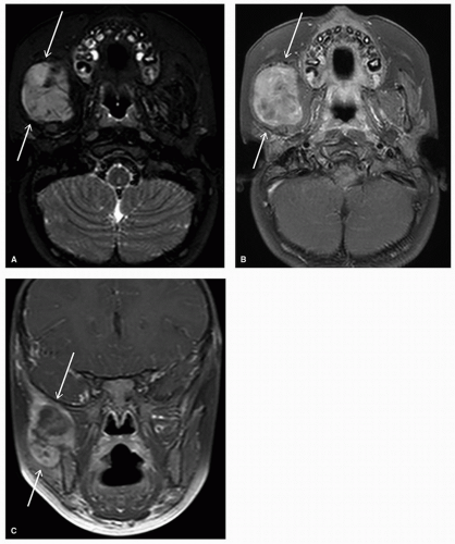
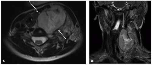
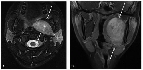
As most masses in the head and neck in pediatric age group are benign, careful clinical history and physical examination with additional workup when appropriate are warranted to diagnose patients with malignant lesions. Conditions to be considered include lesions primarily arising in the head and neck, those that are metastatic to the head and neck, and secondary malignancies. Primary head and neck lesions can include congenital or developmental lesions, inflammatory or infectious masses, benign tumors, or malignant tumors (Table 24.1).

Assessment of a child with a mass in the head and neck should start with a detailed clinical, past medical, and family history, immunization history, and recent and current medications. History should detail the onset, nature, and timing of any changes associated with the mass, along with potential exposures and risk factors for infection (e.g., tuberculosis), travel, and exposure to animals (e.g., zoonoses).

Congenital or developmental lesions may first be evident in the early postnatal period, but in some cases they can manifest later in life, with gradual growth over time or with concomitant secondary infection. Benign tumors can similarly sometimes be brought to medical attention in the setting of concurrent infection. Whereas inflammatory/infectious etiologies are typically first considered for masses that acutely enlarge and are associated with acute fever, tenderness, or skin changes such
as erythema or warmth, red flags should prompt consideration of an underlying malignancy. Potential red flags include firm, fixed/immobile or large (>1.5 to 2 cm) masses; lymph nodes in atypical locations (e.g., supraclavicular) or multiple locations; masses initially queried to be inflammatory or infectious in nature that do not respond to initial empiric management or persist beyond 4 to 6 weeks; or constitutional symptoms such as malaise, weight loss, anorexia, and unexplained fevers or night sweats.


Table 24.1 Sample Differential Diagnoses for Head and Neck Masses


Common Presenting Site


Differential Diagnosesa


Congenita


Inflammatory/Infectious


Benign Tumors


Malignant Tumors


Lateral neck mass


▪ Cervical rib


▪ Branchial cleft cyst


▪ Reactive lymphadenopathy (e.g., viral, bacterial, mycobacterial, granulomatous)


▪ Sialadenitis


▪ Focal myositis


▪ Torticollis


▪ See nonlocalized


▪ Hodgkin lymphoma


▪ Non-Hodgkin lymphoma


▪ Retinoblastoma


Midline neck mass


▪ Thyroglossal duct cyst


▪ Dermoid cyst


▪ Thymic cyst


▪ Lymphatic Malformation


▪ Thyroid disease


▪ Thyroid nodule


▪ Tracheal and endobronchial tumors


▪ Parathyroid adenoma


▪ Paraganglioma


▪ Thyroid carcinoma


▪ Tracheal and endobronchial malignancies


▪ Parathyroid carcinoma


▪ Malignant paraganglioma


Nonlocalized or various


▪ Vascular malformation


▪ Ludwig angina


▪ Abscesses


▪ Hemangioma


▪ Lipoma


▪ Fibroma


▪ Neurofibroma


▪ Lymphangioma


▪ Salivary gland tumors


▪ Hodgkin lymphoma


▪ Non-Hodgkin lymphoma


▪ Thyroid carcinoma


▪ Rhabdomyosarcoma


▪ Neuroblastoma


▪ Nasopharyngeal carcinoma


▪ Germ cell tumor


▪ Desmoid tumor


▪ Salivary gland malignancies


▪ Skin malignancies


▪ NUT midline carcinoma


▪ Metastatic cancer


a Note that variants of some conditions may appear in more than one category.


Relevant clinical and functional impairments to assess on history and to observe on examination may include abnormal eye movements; distorted vision; proptosis; Horner syndrome; cranial nerve palsies; paresthesias; recurrent epistaxis; increasing nasal congestion; evidence of recent suspected/confirmed infections; loss of smell; stridor; dysphagia and odynophagia; impaired gag; trismus; deviated tongue; dental health; change in tooth position; change in voice or hoarseness; otorrhea; progressive tinnitus or hearing loss; otalgia; vertigo; neck tilt or torticollis; facial and/or neck swelling; deviated trachea; increased vascular markings, pulsatility, or overlying skin changes; or features of hypothyroidism or hyperthyroidism.

Physical examination should also include evaluation for movement of the mass with tongue protrusion (e.g., thyroglossal duct cysts, which typically move upward with tongue protrusion) and include a full systemic examination for systemic lymphadenopathy, with particular vigilance for lymph nodes in atypical sites such as the supraclavicular area or that are firm, fixed, and significantly enlarged; ophthalmologic examination; assessment of tonsillar tissue; oral mucosal examination; thyroid examination; cardiorespiratory examination; abdominal examination for masses or hepatosplenomegaly; genitourinary examination; skin examination for evidence of petechiae or purpura or neurocutaneous markings; dysmorphic features; detailed neurologic examination; and note of any current or impending functional impairment from mass effect.

Any concern for potential airway compromise must be immediately evaluated and managed as an oncologic emergency. As tumors in the head and neck may expand into cavities such as the maxillary sinus with relatively limited innervation, pain is not necessarily a dominant symptom and more likely becomes prominent with nerve compression or periosteal impingement.8 Potential intracranial extension, with intraparenchymal brain metastasis or leptomeningeal disease, can also be seen in children and may also be associated with varied acute neurologic symptoms, warranting careful evaluation and management.8,9 Careful clinical and radiologic assessment as appropriate for a potential mediastinal mass should also be pursued, and managed as a medical emergency, given the risk of acute cardiorespiratory decompensation with sedation or suboptimal body positioning. Once oncologic emergencies have been ruled out or definitively managed, a broad differential diagnosis should be considered.

Targeted laboratory studies should be considered to further evaluate suspected conditions based on initial history and physical examination. Evaluations for persistent adenopathy not responsive to empiric antibiotics may include considerations for Bartonella henselae (catscratch), Epstein-Barr virus (EBV), cytomegalovirus, toxoplasmosis, histoplasmosis, tuberculosis, and human immunodeficiency virus as appropriate.

Targeted imaging should also be considered to help with diagnosis and further management planning, with sample modalities outlined in Table 24.2. For children (up to age 14)
with a mass in the neck or adenopathy, the American College of Radiology (ACR) Appropriateness Criteria recommends the use of neck ultrasound as the most appropriate modality, followed by computed tomography (CT) of the neck (with contrast) and magnetic resonance imaging (MRI) of the neck (without and with contrast).10 Although intravenous contrast is typically recommended with cross-sectional imaging to help characterize the margins of the lesions and abnormally enhancing lesions that may not be pathologically enlarged, this should be considered in conjunction with a specialist team that includes a radiologist. For instance, noncontrast CT is recommended in the setting of suspected salivary gland enlargement due to a sialolith,10 whereas for a suspected mass in the thyroid, use of iodinated contrast agents is typically avoided or used judiciously only after discussion as a specialist team, as iodine uptake in the thyroid may affect timing of subsequent diagnostic or therapeutic steps.


Table 24.2 Sample Role of Imaging Modalities for Head and Neck Masses


Imaging Modality


Considerations for Use


Typical Uses


Neck ultrasound (US)


▪ Helpful to assess solid vs. cystic palpable lesions


▪ Can help define location, shape, size


▪ No ionizing radiation


▪ Initial evaluation of most pediatric neck masses, including suspected thyroid nodule10


▪ May be performed as a guidance study to facilitate biopsy


Neck ultrasound with Doppler


▪ Helpful with characterizing vascularity, and blood flow in solid lesions


▪ No ionizing radiation


▪ Queried vascular lesion or malformation


▪ Initial evaluation for queried vascular occlusion


Computed tomography (CT), neck


▪ Typically requested in discussion with specialist team


▪ For suspected thyroid mass, consult specialist team prior to using iodinated contrast media


▪ Noncontrast CT recommended for suspected sialolith10


▪ May require sedation


▪ Preferred evaluation for retropharyngeal mass10


▪ May be performed as a guidance study to facilitate biopsy of potential malignant or nondefinitive lesion in deep or challenging locations


Magnetic resonance imaging (MRI), neck


▪ Typically requested in discussion with specialist team


▪ No ionizing radiation


▪ May require sedation with typically longer examination time than other modalities


▪ Can be helpful for further evaluation of certain lesions, including vascular malformation, after initial ultrasound, or for further soft tissue and perineural delineation


A chest radiograph, which is readily available, can be useful for particular circumstances. These can include situations when one suspects a mediastinal mass, an oncologic emergency that requires care in patient positioning during the evaluation, as well as for suspected lesions extending into the chest or for gross metastatic disease.

Specialized imaging such as 18F-fluorodeoxyglucose positron emission tomography (FDG-PET), CT or magnetic resonance angiography, or other body imaging is typically reserved for confirmed malignancies in the setting of staging or follow-up evaluation. Key considerations for imaging in children with head and neck malignancies are outlined in Table 24.3, with more detailed depictions in the sections and references below.11,12,13,14,15,16,17 In all cases, the concept to “image gently” and expose children to “as low as reasonably achievable” (ALARA) levels of ionizing radiation in imaging should guide practices.16,18

Early consultation with a multidisciplinary specialist team, including providers in pediatric surgery and pediatric hematology/oncology, is warranted for any concerns of potential malignancy.

Fine-needle aspiration (FNA) may be considered for initial tissue evaluation of selected neck lesions of children, with sensitivity ˜90% or higher, and specificity ˜85%, where the most likely diagnosis is reactive lymphadenopathy.19,20 Advantages include its minimally invasive nature compared to an open biopsy, relatively low cost, and potential avoidance of general anesthesia in some children.3,19,20 Anticipatory multidisciplinary planning prior to any potential biopsy is recommended, including at least the surgical, radiology, and pathology teams, as image guidance and rapid on-site processing could critically impact diagnostic yield. With the aid of experienced pediatric cytopathology team members, FNA or core needle/open biopsy specimens can be processed for rapid interpretation with direct smears and cell block preparation for routine hematoxylin and eosin-stained slides, as well as ancillary studies, which can include cultures, special stains for acid-fast bacilli or fungi, flow cytometry, or fluorescent in situ hybridization.20 Core needle or open biopsies are recommended when malignant disease is likely, bony lesions are suspected, or preserved tissue architecture or more complete evaluation of suspected heterogeneous tissue (which may be missed by selective FNA sampling) are required for diagnostic confirmation or where FNA sampling was insufficient, indeterminate, or inconsistent with the clinical impression.8,20


Clinical Management: An Overview

Optimal management of malignant tumors in the head and neck conditions requires a coordinated multidisciplinary team with familiarity and dedicated expertise in working with children with these rare and complex cancers. Inherent in the very location of tumors of the head and neck and their proximity to multiple vital structures, this group of tumors warrants anticipatory evaluation and management of potential airway compromise and special anesthesia needs and potential functional implications of the mass in its natural course, as well as implications secondary to diagnostic and therapeutic procedures, while considering the child’s developmental growth and function.


Table 24.3 Common Imaging Modalities for Selected Primary Malignancies in the Head and Neck


Head and Neck Diagnosis


US


CT


MRI


PET-CT


mIBG


Bone Scan


Other Considerations


Rhabdomyosarcoma


Desmoid tumor


Bone sarcomas


+/-


Hodgkin lymphoma


Non-Hodgkin lymphoma


+/-


Nasopharyngeal carcinoma


+/-


Dental x-ray


Neuroblastoma


Esthesioneuroblastoma


+/-


Retinoblastoma


Salivary gland tumors


Thyroid carcinoma


Radioactive iodine scana


Malignant germ cell tumor


Paraganglioma


Octreoscan


Note: These are potential options for staging and/or monitoring of primary malignancies in the head and neck that have been described in the literature, by consensus groups or in conventional protocols for children; other modalities may be needed as clinically indicated, including modalities and specification of anatomic sites for full metastatic evaluation. Not all options may need to be used for a single patient. Primary presentations of the same diagnosis outside of the head and neck may also preferentially warrant other modalities.


a For select differentiated thyroid carcinoma patients only.


Functional sequelae to be considered and optimally prevented may include more immediate concerns involving compromised swallowing and the risk of aspiration or ophthalmologic complications,21 as well as longer-term implications of the cancer or therapy on orofacial development and dentition,22,23 jaw function,24 cosmesis, and cognitive function. For instance, patients with a history of dysphagia would benefit from clinical bedside swallowing evaluation by speech language pathologist, as well as barium swallow examination if clinically indicated.

Vigilant supportive care is also required to evaluate and address the known toxicities of cancer therapy that may be exacerbated by the location of the tumor of the head and neck, including oral mucositis. Young patients require nutritional and growth monitoring and may benefit from feeding tubes for enteral nutrition as well as for selected administration of medication. As many effects of the cancer therapy may initially become manifest or progress months or years beyond the treatment period, long-term follow-up for potential late effects is warranted.

Updated general guidance for the core personnel and resources essential to provide care for children and adolescents with cancer has been recently articulated by the American Academy of Pediatrics. Outcomes for children with malignancy have been demonstrated to be better when core management is at least initiated by specialist pediatric cancer centers.

Typical specialist multidisciplinary team members engaged closely with families of children with malignant masses in the head and neck should include pediatric physicians, nurses, pharmacists, social workers, and other allied health team members working in hematology/oncology; surgery, otolaryngology, and neurosurgery; ophthalmology; dental oncology and orthodontics; radiation oncology; plastic and reconstructive surgery; radiology and diagnostic imaging; pathology; nutrition; speech/language pathology; and child life. In many cases, the expert consultative input of other pediatric team members in anesthesiology, critical care, endocrinology, genetics, infectious diseases, neurology, ocular prosthesis, palliative care, psychology, rehabilitation specialists including occupational and physical therapy, and team members experienced in late effects of therapy is also invaluable. In all cases, the pediatric patient, the family, and the community-based or primary care team members engaged by the family remain central partners with the multidisciplinary specialist team in decision-making and family-centered care delivery.


Surgical Considerations: An Overview

Surgical management of pediatric age group cancer of the head and neck has undergone many significant changes during the past two decades. Most of these changes have occurred as a result of technologic advances, including better imaging, more precise delivery of radiation, advances in surgical technique, and a better understanding of anatomy. The improved understanding of anatomy has had its greatest impact on the development of better and more reliable reconstructive techniques that allow us to perform surgery that, in the past, would have been impossible. We can now be more certain that the defects that we create surgically can, for the most part, be reliably reconstructed. However, in the pediatric age group, cancer of the head and neck is uniquely affected by the need to account for the growing and developing facial skeleton and associated structures. This has a particular impact in children’s dentition, which owing to its continued growth until skeletal maturity often requires a staged approach to reconstruction.

Evaluation for surgery is first dependent on accurate tissue diagnosis, requiring incisional or excisional biopsies for the vast majority of solid tumors. Exceptions include salivary or thyroid masses, for which fine needle aspiration is an option. Subsequent staging evaluation includes routine laboratory studies (complete
blood count, basic metabolic panel, liver function tests, lactate dehydrogenase [LDH]), CT scan of the chest, and in many cases positron emission tomography (PET) scan. Unlike in cancer in adults, pediatric resections must balance the extent of surgical resection with functional and aesthetic concerns, and thus, radical resection with negative margins (R0) resections is not always possible. Some pediatric tumors, in fact, are so responsive to nonsurgical therapies or are so indolent in nature (e.g., desmoid tumors) as to make resections unnecessary. As with adult malignancies, patients are frequently candidates for either chemotherapy and/or radiation therapy in either a neoadjuvant or an adjuvant setting. Depending on the pathology of the disease, timing of such treatment may vary. Finally, reconstructive concerns share many similarities with cancers in adults, but have specific differences with regard to the developing pediatric facial skeleton and associated structures.


Role of Radiation Therapy: An Overview

Radiotherapy is a critical component of the multimodality management in the pediatric age group of cancer of the head and neck. Although some tumors involving the head and neck are surgically accessible, complete extirpation of lesions occupying anterior craniofacial spaces or the base of the skull is often impossible without significant morbidity necessitating a management plan that relies on nonsurgical modalities either as surgical adjuncts or for definitive tumor control. Microscopic residual disease is a source of local failure that can be addressed via the use of adjuvant radiotherapy or chemoradiotherapy. Clinical studies of pediatric head and neck cancer seek optimal combinations of multimodality therapy in order to reduce late effects while simultaneously increasing rates of long-term disease control.

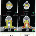
Conformal radiotherapy with or without intensity-modulated radiation therapy has been the primary mechanism of radiation dose delivery during the past two decades.25,26 Advances in software now routinely employ computation-heavy, cost-benefit function analyses of beam modulation to address irregular structures within the body, particularly when they are concentrically contained within or encapsulated by normal organs with dose-limiting sensitivity to radiation effect.27,28 Furthermore, physiologic motion is minimal within the head and neck region, allowing greater confidence in patient setup and targeting the tumor bed. Although some advances, like respiratory gating and breathing management (deep inspiration breath hold), have been developed to address organ and target volume motion, most treatment of the head and neck in the pediatric age group can be accomplished without resorting to these specialized measures.29 Prosthetics in the form of intraoral molds and the use of thermoplastic head and neck immobilization devices frequently serve as positioning aids, both internally and externally for the pediatric oropharynx and head/neck, respectively.30,31 Fiducial markers can also be implanted to further aid in targeting when extraordinarily high precision is required to spare critical structures skirting radiation dose thresholds.

Frequently, these tumors afflict young children and infants requiring sedation to achieve adequate immobilization suitable for treatment. Most institutions choose to sedate with propofol and avoid airway management when possible, although occasional management of the airway is required to ensure patient safety through a full course of radiation, as determined by the anesthesiologist.32

Proton therapy has recently become central to the discussion of pediatric radiotherapy due to the promise of reduced toxicity; larger-scale clinical trials designed to demonstrate the potential advantages are ongoing. Although a major objective of proton therapy is reduction of toxicity, protons do not inherently promise a more efficacious means of disease control (unless normal tissue sparing through improved tumor targeting is enhanced such that dose escalation of residual tumor can result in improved disease control).33 At this time, precision targeting with proton beam is still developing as a routine component of clinical practice due to various factors, including cost and accessibility. Proton therapy requires prospective study design to provide evidence for the appropriate indications and degree of benefit that may be obtainable.


DISEASE-SPECIFIC OVERVIEW: DIAGNOSIS, STAGING, TREATMENT, AND PROGNOSIS


Rhabdomyosarcoma

Rhabdomyosarcoma is the most common pediatric soft tissue sarcoma, with an overall annual incidence in the United States of 4.5 cases per million children and adolescents, with the majority occurring in children younger than 10 years.34 It arises as a primary malignancy in the head and neck in ˜35% to 40% of cases, most commonly in the orbit.3,4,8,21 Cooperative group efforts, including the Intergroup Rhabdomyosarcoma Studies (IRS), have contributed to the advances in management. The mainstay of therapy is multimodal, including multiagent systemic chemotherapy for all patients and either radiation therapy, surgery, or both, as local control for most patients. Across the pediatric age spectrum, outcomes for patients who have rhabdomyosarcoma have improved since 1975, with 5-year survival rates increasing by more than 10% to ˜65% or higher for patients younger than 15 years old and to ˜50% for patients 15 to 19 years old.35

Rhabdomyosarcoma encompasses two main histologic subtypes: embryonal and alveolar. The embryonal subtype is the most common among children, noted in approximately two-thirds of the patients, with common locations including the head and neck and the genitourinary tract. Botryoid and spindle cell variants of embryonal rhabdomyosarcoma have a particularly favorable prognosis.

Within the head and neck, rhabdomyosarcomas are further classified as originating from the orbit (including eyelid), parameningeal (including middle ear/mastoid, nasopharyngeal/nasal cavity, paranasal sinus, parapharyngeal region, and pterygopalatine/infratemporal fossa), or nonparameningeal sites (including the scalp, ear lobe, parotid, oropharynx, and hypopharyngeal region).8,36 Key prognostic factors for pediatric patients with rhabdomyosarcoma are used by the Children’s Oncology Group (COG) to categorize patients into three risk groups (low, intermediate, and high) based on risk of disease recurrence. Standard prognostic factors include age (best age 1 to 9 years), primary site (favorable sites including orbit, nonparameningeal head and neck, genitourinary sites excluding bladder and prostate, and biliary tract), tumor size (smaller, ≤5 cm better), tumor resectability, disease spread (better prognosis with localized disease without regional lymph node involvement or distant metastases), and tumor histopathology (embryonal more favorable than alveolar subtype).36 In recent years, ˜80% of alveolar
rhabdomyosarcomas have been found to harbor a fusion of the FOXO1 gene (on chromosome 13) with either the PAX3 or PAX7 genes (on chromosome 2). As the presence of FOXO1 gene fusion status is more closely associated with adverse outcomes, in upcoming COG trials, FOXO1 fusion status is anticipated to replace histopathology classification in risk stratification criteria.37

Presentation of rhabdomyosarcomas can vary greatly depending on the primary site and typically require an open or core needle biopsy to confirm histopathologic and molecular diagnosis. Radiologic staging has typically included a baseline chest radiograph, chest CT, bilateral bone marrow aspirates and biopsies, and bone scan (with some centers opting for PET-CT in lieu of bone scan). For patients with parameningeal primary tumors, in particular, an MRI of the brain and skull base is required, and baseline lumbar puncture for malignant cells in the cerebrospinal fluid should also be obtained. MRI with MRA/MRV may be used to evaluate the relationship of the tumor to nearby vascular structures. Suspected spinal cord involvement can be investigated with MRI including contrast. Rhabdomyosarcomas of the head and neck have a variable imaging appearance but are often heterogeneous and may contain areas of necrosis and/or hemorrhage. They often erode adjacent bone and may extend intracranially. There are no pathognomonic imaging findings (Figs. 24.1 and 24.2).38

A recent review of more than 1,600 children with rhabdomyosarcoma from 1991 to 2004 on intergroup studies suggested that staging procedures can be adapted in an algorithmic fashion based on presenting clinical factors, such that patients at low risk of distant metastatic disease—approximately one-third of patients—may be spared bone marrow studies and bone scans.39

In addition to standard cross-sectional imaging, PET-CT may also be a helpful adjunct to detect regional or distant metastatic disease, help evaluate therapeutic response, and identify patients who may benefit from therapy intensification for regional control.40,41,42 Assessment of regional lymph nodes and potential biopsy where feasible may also guide management,43 although neither PET scans or lymph node assessments are currently included as requirements in COG treatment protocols.


Figure 24.1. Axial short T1 inversion recovery (STIR) (A) and coronal postgadolinium fat-saturated T1-weighted (B) images through the face of a 17-year-old patient demonstrate an irregularly shaped mass (large arrows) that involves the central skull base, ethmoid region, and portions of the orbits with intracranial epidural extension (small arrows). A biopsy confirmed the diagnosis of alveolar rhabdomyosarcoma.

Comprehensive staging of rhabdomyosarcoma in children typically involves designating a stage (Table 24.4), a surgical-pathologic diagnosis, local tumor group (Table 24.5), and finally a risk group (Table 24.6).

The nature and intensity of treatment, and subsequent outcomes, depend on appropriate staging and pathologic delineation of rhabdomyosarcomas, including tumor biology.

In comparison with children and adolescents with rhabdomyosarcoma, adult patients are more likely to present with disease in unfavorable primary sites, and exhibit pleomorphic histology, which is rarely seen in the pediatric population.44

Although the majority of cases of rhabdomyosarcoma are apparently sporadic in origin, reported associated familial and genetic conditions include Li-Fraumeni syndrome (with TP53 mutations), Costello syndrome, Beckwith-Wiedemann syndrome, neurofibromatosis, and Noonan syndrome.34,45 Additional studies are needed to evaluate the role of environmental factors, such as immune function and atopic exposure as potential protective factors in pediatric rhabdomyosarcoma.46

Standard chemotherapy consists typically of vincristine, actinomycin-D (also known as dactinomycin), and cyclophosphamide, in a regimen commonly known as VAC.47 Additional agents have not convincingly altered the outcomes of this therapeutic backbone.48 Focus continues to hone in on advancing cure rates while minimizing potential late effects of therapy. For patients who initially present with localized rhabdomyosarcoma, preventing local recurrence with surgery and/or radiotherapy remains vital.

Local therapy for rhabdomyosarcoma follows an approach of risk-adapted therapy with selection of modality (surgery vs. radiotherapy) designed to limit morbidity while optimizing local control. Maximal safe resection is performed at diagnosis when possible, followed by systemic therapy and additional local control measures including radiation therapy and
additional surgery as determined by tumor histology, resectability, and the status of the surgical margins. Second-look surgery is an acceptable approach in order to balance outcome with toxicity. This approach enhances local control while minimizing long-term late effects. Defining appropriate patients for postoperative adjuvant radiotherapy remains somewhat controversial. Historically, patients have been selected for adjuvant radiotherapy based on histology (alveolar vs. embryonal). The basis for this approach derives from subset analyses of IRSG III to IV trials that have suggested no decrement to local control in children with alveolar rhabdomyosarcoma managed with surgery alone.49


Figure 24.2. Axial STIR (A) and axial (B) and coronal (C) postgadolinium fat-saturated T1-weighted images through the face of a 2-year-old child reveal a heterogeneous mass (arrows) involving the right masticator space. Biopsies of the mass demonstrated embryonal rhabdomyosarcoma.

Primary surgical resection can be considered prior to chemotherapy, but only if this would be safely achieved without
adverse functional or cosmetic outcomes. Aggressive primary surgical approaches are not recommended, as attempts to debulk the tumor (with gross residual tumor) are not associated with improved outcomes compared to biopsy alone.50 Particularly for head and neck rhabdomyosarcomas, typically only a biopsy is performed prior to chemotherapy; hence, most patients will have gross residual (group III) tumor and receive radiotherapy as primary means of local control.36 Orbital rhabdomyosarcomas should be biopsied only; upfront exenteration should not be performed, and later orbital exenteration should only be considered for those few patients with persistent or recurrent disease after chemotherapy and radiation therapy, which can achieve survival rates of 90% and above. For nonparameningeal and nonorbital rhabdomyosarcomas of the head and neck, such as those with superficial facial tumors, primary tumor excision (accepting narrow margins due to anatomy) and ipsilateral neck sampling of clinically concerning nodes can be considered.8,51 Selected patients may be considered for secondlook surgery to remove residual tumor (i.e., delayed primary excision) after initial therapy or in cases where this may modify the radiation dose required or have anticipated net positive impact on a given patient’s functional outcome. The detection of viable tumor at the time of second-look surgery has not been found to impact overall survival.49


Table 24.4 Children’s Oncology Group: Pretreatment Staging System for Rhabdomyosarcoma


Stage


Sites of Primary Tumor


T Stage


Tumor Size


Regional Lymph Nodes


Distant Metastasis


1


Favorable site (Orbit; nonparameningeal head and neck; genitourinary tract other than kidney, bladder, and prostate; biliary tract)


T1 or T2


Any size


N0 or N1 or NX


M0


2


Unfavorable sites (any site other than favorable)


T1 or T2


a, ≤ 5 cm


N0 or NX


M0


3


Unfavorable sites (any site other than favorable)


T1 or T2


a, ≤ 5 cm


b, > 5 cm


N1


N0 or N1 or NX


M0


4


Any site


T1 or T2


Any size


N0 or N1 or NX


M1


N0, absence of nodal spread; N1, presence of regional nodal spread beyond the primary site; X, unknown N status; M0, absence of metastatic spread; M1, presence of metastatic spread beyond the primary site and regional lymph nodes; T1, tumor confined to anatomic site of origin (noninvasive); T2a, tumor extension and/or fixation to surrounding tissue (invasive), tumor ≤5 cm in maximum diameter; T2b, tumor extension and/or fixation to surrounding tissue (invasive), tumor >5 cm in maximum diameter. From National Cancer Institute: PDQ® Childhood Rhabdomyosarcoma Treatment. 2014.


Table 24.5 Children’s Oncology Group: Surgical-Pathologic Group Assignment System for Rhabdomyosarcoma


Group


Incidence


Definition


I


˜13%


Localized tumor, completely removed with microscopically clear margins, and no regional lymph node involvement. Lymph node biopsy or sampling is encouraged if lymph nodes are clinically or radiographically suspicious.


II


˜20%


Localized tumor, completely removed with (a) microscopic disease at the margin; (b) regional disease with involved, grossly removed regional lymph nodes without microresidual disease; or (c) regional disease with involved nodes, grossly removed but with microscopic residual and/or histologic involvement of the most distal node from the primary tumor


III


˜48%


Localized tumor, incompletely removed with gross, residual disease after (a) biopsy only or (b) gross major resection of the primary tumor (>50%)


IV


˜18%


Distant metastases are present at diagnosis. This category includes (a) radiographically identified evidence of tumor spread and (b) positive tumor cells in cerebral spinal fluid, pleural, or peritoneal fluids, or implants in these regions


From National Cancer Institute: PDQ® Childhood Rhabdomyosarcoma Treatment. 2014.


Table 24.6 Children’s Oncology Group: Risk Group Classification for Rhabdomyosarcoma


Risk Group


Histology


Stage


Group


Low risk


Embryonal


Embryonal


1


2, 3


I, II, III


I, II


Intermediate risk


Embryonal


Alveolar


2, 3


1, 2, 3


III


I, II, III


High risk


Embryonal or alveolar


4


IV


From National Cancer Institute: PDQ® Childhood Rhabdomyosarcoma Treatment. 2014.


As only an estimated 15% of pediatric rhabdomyosarcoma patients have completely resected (group 1) disease, radiation therapy is included for local control in most cases.36

Recent data suggest the proton radiotherapy may be a comparably effective modality to photon radiotherapy for selected pediatric rhabdomyosarcoma patients, with future larger studies with sustained follow-up awaited to clarify whether it may indeed definitively reduce late effects.52

With modern therapy, typical survival rates for pediatric patients with rhabdomyosarcoma in the orbit are in the range
of 90% or greater and 70% or greater for nonparameningeal head and neck rhabdomyosarcoma. Patients with parameningeal rhabdomyosarcoma are typically associated with poorer prognosis.8,53 Cranial nerve palsies or skull base erosion is noted in approximately a third of children with parameningeal rhabdomyosarcoma and is associated with increased risk of recurrence. Urgent management typically includes earlier initiation of radiation therapy, with incomplete or longer-term clinical neurologic recovery from cranial nerve palsies possible.8,54

Generally, patients with recurrent and refractory rhabdomyosarcoma have a guarded long-term prognosis, but selected patients can achieve complete remission and more favorable 5-year survival rates with intensive salvage therapy, including those with initially localized presentation in favorable sites such as the orbit.55,56


Other Soft Tissue Sarcomas and Related Tumors

A number of soft tissue sarcomas constitute the varied group of sarcoma malignancies, apart from rhabdomyosarcoma, that can arise in the head and neck. The various nonrhabdomyosarcomatous soft tissue sarcomas (NRSTSs) altogether constitute <5% of pediatric malignancies; most pose challenges for local management, with infrequent lymph node or distant metastatic involvement.57

Synovial sarcoma is the most common NRSTS, with head and neck primary involvement in ˜6%.58 Synovial sarcomas are frequently small and, particularly when they present in the head and neck region, are often missed as a malignant entity due to its slow growth and well-circumscribed appearance.59 Immunohistochemical staining typically demonstrates reduced INI1 nuclear reactivity, with a typical and specific chromosomal translocation, t(X;18)(p11.2;q11.2), associated with this diagnosis in 95% of cases, leading to rearrangement of the SYT gene (located on chromosome 18) with SSX1 or SSX2 (on chromosome X).59 Better outcomes have generally been described among children than in adults, with SEER database analysis of 5-year survival estimates of 83% for children and adolescents and 62% for adults.58 Typical management approaches are centered on surgical resection, with ongoing need for prospective data to definitively delineate the contribution of radiation therapy and chemotherapy.59,60 Although various approaches have been used, a common chemotherapy regimen considered for NRSTSs selected for systemic therapy is doxorubicin with ifosfamide.59,60


Figure 24.3. Axial (A) and coronal (B) T2-weighted images through the neck of a 7-year-old child show a heterogeneous, fairly wellmarginated mass (arrows) involving the retrostyloid parapharyngeal space. The lesion was subtotally resected and found to be a malignant peripheral nerve sheath tumor (MPNST).

In a recent analysis of the National Cancer Institute’s SEER database, among 1,244 pediatric cases, MFH was among the most common diagnoses reported.4 Whereas MFH is a common soft tissue sarcoma in adult patients, it has been inconsistently recognized and reported in the pediatric literature.61,62 Although the nomenclature and classification of MFH and related tumors were revised by the World Health Organization in recent years, including in 1994 and 2002, studies reporting on MFH unfortunately have often aggregated MFH with other less aggressive subtypes, such as angiomatoid fibrous histiocytoma and plexiform fibrohistiocytic tumors,61 with caution thus warranted in interpreting studies that may not have had the benefit of MFH as defined consistently on contemporary pathology review. One institutional retrospective study from 1971 to 2000 identified 28 patients initially diagnosed with MFH, including 6 in the head and neck; 10 were reclassified as angiomatoid fibrous histiocytoma and one with plexiform fibrohistiocytic tumor; among the 17 patients with confirmed MFH, 5-year event-free survival is ˜71%, compared to 100% for those reclassified with angiomatoid fibrous histiocytoma and plexiform fibrohistiocytic tumor. Wide local excision was favorably associated with event-free survival for localized tumors.61

Malignant peripheral nerve sheath tumors (MPNSTs) constitute another relatively common diagnosis within this rare group of pediatric head and neck NRSTSs.57,63 A recent SEER analysis64 demonstrated that in pediatric MPNST patients, the incidence was higher among adolescents; localized disease and surgical resection were positive prognostic factors, and median overall survival was only 30 months, consistent with other reports suggesting less favorable outcomes for MPNST among challenging NRSTS cases.63 Unfortunately, MPNSTs often present with large tumors in axial locations, both of which are clinical factors reported to adversely impact outcome63 (Figs. 24.3 and 24.4).


Figure 24.4. Axial fat-saturated T2-weighted (A) and coronal postgadolinium fat-saturated T1-weighted (B) images of the neck of a 27-year-old survivor of childhood neuroblastoma demonstrate a mildly heterogeneous well-defined mass in the left parapharyngeal space. The lesion was resected and determined to be an MPNST.

Other reported NRSTSs presenting in the head and neck in children and adolescents have included alveolar soft part sarcoma, epithelioid sarcoma, dermatofibrosarcoma protuberans, malignant hemangiopericytoma, leiomyosarcoma, fibrosarcoma, myofibroblastic sarcoma, inflammatory myofibroblastic tumor, and undifferentiated sarcoma.8,57,62,63,65 Definitive management is predicated on accurate diagnosis and coordinated multidisciplinary care. In the largest single-institutional retrospective review to date, Federico et al. described 58 pediatric and young adult patients with head and neck NRSTSs from 1964 to 2003.57 Most patients were found to have small, highgrade tumors. Rates for event-free and overall survival at 10 years were 53% and 63% respectively. High grade, large size (>5 cm), invasiveness, and gross residual disease after surgery were associated with worse prognosis.57 Novel approaches and further prospective data are needed to define optimal standardized management for children with NRSTSs.


Bone Sarcomas and Related Tumors

Pediatric bone sarcomas of the head and neck are generally rare and typically are localized at diagnosis.66 Although osteosarcoma represents the most common pediatric bone sarcoma, only ˜6% to 13% arise in the head and neck.67 The maxilla and mandible are the most common sites and are also associated with better prognosis than are other, extragnathic sites in the head and neck.67,68 Osteosarcomas in the head and neck appear distinct in a number of ways from the typical osteosarcoma that presents in the extremities, with variable histology, more often involving low or intermediate tumor grade, higher risk for local recurrence and less risk for distant metastatic disease compared to extremity osteosarcomas.8,68 On CT scan, the tumor appears expansile with a prominent periosteal reaction and/or mineralization associated with osteoid tumor matrix69 (Fig. 24.5A and B). MRI demonstrates a heterogeneous mass with low-signal intensity areas on both T1- and T2-weighted images, consistent with osseous lesion components, and intermediate- (T1-weighted) to high-signal (T2-weighted) tumor components that correspond to soft tissue (Fig. 24.5C and D). Complete surgical resection remains the foundation for improved failure-free and survival outcomes.66,68 Prospective clinical data are warranted to better verify and define how prognostic factors, such as posttherapy tumor necrosis, as well as treatment approaches utilized for extremity osteosarcomas, including radiation therapy and chemotherapy, should be best interpreted and applied in head and neck osteosarcomas.8,70 In addition to primary osteosarcoma, secondary osteosarcoma should be considered in older children or adolescents with a prior history of irradiation of the head and neck or prior hereditary retinoblastoma, with potential for similar outcomes as those with primary osteosarcoma.68

Only gold members can continue reading. Log In or Register to continue

Stay updated, free articles. Join our Telegram channel

Dec 18, 2016 | Posted by in ONCOLOGY | Comments Off on Cancer of the Head and Neck in the Pediatric Population

Full access? Get Clinical Tree

Get Clinical Tree app for offline access