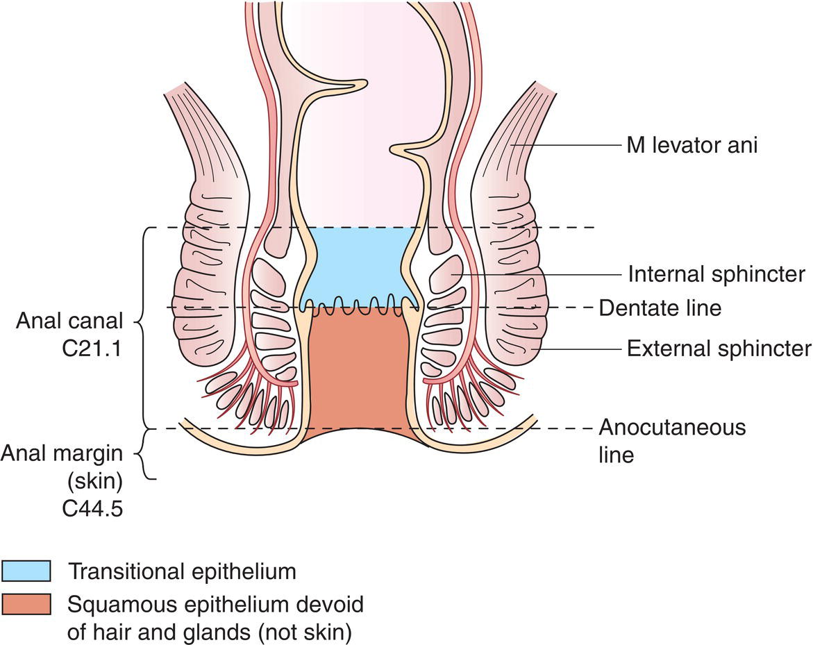
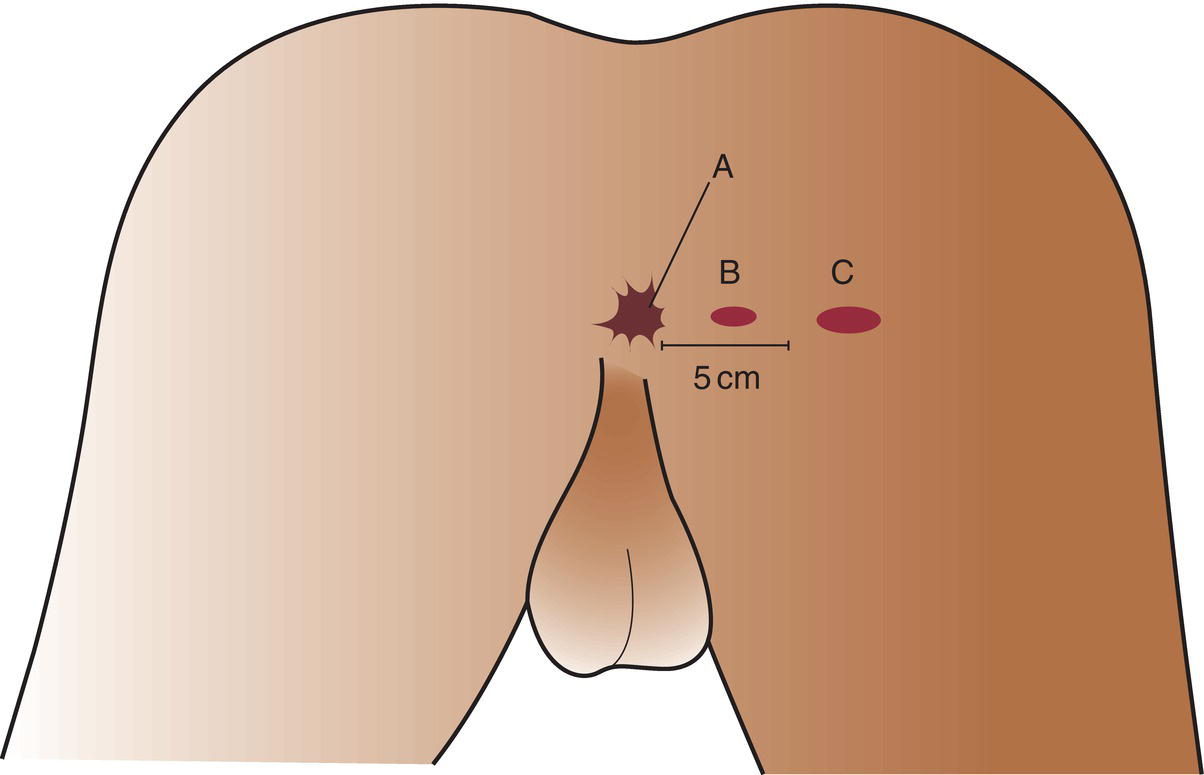
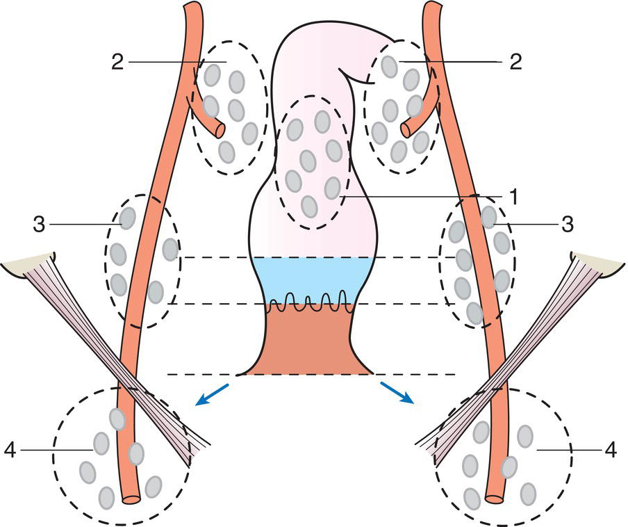
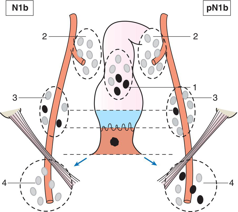
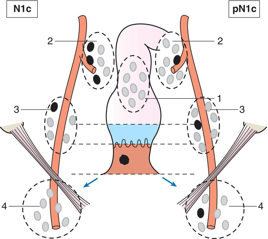
The anal canal (Fig. 189) extends from rectum to perianal skin (to the junction with hair‐bearing skin). It is lined by the mucous membrane overlying the internal sphincter, including the transitional epithelium and dentate line. Tumours of anal margin and perianal skin defined as within 5 cm of the anal margin (ICD‐O‐3 C44.5) are now classified with carcinomas of the anal canal (Fig. 190). The classification applies only to carcinomas. There should be histological confirmation of the disease and division of cases by histological type. The regional lymph nodes are the perirectal (1), internal iliac (2), external iliac (3) and inguinal lymph nodes (4). Source: From Zinner MI, Ashley SW (2012) Maingot’s Abdominal Operations, 12th edition, McGraw Hill Education. © 2012 McGraw Hill Education. Note * Direct invasion of the rectal wall, perianal skin, subcutaneous tissue or the sphincter muscle(s) alone is not classified as T4. The pT and pN categories correspond to the T and N categories. Note pM0 and pMX are not valid categories.
ANAL CANAL (ICD‐O‐3 C21, ICD‐O‐3 C44.5)
Rules for Classification
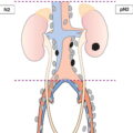
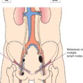
Regional Lymph Nodes (Fig. 191)
TNM3 Clinical Classification
T0
No evidence of primary tumour
Tis
Carcinoma in situ, Bowen disease, high‐grade squamous intraepithelial neoplasia (HSIL), anal intraepithelial neoplasia (AIN II–III)
T1
Tumour 2 cm or less in greatest dimension (Fig. 192)
T2
Tumour more than 2 but not more than 5 cm in greatest dimension (Fig. 193)
T3
Tumour of more than 5 cm in greatest dimension (Fig. 194)
T4
Tumour of any size invades adjacent organ(s), e.g., vagina, urethra, bladder* (Fig. 195) 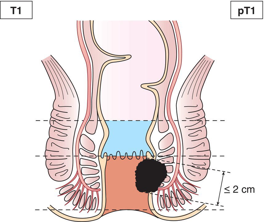
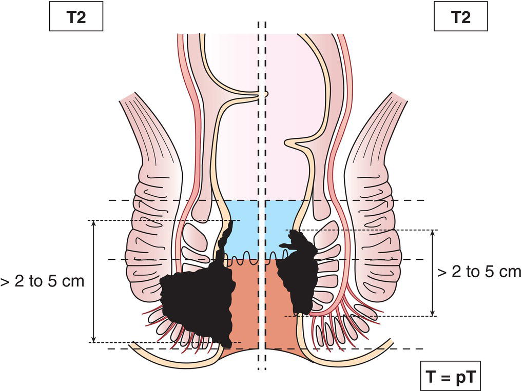
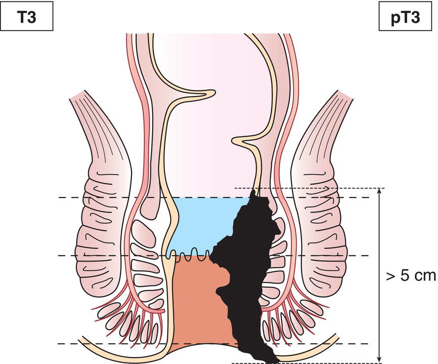
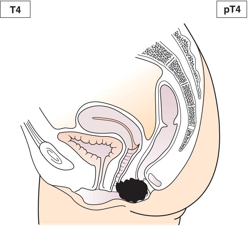
N – Regional Lymph Nodes
NX
Regional lymph nodes cannot be assessed
N0
No regional lymph node metastasis
N1a
Metastases in inguinal, mesorectal and/or internal iliac nodes (Fig. 196)
N1b
N1c
Metastases in external iliac nodes (Fig. 197)
Metastases in external iliac and in inguinal, mesorectal and/or internal iliac nodes (Fig. 198)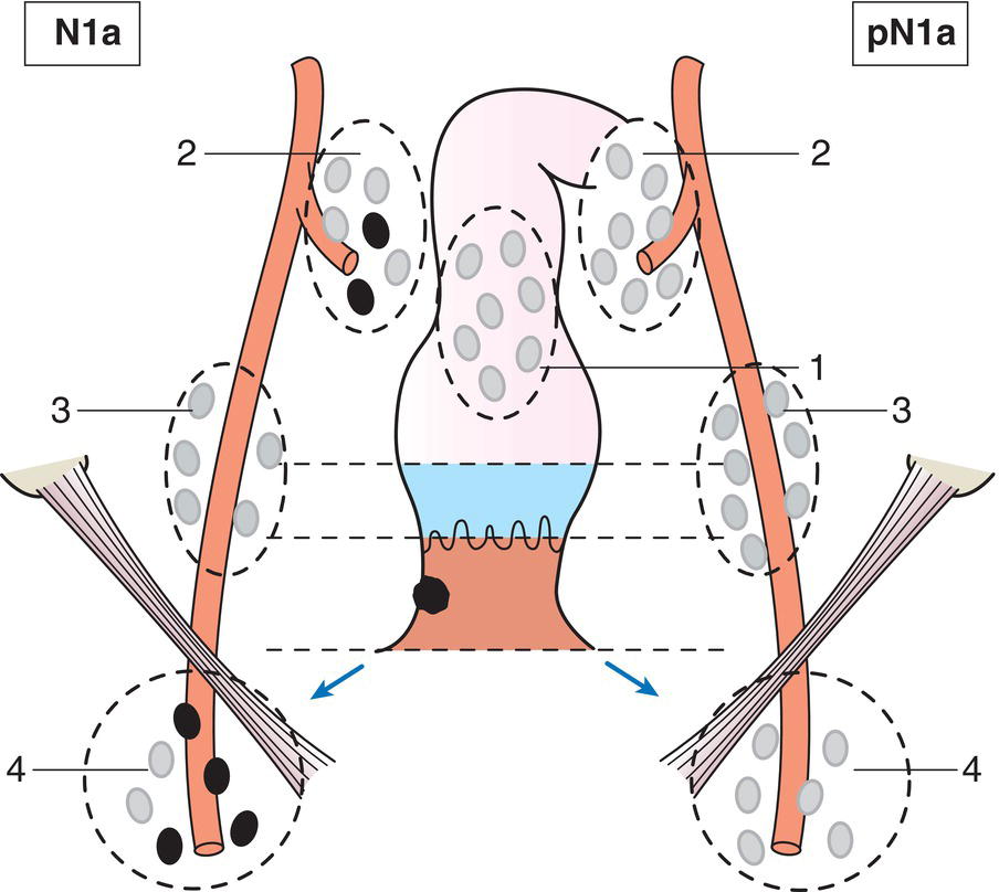
M – Distant Metastasis
M0
No distant metastasis
M1
Distant metastasis
TNM Pathological Classification
pM1
Distant metastasis microscopically confirmed
pN0
Histological examination of a regional perirectal/pelvic lymphadenectomy specimen will ordinarily include 12 or more lymph nodes; histological examination of an inguinal lymphadenectomy specimen will ordinarily include 6 or more lymph nodes. If the lymph nodes are negative, but the number ordinarily examined is not met, classify as pN0.
Summary
Stay updated, free articles. Join our Telegram channel

Full access? Get Clinical Tree


