Fig. 8.1
Historical perspective of selected hallmarks on the evolution of microRNA history
MicroRNAs: The Biogenesis
Various approaches have provided a basic understanding of the molecular details of miRNA biogenesis (Fig. 8.2) and it has long been viewed as linear and universal to all mammalian miRNAs. To understand miRNA biogenesis at a molecular level, we have classified miRNA biogenesis into the following three subheadings:
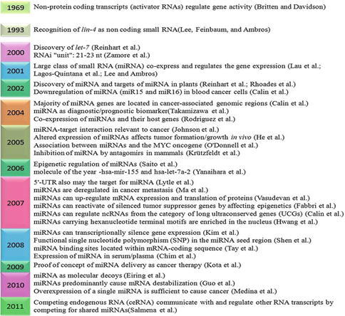
Fig. 8.2
MicroRNA processing and activity. Depicts the formation of long primary microRNA (pri-miRNA) in the nucleus which is processed by the microprocessor complex (Drosha, an RNAse III enzyme, and Pasha, a double-stranded RNA-binding protein) into precursor microRNA (pre-miRNA) (70 nt stem-loop structure) and transported to the cytoplasm by Exportin-5-mediated export, where Dicer, an RNase II enzyme, cleaves it to 20–25 nt mature miRNA that integrates it into the miRNA-inducing silencing complex (miRISC), a complex of proteins that is responsible for regulation of gene expression either by translational inhibition or by target mRNA degradation
Nuclear Processing by Drosha
This canonical maturation includes the production of the primary miRNA transcript (pri-miRNA) by RNA polymerase II or III and cleavage of the pri-miRNA by the microprocessor complex Drosha–DGCR8 (Pasha) in the nucleus [13]. A primary transcript RNA (pri-miRNA) transcribed from a miRNA gene by RNA polymerase II or III is first processed into a stem-loop structure of about 70–80 nucleotides known as precursor miRNA (pre-miRNA) by a microprocessor enzyme comprising of a double-strand (ds)-RNA-specific ribonuclease, Drosha, with the help of its binding partner DGCR8.
Nuclear Export of Pre-miRNAs
The resulting precursor hairpin, the pre-miRNA, is exported from the nucleus by Exportin-5–Ran-GTP [13]. Exportin-5 recognizes the pre-miRNA independently of its sequence or the loop structure. A defined length of the double-stranded stem and the 3′ overhangs are important for the successful binding to Exportin-5, ensuring the export of only correctly processed pre-miRNAs [13].
Cytoplasmic Processing by Dicer
In the cytoplasm, the RNase Dicer in complex with the double-stranded RNA-binding protein TRBP cleaves the pre-miRNA hairpin to its mature length. The functional strand of the mature miRNA is loaded together with Argonaute (Ago2) proteins into the RNA-induced silencing complex (RISC), where it guides RISC to silence target mRNAs through mRNA cleavage, translational repression, or deadenylation, whereas the passenger strand is degraded [13].
MicroRNA Genes and Their Transcription
MicroRNA genes reside in regions of the genome as distinct transcriptional units as well as in clusters of polycistronic units—carrying the information of several microRNAs [10, 14–16]. Studies suggest that approximately half of known microRNA reside in non-protein-coding RNAs (intron) or within the intron of protein-coding genes [17].
The understanding of microRNA transcription is very important for determining their regulators as well as the specific role they may play in signaling cascades. The understanding of microRNA transcriptional regulation has great public health significance. The ability to understand how these post-transcriptional gene regulators function in cellular networks may provide new molecular targets for cures or therapies to a variety of human diseases.
Transcription of miRNA Genes
Little is known about the transcriptional regulation of these intergenic miRNAs, although RNA polymerase II appears to be involved in the process [18]. This suggests that miRNAs may have active promoter regions that contain cis-regulator elements similar to coding genes. miRNA genes are currently believed to be transcribed by RNA polymerase II (Pol II), [18] although a few may be transcribed by RNA polymerase III [19]. RNA polymerase II transcribes miRNA genes, generating long primary transcripts (pri-miRNAs) [20]. Subsequently, the process to yield mature miRNAs involves two steps involving RNase III enzymes and companion double-stranded RNA-binding domain (dsRBD) proteins. There are two different classes of miRNAs with respect to transcription mechanism—those found within annotated genes (intronic miRNAs) and those found in intergenic regions of the genome (intergenic miRNAs). It is presently believed that all intronic miRNAs are co-transcribed along with their host gene; this has been shown in both expression correlation studies [21] as well as PCR-based biochemical verification [17]. Intergenic miRNAs have been postulated to come from transcripts of up to 50 kb in length, allowing for the co-transcription of neighboring miRNAs (polycistronic miRNA clusters) [21]. Identification of the method by which miRNA genes are transcribed can lead to the identification of the factors that are responsible for their regulation.
MicroRNAs in Breast Cancer
MicroRNAs have been concerned with an increasing number of neoplasia and biological process. The latest studies have shown a contribution for these regulatory molecules in breast cancer. For instance, microRNA profiling studies have identified microRNAs that are deregulated in breast cancer. Moreover, functional studies have revealed their roles in breast cancer as both oncogenes (e.g., hsa-miR-21) and cancer suppressor genes (e.g., hsa-miR-335). MicroRNAs deregulated in breast cancer control the translational regulation of entrenched regulative molecules, such as estrogen receptor-α, which are regulated by novel cancer-related molecules and miR-206 whose functions are not yet completely understood.
MicroRNA in Cancer in General
miRNAs have been associated with the regulation of differentiation, proliferation, apoptosis, and even exocytosis [22]. Volinia et al. has demonstrated that the predicted targets for the differentially expressed miRNAs are significantly enriched for protein-coding tumor suppressors and oncogenes [22]. There is also confirmation to suggest that these miRNAs function in concert with classical tumor suppressors and oncoproteins to regulate key pathways involved in cellular growth control [23, 24]. miRNA profiling has the potential not only to classify tumors but also to augur patient outcome with high accuracy, but this approach needs more validation and detailed studies by using clinical samples [25]. Jian et al. showed that profile analysis with a probe set of 201 miRNAs achieved the similar discriminative potential as traditional gene array with 8,000 mRNA probes [26]. Consequently, this would mean that classification of tumors can be achieved with a more manageable amount of data and could potentially diminish the disparity that is often seen with mRNA-based classifier systems.
MicroRNAs Study in Breast Cells and Tissues
Since the study of mRNA, various technologies exist that allow the investigation of the expression of either profiling of a large number of miRNAs or individual miRNAs simultaneously. In general, these observational approaches have implicated individual or groups of miRNAs in pathological or physiological processes, as a result of the detection of changes in their expression, while additional functional experiments are required to gain further knowledge of their current roles. A few miRNA profiles have been developed using a large number of single miRNA detection experiments, such as Northern blotting [14], and these technologies remain the standard against which newer profiling methods are primarily compared. Nevertheless, oligonucleotide microarray-based detection platforms, with their associated ease of use and high-throughput nature, have largely supplanted this technique [27]. Microarrays have been used for miRNAs profiling from a wide range of breast tissue types and cell lines, including formalin-fixed paraffin-embedded (FFPE) clinical samples. It is essential to note that, due to the small size of miRNAs, they are comparatively insensitive to the damage that typifies mRNAs within FFPE. Accordingly, miRNAs present an invaluable new target for studies using archival clinical samples, which can often be linked to extensive clinical background and, more importantly, follow-up data or meta-analysis [28]. Multiplex real-time RT-PCR and liquid bead-based technologies are current alternative strategies for miRNA profiling, and it is claimed that they may have a higher sensitivity and specificity [29, 30]. Methodologies based on deep sequencing of small RNA libraries obtained from tissues may also allow miRNA profiling, with the supplementary advantage that these techniques are unbiased with respect to target sequences and may permit detection of novel miRNAs [31].
Profiling Data of miRNA in Breast Cancer
The expression of miRNAs has been investigated in an extensive range of breast cancer cell lines, tissues, and clinical normal. These metadata hint toward functional roles of various miRNAs by association with cellular behavior or particular molecular markers. To gain the best insight into breast cancer, it is also essential to understand miRNA function in normal mammary gland development, and work is underway to address this question in detail [32]. A potentially powerful empirical approach is to compare miRNA expression in normal breast versus breast cancer and thereby to differentiate those miRNAs expressed at different levels. Table 8.1 lists miRNAs identified as playing a role in breast cancer and their potential targets. Iorio et al. reported for 76 breast samples diagnosed for the expression of 246 miRNAs, out of which 29 miRNAs expression levels were found to be significantly different (i.e., p <0.05) in cancer versus normal clinical tissue. The majority consistently downregulated were has-miR-10b, has-miR-125b, and has-miR-145, while has-miR-21 and has-miR-155 were upregulated, suggesting that these may act as oncogenes or tumor suppressor genes, respectively [32]. They went on to examine whether the expression profile varied according to conventional clinical aspects: ER+ /ER−, PR+ /PR−, HER2+ /HER2−, positive and negative lymph node status, presence and absence of vascular invasion, high and the low proliferation index, and ductal/lobular histopathological subtype. The majority of comparisons discovered a small number of differentially expressed miRNAs, indicating that miRNAs may have roles in defining the differences between these pathological and molecular profiles. Yet, comparison between ductal/lobular carcinomas and HER2+ and HER2− tumors did not reveal differentially expressed miRNAs.
Table 8.1
miRNAs whose expression is deregulated in breast cancer and their potential targets
MicroRNA | Expression pattern | Target |
|---|---|---|
hsa-let-7 family | Down | IL-6, ESR1 (ER) |
hsa-miR-101 | Down | EZH2 |
hsa-miR-10b | Down | HOXD10, Tiam1 |
hsa-miR-1226 | Up | MUC1 |
hsa-miR-122a | Up | |
hsa-miR-125 | Down | HuR, ERBB2, ERBB3, BAK1, BMPR1B, CYP24, MUC1 |
hsa-miR-126 | Down | IRS1 |
hsa-miR-128 | Up | TGFβR1 |
hsa-miR-136 | Up | |
hsa-miR-143 | Down | |
hsa-miR-145 | Down | MUC1, RTKN, ESR1 |
hsa-miR-146a | BRCA1, BRCA2 | |
hsa-miR-146 | IRAK1, TRAF6 | |
hsa-miR-149 | Up | |
hsa-miR-150 | Up | c-MYB |
hsa-miR-155 | Up | FOXO3, SOCS1, RHOA |
hsa-miR-16 | Down | MYB, WIP1 |
hsa-miR-17/92 cluster | Deleted | BRCA1, IL-8, CCND1, HBP1, AIB1, ESR1 (ER), ESR2 (ER), HIF1, STAT3 |
hsa-miR-185 | Down | SIX1 |
hsa-miR-191 | UP | |
hsa-miR-196 | UP | ANXA1 |
hsa-miR-200 | Down | ZEB1, ZEB2, FTH1, PLCG1, BMI1, FN1, NTRK2, QKI |
hsa-miR-202 | Up | |
hsa-miR-203 | Up | |
hsa-miR-204 | Down | |
hsa-miR-205 | Down | ERBB3, ZEB1, HER3, VEGF-A |
Similarly, specific miRNA profiles have been associated with breast cancer subgroups of distinct patterns of molecular marker expression. Profiling of 204 miRNAs has been shown sufficient to allow unsupervised clustering to be used to distinguish HER2+ /ER−, HER2+ /ER−, and HER2−/ER+ breast cancers within a cohort of 20 tumors. While this in itself is not an advance, since these cancers are routinely defined using immunohistochemistry, further supervised analysis of the profiles allowed distinct miRNA subsets to be identified that distinguished HER2+ from HER2− and ER+ from ER− breast cancers, independent of other clinically important parameters. Restricted subsets of miRNAs specific to HER2 status (let-7f, let-7g, miR-107, miR-10b, miR-126, miR-154, and miR-195) and specific to ER/PR status (miR-142-5p, miR-200a, miR-205, and miR-25) have been also established [33].
Functional Studies: miRNA in Breast Cancer
The functional activity of only a few miRNAs has been practically modeled in the perspective of breast cancer, and it is clear that most of this type of work remains to be done. Potential functions of miRNAs in carcinogenesis into potential oncogenes and tumor suppressor genes have been established in regulating immune system, cell proliferation, differentiation and development, cancer and cell cycle.
Breast Cancer Oncomirs
Table 8.2 summarizes examples of miRNAs that have apparent oncogenic activity in breast cancer. Oncogenic miRNAs, commonly known as oncomirs, may act by hindering the expression of tumor suppressor genes and/or genes responsible for apoptosis and differentiation. Recent studies have identified functional oncogenic role(s) for miRNAs, their individual manipulation in breast cancer cell line models, and subsequent assessment of associated phenotypic changes. Iorio et al. showed that miRNAs aberrantly expressed in human breast cancer compared with normal breast tissue, with the most significantly downregulated miRNAs being miR-10b, miR-125b, and miR-145, whereas the most significantly upregulated miRNAs being miR-21 and miR-155 [33]. Si et al. found that the anti-miR-21-mediated cell growth inhibition associated with increased apoptosis and decreased cell proliferation [34]. Their results suggested that miR-21 functioned as an oncogene and modulated tumorigenesis through regulation of genes such as Bcl-2. miR-301 has been identified as a novel oncomir in human breast cancer, which promotes growth, proliferation, invasion, and metastases, mediated at least by FOXF2, BBC3, and PTEN genes [35]. miR-221/miR-222 activate β-catenin and contributed to estrogen-independent growth, whereas TGFβ-mediated growth inhibition was repressed by the two miRNAs [36]. miR-21, miR-210, and miR-221 expressions play a significant role in triple-negative primary breast cancers [37].
Table 8.2
Oncogenic miRNAs involved in breast cancer and their potential targets
MicroRNA | Targets identified |
|---|---|
hsa-miR-21 | Bcl-2, PDCD4, PTEN, TPM1, TIMP3, HER2, maspin |
hsa-miR-155 | Caspase 3, SOCS1, RhoA, FOXO3a |
hsa-miR-27a | ZBTB10, FOXO1 |
hsa-miR-96 | FOXO1 |
hsa-miR-182 | FOXO1, CBS7, DOK4, NMT2, EGR1 |
hsa-miR-128a | TGFβR1 |
hsa-miR-10b | RhoC, HOXD10 |
hsa-miR-373 | CD44 |
hsa-miR-520c | CD44 |
hsa-miR-221 | TRPS1 |
hsa-miR-222 | TRPS1 |
hsa-miR-375 | RASD1 |
hsa-miR-224 | RKIP |
hsa-miR-135a | HOXA10 |
hsa-miR-183 | CBS7, DOK4, NMT2, EGR1 |
Breast Cancer Tumor Suppressor miRNAs
In contrast to oncomirs, if the expression of an miRNA is lowered in cancer cells compared to normal cells, it is regarded as a tumor suppressor (oncosuppressor). Such miRNAs are associated with tumor-suppressive activity, because they operate by inhibiting genes that promote tumorigenesis (oncogenes) and control cellular differentiation and/or apoptosis. Accordingly, the dysfunction of an oncosuppressor may ultimately lead to the development of malignant cells. Table 8.3 summarizes examples of such miRNAs, which support their role as tumor suppressors in breast cancer. The expression of miR-125b, miR-145, miR-21, and miR-155 has been shown significantly reduced in breast cancer tissues [33]. Studies have shown that miR-204 exerts its function by targeting genes involved in tumorigenesis and the genomic loci encoding miR-204 are frequently lost in multiple cancers, including breast cancers, ovarian cancers, and pediatric renal tumors [38]. miR-34b is recognized as an oncosuppressor that targets cyclin D1 and Jagged-1 (JAG1) in an ER+/wild-type p53 breast cancer cell line (MCF-7), as well as in ovarian and endometrial cells, but not in ER-negative or mutant p53 breast cancer cell lines (T47D, MBA-MB-361, and MDA-MB-435) [39].
Table 8.3
Tumor-suppressive miRNAs involved in breast cancer and their potential targets
microRNA | Targets identified |
|---|---|
hsa-miR-125a | HER2, HER3, HuR |
hsa-miR-125b | HER2 HER3, c-Raf |
hsa-miR-205 | HER2, HER3, VEGF-A |
hsa-miR-27b | CYP1B1 |
hsa-miR-17–5p | AIB1, CCND1, E2F1 |
hsa-miR-17/20 | Cyclin D1 |
hsa-miR-206 | ESR1 |
hsa-miR-145 | RTKN, ER-alpha |
hsa-miR-200 | ZEB1, ZEB2, PLCG1, BMI1 |
hsa-miR-146 | NF-κB
Related posts: Gynecologic Considerations for Women with Breast Cancer
In Silico Disease Models of Breast Cancer
Molecular Diagnosis of Metastasizing Breast Cancer Based Upon Liquid Biopsy Gynecologic Considerations for Women with Breast Cancer
In Silico Disease Models of Breast Cancer
Molecular Diagnosis of Metastasizing Breast Cancer Based Upon Liquid Biopsy
 Breast Circulating Tumor Cells: Potential Biomarkers for Breast Cancer Diagnosis and Prognosis Evaluation Breast Circulating Tumor Cells: Potential Biomarkers for Breast Cancer Diagnosis and Prognosis Evaluation
 Breast Cancer Gene Therapy Breast Cancer Gene Therapy
 Exhaled Volatile Organic Compounds as Noninvasive Markers in Breast Cancer Exhaled Volatile Organic Compounds as Noninvasive Markers in Breast Cancer
Stay updated, free articles. Join our Telegram channel
Full access? Get Clinical Tree
 Get Clinical Tree app for offline access

|