10 Breast cancer
Aetiology
The reported risk factors include hormonal, genetic, dietetic and radiation. Table 10.1 shows these risk factors and protective factors for breast cancer. The effect of hormones, notably oestrogen, is the most significant aetiological factor in breast cancer. Genetic factors in breast cancers are dealt with in Chapter 5 (p. 47). BRCA1 mutation associated cancers tend to occur at an early age, are highly aggressive and are typically negative for oestrogen (ER), progesterone (PgR) and human epithelial growth factor receptor 2 (HER2/neu (’Triple negative’). BRCA2 mutations account for 1% of breast cancers which are often ER and PgR positive.
Table 10.1 Risk factors of breast cancer
| Relative risk | |
|---|---|
| Early menarche (before 11 years) | 3 |
| Late menopause (after 54 years) | 2 |
| First pregnancy after 40 years | 3 |
| Nulliparity | 3 |
| HRT | 1.7 |
| Oral contraceptive | 1.2 |
| One maternal first degree relative | 1.5–2 |
| Two first degree relatives | 3–5 |
| First degree relative diagnosed before 40 years | 3 |
| Bilateral breast cancer | 4 |
| Alcohol | 1.3 |
| Protective factors | |
| Artificial menopause before 35 years | 0.5 |
| Increased parity | 0.5–0.8 |
| Age at first pregnancy less than 30 years | 0.6–0.8 |
| Breast feeding | 0.8 |
Pathogenesis and pathology
Breast cancer arises from the epithelial cells lining the terminal duct lobular unit. Development of invasive breast cancer is thought to be due to a multi-step process. WHO classification of breast cancer is shown in Box 10.1.
Malignant breast lesions
The postoperative pathology report should include: number of tumours, maximum diameter of largest tumour, histologic type and grade, circumferential excision margin and minimal margin, vascular invasion, number of nodes retrieved, number of nodes involved and extent of involvement (e.g. micrometastasis or metastasis), presence of DCIS and immunohistochemical status of ER and PgR and HER2. Patients with an ambiguous HER2 (2+) status on immunohistochemistry, require fluorescent in situ hybridization (FISH) to look for gene amplification (Box 10.2).
Box 10.2
Grading and scoring systems in breast cancer
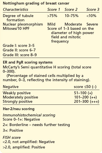
ER and PgR scoring systems
McCarty’s Semi quantitative H scoring (total score 0–300).
(Percentage of stained cells multiplied by a number, 0–3, reflecting the intensity of staining).
| Negative | score ≤50 (−) |
| Weakly positive | 51–100 (+) |
| Moderately positive | 101–200 (++) |
| Strongly positive | 201–300 (+++) |
Surgical anatomy of breast and lymphatics (Figure 10.1)
The adult breast extends from the second rib superiorly to the sixth rib inferiorly and from the lateral edge of the body of the sternum medially to mid-axillary line laterally. There are approximately 20–30 axillary lymph nodes which are surgically defined as:
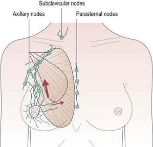
More than 75% of all lymphatic drainage passes through the axillary nodes and the remainder through the internal mammary nodes. There is a higher risk of internal mammary node involvement with medially located tumours and in those with extensive axillary nodes. Apical axillary node involvement leads to blockage of lymphatics and subsequent retrograde spread of cancer to supraclavicular nodes (see Figure 10.1).
Presentation
Example Box 10.1
Breast cancer: key points for a case presentation
All patients
Patient with local relapse
Many patients with early breast cancer are detected during screening by mammography.
Initial assessment
‘Triple’ assessment of patients with suspected breast cancer includes a combination of clinical examination, breast imaging (mammography and/or ultrasound) and pathologic evaluation (core biopsy). With this approach, a definite diagnosis of breast cancer can be made in 99% of cases.
Breast imaging
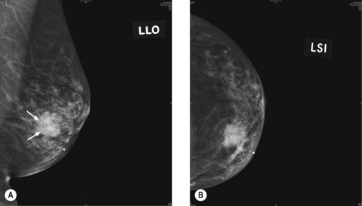
Mammogram
The commonest mammographic abnormality of DCIS is micro-calcification (in 50% cases). Other features include asymmetric density and mass. High-grade DCIS is associated with linear and branching calcification, whereas low-grade DCIS is associated with fine and granular calcification. Invasive cancer appears as a speculated mass (Figure 10.2). Although calcification can occur in various conditions, a linear small (<1 mm in diameter), non-uniform size and clustered calcification is suggestive of malignancy.
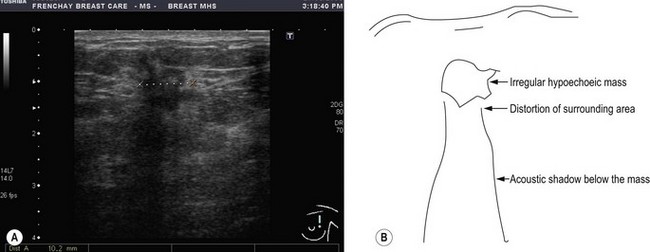
Ultrasound
Ultrasound examination of the breast helps to distinguish between solid and cystic lesions of more than 1 cm in size. Malignancy appears as a hypoechoic lesion with associated distortion of the surrounding tissues and an acoustic shadow on the breast tissue below it (Figure 10.3). It is also useful to assess axillary nodal status as well as to aid in guided FNA and core biopsy. Ultrasound is less sensitive than mammography for early detection and hence is not used for screening.
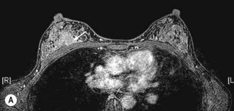
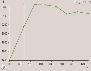
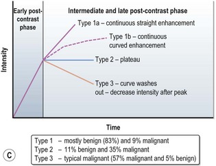
MRI scan
MRI can more accurately define the size of the tumour compared with mammography and is better in delineating intraductal disease and multifocal disease. MRI has a high sensitivity in detecting cancers (almost 100%) and lower sensitivity in detecting DCIS (80%), but has a higher false positivity. MRI is more useful for young women who have dense breast tissue (Figure 10.4). MRI is particularly useful in screening women less than 40 years with a high risk of breast cancer, to rule out multifocal disease prior to conservation, imaging women with implants and to distinguish scars from tumour recurrence.
Staging
TNM staging of breast cancer is given in Box 10.3.


