Biologic and Pharmacologic Basis of Cancer Chemotherapy
Roland T. Skeel
I. GENERAL MECHANISMS BY WHICH CHEMOTHERAPEUTIC AGENTS CONTROL CANCER
The purpose of treating cancer with chemotherapeutic agents is to prevent cancer cells from multiplying, invading, metastasizing, and ultimately killing the host (patient). Most traditional chemotherapeutic agents currently in use appear to exert their effect primarily on cell proliferation. Because cell multiplication is a characteristic of many normal cells as well as cancer cells, most nontargeted cancer chemotherapeutic agents also have toxic effects on normal cells, particularly those with a rapid rate of turnover, such as bone marrow and mucous membrane cells. The goal in selecting an effective drug, therefore, is to find an agent that has a marked growth-inhibitory or controlling effect on the cancer cell and a minimal toxic effect on the host. In the most effective chemotherapeutic regimens, the drugs are capable not only of inhibiting but also of completely eradicating all neoplastic cells while sufficiently preserving normal marrow and other target organs to permit the patient to return to normal, or at least satisfactory, function and quality of life.
Ideally, the cell biologist, pharmacologist, and medicinal chemist would like to look at the cancer cell, discover how it differs from the normal host cell, and then design a chemotherapeutic agent to capitalize on that difference. Until the last decade, less rational means were used for most of the chemotherapeutic agents that are now in use. The effectiveness of agents was discovered by treating either animal or human neoplasms, after which the pharmacologist attempted to discover why the agent worked as well as it did. With few exceptions, the reasons that traditional chemotherapeutic agents are more effective against cancer cells than against normal cells have been poorly understood. With the rapid expansion of information about cell biology and the factors
within the neoplastic cell that control cell growth, the strictly empiric method of discovering effective new agents has changed. For example, antibodies against the protein product of the overexpressed HER2/neu oncogene have been demonstrated to be effective in controlling metastatic breast cancer and reducing recurrences after primary therapy in patients whose tumors overexpress this gene. Discovery of the constitutively activated Bcr-Abl tyrosine kinase created as a consequence of the chromosomal translocation in chronic myelogenous leukemia has led to a burgeoning era of orally administered small molecular inhibitors of antibodies targeting critical molecular changes in cancer cells and their environment. These sentinel events have presaged the development of a host of new therapeutic agents that are directed at known specific targets within and around the cancer cell. These targets have been selected because they are altered in the cancer cell and are critical for cancer cell growth, invasion, and metastasis. This increased understanding of cancer cell biology has already provided more specific and selective ways of controlling cancer cell growth in several human cancers and will continue to dominate systemic therapy drug development in the decades to come.
Inhibition of cell multiplication and tumor growth can take place at several levels within the cell and its environment:
Macromolecular synthesis and function
Cytoplasmic organization and signal transduction
Cell membrane and associated cell surface receptor synthesis, expression, and function
Environment of cancer cell growth.
A. Classic chemotherapy agents
Most agents currently in use, with the exception of immunotherapeutic agents, other biologic response modifiers, and molecular targeted therapies appear to have their primary effect on either macromolecular synthesis or function. This effect means that they interfere with the synthesis of DNA, RNA, or proteins or with the appropriate functioning of the preformed molecule. When interference in macromolecular synthesis or function in the neoplastic cell population is sufficiently great, a proportion of the cells die. Some cells die because of the direct effect of the chemotherapeutic agent. In other instances, the chemotherapy may trigger differentiation, senescence, or apoptosis, the cell’s own mechanism of programmed death.
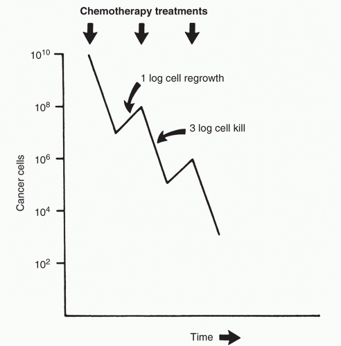
Cell death may or may not take place at the time of exposure to the drug. Often, a cell must undergo several divisions before the lethal event that took place earlier finally results in the death of the cell. Because only a proportion of the cells die as a result of a given treatment, repeated doses of chemotherapy must be used to continue to reduce the cell number (Fig. 1.1). In an ideal system, each time the dose is repeated, the same proportion of cells—not the same absolute number—is killed. In the example shown in
Figure 1.1, 99.9% (3 logs) of the cancer cells are killed with each treatment, and there is a 10-fold (1-log) growth between treatments, for a net reduction of 2 logs with each treatment. Starting at 1010 cells (about 10 g or 10 cm3 leukemia cells), it would take five treatments to reach fewer than 100, or 1, cell. Such a model makes certain assumptions that rarely are strictly true in clinical practice:
All cells in a tumor population are equally sensitive to a drug.
Drug accessibility and cell sensitivity are independent of the location of the cells within the host and of local host factors such as blood supply and surrounding fibrosis.
Cell sensitivity does not change during the course of therapy.
The lack of curability of most initially sensitive tumors is probably a reflection of the degree to which these assumptions do not hold true.
B. Biologic response modifiers and molecular targeted therapy
Within individual cells and cell populations are intricate inter-related mechanisms that promote or suppress cell proliferation, facilitate invasion or metastasis when the cell is malignant, lead to cell differentiation, promote (relative) cell immortality, or set the cell on the path to inevitable death (apoptosis). These activities are controlled in large part by normal genes and, in the case of cancer, by mutated cancer promoter genes, tumor suppressor genes, and their products. Included in these products are a host of cell growth factors that control the machinery of the cell. Some of these factors that affect normal cell growth have been biosynthesized and are now used to enhance the production of normal cells (e.g., epoetin alfa and filgrastim) and to treat cancer (e.g., interferon).
The recent expansion of our understanding of the biologic control of normal cells and tumor growth at the molecular level has only begun to offer improved therapy for cancer, though it has helped to explain differences in response among populations of patients. New discoveries in cancer cell biology have provided insights into apoptosis, cell cycling control, angiogenesis, metastasis, cell signal transduction, cell surface receptors, differentiation, and growth factor modulation. New drugs in clinical trials have been designed to block growth factor receptors, prevent oncogene activity, block the cell cycle, restore apoptosis, inhibit angiogenesis, restore lost function of tumor suppressor genes, and selectively kill tumors containing abnormal genes. Further understanding of each of these holds a great potential for providing powerful and more selective means to control neoplastic cell growth and may lead to more effective cancer treatments in the next decade. The fundamental principles related to this group of antineoplastic agents is discussed in Chapter 2.
II. TUMOR CELL KINETICS AND CHEMOTHERAPY
Cancer cells, unlike other body cells, are characterized by a growth process whereby their sensitivity to normal controlling factors has been partially or completely lost. As a result of this uncontrolled growth, it was once thought that cancer cells grew or multiplied faster than normal cells and that this growth rate was responsible for the sensitivity of cancer cells to chemotherapy. Now it is known that most cancer cells grow less rapidly than the more active normal cells such as bone marrow. Thus, although the growth rate of many cancers is faster than that of normal surrounding tissues, growth rate alone cannot explain the greater sensitivity of cancer cells to chemotherapy.
A. Tumor growth
The growth of a tumor depends on several interrelated factors.
1. Cell cycle time, or the average time for a cell that has just completed mitosis to grow, redivide, and again pass through mitosis, determines the maximum growth rate of a tumor but probably
does not determine drug sensitivity. The relative proportion of cell cycle time taken up by the DNA synthesis phase may relate to the drug sensitivity of some types (synthesis phase-specific) of chemotherapeutic agents.
2. Growth fraction, or the fraction of cells undergoing cell division, contains the portion of cells that are sensitive to drugs whose major effect is exerted on cells that are dividing actively. If the growth fraction approaches 1 and the cell death rate is low, the tumor-doubling time approximates the cell cycle time.
3. Total number of cells in the population (determined at some arbitrary time at which the growth measurement is started) is clinically important because it is an index of how advanced the cancer is; it frequently correlates with normal organ dysfunction. As the total number of cells increases, so does the number of resistant cells, which in turn leads to decreased curability. Large tumors may also have greater compromise of blood supply and oxygenation, which can impair drug delivery to the tumor cells as well as impair sensitivity to both chemotherapy and radiotherapy.
4. Intrinsic cell death rate of tumors is difficult to measure in patients but probably makes a major and positive contribution by slowing the growth rate of many solid tumors.
B. Cell cycle
The cell cycle of cancer cells is qualitatively the same as that of normal cells (Fig. 1.2). Each cell begins its growth during a postmitotic period, a phase called G1, during which enzymes necessary for DNA production, other proteins, and RNA are produced. G1 is followed by a period of DNA synthesis, in which essentially all DNA synthesis for a given cycle takes place. When DNA synthesis is complete, the cell enters a premitotic period (G2), during which further protein and RNA synthesis occurs. This gap is followed immediately
by mitosis, at the end of which actual physical division takes place, two daughter cells are formed, and each cell again enters G1. G1 phase is in equilibrium with a resting state called G0. Cells in G0 are relatively inactive with respect to macromolecular synthesis and are consequently insensitive to many traditional chemotherapeutic agents, particularly those that affect macromolecular synthesis.
C. Phase and cell cycle specificity
Most classic chemotherapeutic agents can be grouped according to whether they depend on cells being in cycle (i.e., not in G0) or, if they depend on the cell being in cycle, whether their activity is greater when the cell is in a specific phase of the cycle. Most agents cannot be assigned to one category exclusively. Nonetheless, these classifications can be helpful for understanding drug activity.
1. Phase-specific drugs. Agents that are most active against cells in a specific phase of the cell cycle are called cell cycle phase-specific drugs. A partial list of these drugs is shown in Table 1.1.
a. Implications of phase-specific drugs. Phase specificity has important implications for cancer chemotherapy.
(1) Limitation to single-exposure cell kill. With a phase-specific agent, there is a limit to the number of cells that can be killed with a single instantaneous (or very short) drug exposure because only those cells in the sensitive phase are killed. A higher dose kills no more cells.
TABLE 1.1 Examples of Cell Cycle Phase-Specific Chemotherapeutic Agents
Phase of Greatest Activity
Class
Type
Characteristic Agents
Gap 1 (G1)
Natural product
Enzyme
Asparaginase
Hormone
Corticosteroid

Stay updated, free articles. Join our Telegram channel

Full access? Get Clinical Tree

 Get Clinical Tree app for offline access


