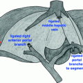
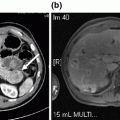
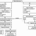
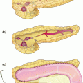
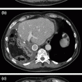
Fig. 17.1
MRI showing loss of confluence.
Surgical treatment of bile duct injury is indicated when loss of duct continuity is found and an endoscopic and/or radiological approach is ruled out [1]. Roux-en-Y hepaticojejunostomy has been proven to be the best treatment option by several groups [2–5]. A high-quality bilioenteric anastomosis, defined as a tension-free, wide, with adequate suture material, done in healthy, non-scarred non-ischemic ducts that are anastomosed to a defunctionalized Roux-en-Y jejunal limb, offers the best results [6]. There are several technical maneuvers that can be done in order to reach this goal, including the anterior opening of the confluence and the left duct, as well as partial removal of hepatic segments IV and V [7].
Our group has shown that an anastomosis performed in a patient with preserved confluence offers the best results [8]. These results can also be optimized if the patient has no stones or biliary sludge, which are usually developed as a result of bacterial colonization.
Loss of confluence, classified as a Bismuth IV [9] or a Strasberg E-4, [3] is a technical challenge for the surgeon. This is also, in our experience, the most undesirable scenario for repair and long-term results are unpredictable. In some cases, the anatomical variation of a low confluence results in a higher rate of bile duct injury [10]. After section and ablation of the duct, two separated lumens can be observed.
In other situations, ischemic damage due to thermal energy may be the cause of the injury, secondary to the heuristic error in which the common bile duct is mistaken with the cystic duct. Also, the presence of a biloma (infected or not), as well as the anatomical deformity contribute to the ductal damage. The most common cause for biliary injury in our series is a technically deficient repair attempt, usually performed by the primary-care surgeon.
Intraoperative Technical Pearls
Nearby organs should be methodically separated and, in patients who have previously undergone biliodigestive derivation, it is particularly important to free the small intestine in order to determine whether the anastomosed loop is not obstructed or defunctionalized, in case there is an enteral anastomotic variant (Omega loop with Braun anastomosis, Nakayama Beta-anastomosis), or if there is an abnormal positioning of the loop compromising its appropriate function.
In our center’s experience, longitudinally sectioning the anterior aspect of the duct (considering circulation is located on the lateral aspects) and directing this section toward the left duct without moving its posterior aspect makes creating the confluence a simpler task. Every section on the anterior aspect measures approximately 2–3 mm, having thoroughly verified and certified the direction of the ducts.
In order to expose the hepatic hilum, the base of segment IV is removed with a 3 × 3 × 3 cm wedge. The small parenchymal vessels bleeding are controlled mainly through compression and, in some cases, using transfictive 5-0 sutures. This maneuver adequately exposes the left duct that follows an extrahepatic trajectory, from the confluence to the round ligament’s end.
The loss of confluence can be easily diagnosed nowadays with the aid of MRCP. Endoscopic management can be a suitable therapeutic procedure by placing a percutaneous biliary drain or stents in order to maintain the function of the ducts. When endoscopic treatment is not an option, the surgical alternatives available for complex biliary injuries are: Portoenterostomy, double barrell anastomosis, construction of a neoconfluence, partial hepatectomy, and liver transplantation.
Portoenterostomy
This is the adult variant of the Kasai procedure [11]. It is the least desired option hence it has presented a high failure rate in our center [12]. We suggest its usage when very small, joined ducts are found and the construction of a high-quality bilioenteric anastomosis (wide, tension free, with appropriate epithelization of mucosae, done in healthy ducts using adequate suture material) is not feasible. In some cases, it is possible to place percutaneous stents during the preoperative or postoperative period in order to advance them to the intestinal lumen at the time of portoenterostomy. Along with periodical changes of the percutaneous stents, this option allows the patient to maintain an acceptable quality of life (without jaundice and cholangitis, with the evident disadvantage of having an indwelling catheter for a long period of time).
When the stents are removed, failure of the patency of the ducts is almost constant. In our hands, several of these cases are enrolled into the liver transplant waiting list.
Double Barrell Anastomosis
This variant can be performed when the ducts are widely separated (more than 1 cm). The right duct anastomosis is technically demanding and it anticipates a high chance of long-term dysfunction. Even after stenting, the final outcome after removal is unpredictable.
The anastomosis to the left duct can usually be done with a moderate level of difficulty by extending the incision to the anterior aspect of the duct. We have less than 10 cases repaired with this surgical approach. Complications such as secondary biliary cirrhosis may arise (with or without cholangitis, which in some conditions is severe). A couple of cases in our series have been treated by means of unilobar portal vein embolization with the objective of inducing atrophy of the affected liver lobe. Segmentary portal vein embolization does not offer good results.
Alternative Approaches and Controversies
There are unfortunate isolated cases in which an appropriate anastomosis is impossible to perform. The anastomosis of the jejunal opening to the hepatic parenchyma and the need of stents in every duct included in the anastomosis technically leads to a portoenterostomy, similar to the one described by Kasai [13]. In our experience and that of others, these are very infrequent cases that often require prolonged stenting, and may also develop acute cholangitis episodes, and thus should be considered for liver transplant.
Endoscopic access is not a simple procedure in these types of patients since it is challenging to insufflate the intestinal loop. If this is feasible, there is also the problem of identifying the anastomosis that oftentimes is punctiform and/or obstructed.
Stay updated, free articles. Join our Telegram channel

Full access? Get Clinical Tree


