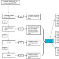
The advent of imatinib has been a major breakthrough in chronic myeloid leukemia (CML) treatment. A few patients treated with imatinib are either refractory to imatinib or eventually relapse. Resistance is frequently associated with mutations in the kinase domain of BCR-ABL. Over 100 point mutations coding for single amino acid substitutions in the BCR-ABL kinase domain have been isolated from CML patients resistant to imatinib treatment. Most reported mutants are rare, whereas 7 mutated residues comprise two-thirds of all mutations detected. BCR-ABL mutations affect amino acids involved in imatinib binding or in regulatory regions of the BCR-ABL kinase domain, resulting in decreased sensitivity to imatinib while retaining aberrant kinase activity. The early detection of BCR-ABL mutants during therapy may aid in risk stratification as well as molecularly based treatment decisions.
Crystallographic analysis has shown that imatinib binds to and stabilizes an inactive conformation of ABL, in which the centrally located activation loop is not phosphorylated and thus in a closed position. Before first clinical reports of resistance due to kinase mutations, biochemical studies guided by the crystallographic analysis predicted mutant BCR-ABL with retained kinase activity, but reduced or lost drug affinity to imatinib as a potential mechanism of resistance. Consistent with preclinical analysis, Gorre and colleagues found that imatinib resistance was associated with the reactivation of BCR-ABL signal transduction in a cohort of relapsed patients. The exchange of the amino acids threonine and isoleucine at position 315 (T315I) of the BCR-ABL protein was the first mutation detected in imatinib resistant chronic myeloid leukemia (CML) patients. Sequencing of cloning products revealed the T315I mutation in 6 of 9 resistant patients. Based on the known crystal structure of the ABL kinase domain, this substitution was predicted to abrogate affinity for the drug. Since this original report, many investigators have reported kinase domain mutations that impart resistance to imatinib.
A bacterial mutagenesis screen revealed a set of mutations that grow out in the presence of imatinib. These mutations largely reflected the findings in clinical trials. Mutations may be categorized into 4 groups, based upon the crystallographic structure of ABL: (1) those that directly impair imatinib binding to the catalytic domain of the oncogenic protein; (2) those within the P-loop; (3) those within the activation loop, preventing the kinase from achieving the inactive conformation required for imatinib binding; and (4) those within the catalytic domain with impact on the activity of the tyrosine kinase. Even mutations outside the kinase domain can lead to enhanced autophosphorylation of the kinase, thereby stabilizing the active conformation that resists imatinib binding. Mutant BCR-ABL alleles retain biologic activity but show varying degrees of resistance to imatinib in biochemical and cellular assays. However, the level of resistance against imatinib in vitro cannot always explain resistance seen in patients.
Methods of BCR-ABL mutation detection
Several techniques have been described for the detection of BCR-ABL kinase domain mutations, but there is currently no consensus concerning which technique should be used ( Table 1 ). Mutations can be reliably and sensitively detected by selection and expansion of specific clones followed by DNA sequencing. This procedure, however, is cumbersome and not eligible for clinical routine. Alternatively, sequencing of nested polymerase chain reaction (PCR)-amplified BCR-ABL products has been widely used to search for known and unknown BCR-ABL kinase domain mutations. A potential limitation of direct sequencing is the sensitivity of only 10% to 20%. Sensitivities of 1% to 5% could be obtained by double-gradient denaturing electrophoresis, pyrosequencing, high-resolution melting, or array based assays. More sensitive methods include peptide nucleic acid-based PCR clamping and allele-specific oligonucleotide (ASO-) PCR. However, these techniques are specific and cannot be applied for screening of unknown mutations. Denaturing high-performance liquid chromatography (D-HPLC) has been described as a highly sensitive screening method for BCR-ABL kinase domain mutations, even when the site of the mutation is unknown.
| Sensitivity | Method | References |
|---|---|---|
| 10%–20% | Selection of clones plus DNA sequencing Nested polymerase chain reaction (PCR) plus DNA sequencing | |
| 1%–5% | Denaturing high-performance liquid chromatography (D-HPLC) | |
| Double gradient denaturing electrophoresis | ||
| Pyrosequencing | ||
| High-resolution melting (HRM) | ||
| MALDI-TOF mass spectrometry | ||
| Nanofluidic array | ||
| <1% | Fluorescence PCR and PNA clamping Allele-specific oligonucleotide (ASO-) PCR |
Use of various techniques for the detection of BCR-ABL mutations has led to different frequencies of mutations that are reported. Furthermore, the pattern of individual mutations reported seems to depend on the specific method used for mutation detection. Standardized techniques and protocols for the detection of BCR-ABL mutations will be necessary to obtain comparable mutation results within clinical studies. A recent cooperative evaluation of different methods of detecting BCR-ABL kinase domain mutations in 3 European laboratories has revealed major differences particularly in the detection of low-level mutations (ie, mutations below the sensitivity of standard direct sequencing). Further studies toward standardized techniques and protocols are necessary and underway.
BCR-ABL mutation types
BCR-ABL Point Mutations
More than 70 amino acid residues within the kinase domain have been reported as targets of about 100 different mutations. Recently published review articles have summarized the distribution and relative sensitivities to imatinib in vitro. Most reported mutants are rare, whereas 7 mutated sites comprise two-thirds of all mutations detected: G250, Y253, E255, T315, M351, F359, H396. Different amino acid substitutions occur at the same residue (eg, F317C, F317L, and F317V) and confer different imatinib sensitivities. The functional importance of individual amino acid substitutions with regard to oncogenicity of BCR-ABL is difficult to determine. Besides being gain of function or loss of function mutations, they might simply be regarded as surrogate markers of the increased genetic instability associated with advanced phase disease. Griswold and colleagues studied the transformation potential, kinase activity, and substrate specificity of five of the most frequent mutations—Y253F, E255K, T315I, M351T, and H396P. Compared with unmutated BCR-ABL, Y253F was noted to produce enhanced kinase activity (gain of function), with E255K producing comparable activity. The remaining 3 mutations decreased enzymatic function (loss of function). Transforming potency did not always correlate with kinase activity. However, clear differences did exist in the pan-tyrosine phosphorylation patterns of the Ba/F3 cells expressing the various mutants, which would be consistent with differences in substrate use and signaling pathway activation. Phosphoproteomic studies on mutant T315I also support the notion of mutation-dependent oncogenic potential of individual substitutions. Skaggs and colleagues showed that the intramolecular tyrosine phosphorylation pattern is altered, indicating modified biologic activity of mutant BCR-ABL.
BCR-ABL Deletion Mutants
Some imatinib resistant patients express deletion mutants of BCR-ABL, apparently due to mis-splicing. Most commonly these deletion mutants lack a significant proportion of the kinase domain that includes the P-loop. The L248V mutation was shown in 2 patients to additionally induce the alternative splicing of a shorter variant by introducing a donor splice site within ABL exon 4 lacking amino acids 248 to 274. Sherbenou and colleagues performed a screen for such mutations in 95 patients with CML and identified 14 patients (15%) with a total of six different deletion mutations. Deletion mutants detected most frequently involved splice junctions. Functional studies further demonstrated that such deletion mutations are not oncogenic and are catalytically inactive. The authors hypothesized that coexpressing BCR-ABL deletion mutants have a dominant-negative effect on the native form through heterocomplex formation. However, the prognostic impact of coexpression of deletion mutants in CML patients during imatinib treatment is unknown.
ABL Polymorphisms
The BCR-ABL K247R change is based on a rare single-nucleotide polymorphism (SNP) of ABL occurring likewise in healthy controls and nonhematologic cell types. Despite its juxtaposition to the P-loop, functional analysis showed no alteration compared with nonmutated BCR-ABL. To investigate if other changes in the BCR-ABL kinase domain should be considered as SNPs rather than acquired mutations, 911 CML patients after failure or suboptimal response to imatinib were screened for BCR-ABL kinase domain mutations. SNP analysis was based on the search for nucleotide changes in corresponding normal, nontranslocated ABL alleles by ABL allele-specific PCR following mutation analysis. In addition to the K247R polymorphism five new SNPs within the BCR-ABL kinase domain were uncovered; two of them led to amino acid changes ( Table 2 ). SNPs theoretically could modify the primary response to tyrosine kinase inhibitors, and must therefore be distinguished from acquired mutations that govern the evolution of secondary resistance. Novel point mutations should be confirmed by analyzing the normal ABL alleles to exclude polymorphisms.
| Nucleotide Position a | Nucleotide Polymorphism | Amino Acid Change b | N c | Allele Frequency (%) |
|---|---|---|---|---|
| 58,758 | A | T240T | 1 | 0.1 |
| 58,778 | G | K247R | 9 | 1.0 |
| 68,708 | G | F311V | 2 | 0.2 |
| 68,722 | G | T315T | 1 | 0.1 |
| 68,736 | G | Y320C | 1 | 0.1 |
| 74,901 | G | E499E | 73 | 8.0 |
a Nucleotide positions according to GenBank accession number U07563 for the ABL 1a splice variant.
b Amino acid residues are denoted with the single letter code and correspond to the ABL 1a variant.
c Total number of patients harboring the respective polymorphism.
Incidence of BCR-ABL mutations
The frequency of BCR-ABL mutations in resistant patients was reported to range from 42% to 90% depending on the methodology of detection, the definition of resistance, and the phase of the disease. Mutations are found more frequently in accelerated phase or blast crisis. A twofold increase of the BCR-ABL transcript level might be a diagnostic red flag to detect patients carrying resistance mutations. The identification of the specific type of mutation in relapsed patients is of prognostic relevance, since the different types of mutation correlate with the risk of evolution of relapsed patients. However, mutant Ph+ subclones may remain at low levels, may be transient or unstable, or may not be consistently associated with subsequent relapse. In many cases, mutations have been detected in samples that were collected during imatinib treatment, but in several cases mutations were also traced back to samples collected prior to treatment, especially in patients who consecutively developed accelerated phase or blast crisis. Using more sensitive techniques, mutations were also found in imatinib-naïve patients and in patients having achieved complete cytogenetic remission. It is important to note that Ph+ primitive cells have been reported to be less sensitive to imatinib in vitro and in vivo, to harbor BCR-ABL mutations even prior to imatinib exposure, and to rapidly develop mutations under imatinib-induced selection pressure. Various BCR-ABL mutations show different biochemical and clinical properties. The biochemical and cellular impact of different mutations is heterogeneous, ranging from a minor increase of the median inhibitory concentrations of imatinib to a virtual insensitivity of imatinib. The T315I mutation and some mutations affecting the so-called P-loop of BCR-ABL confer a greater level of resistance, whereas the biochemical resistance of other mutations can be overcome by dose increase, and others seem functionally irrelevant. Thus, the detection of a kinase domain mutation must be interpreted within the clinical context.
Stay updated, free articles. Join our Telegram channel

Full access? Get Clinical Tree


