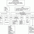
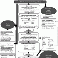
Chapter 33 Amrana Qureshi Paediatric Haematology and Oncology Children’s Hospital, John Radcliffe Hospital, Oxford, UK Causes of thrombocytopenia are varied in paediatric and neonatal intensive care [1]. Causes of thrombocytopenia may be primary (e.g. immune thrombocytopenic purpura) or part of an existing condition and the reason for admission to ITU (e.g. leukaemia) or secondary to sepsis/disseminated intravascular coagulation (DIC). Broadly, all causes may be divided into those with a predominant immune or non-immune aetiology. Although there is overlap of causes of thrombocytopenia in adults and children, many conditions are either specific to neonates and children, such as prematurity, neonatal allo- and autoimmune thrombocytopenia and congenital causes, or have a different natural history, such as immune thrombocytopenia, which is usually self-limiting. In this chapter, the approach to thrombocytopenia for patients developing thrombocytopenia in neonatal and paediatric intensive care is reviewed with particular focus on practical approaches to diagnosis and management. The mainstay of management remains transfusions of platelets. A specific section at the end describes an approach to use of platelets in neonatal and paediatric critical care. This will provide practical guidelines on transfusion practice for both prophylaxis prior to procedures in patients with thrombocytopenia and for (less commonly) bleeding in association with thrombocytopenia. For certain clinical conditions, platelet transfusions are not indicated, for example, thrombotic thrombocytopenic purpura (TTP) in childhood. In the face of major bleeding and if the predominant mechanism for thrombocytopenia is immune (ITP), platelet transfusions are likely to be less effective, and immunomodulatory therapy with steroids and immunoglobulins will be warranted. In the ITU setting, sepsis is the most common cause of thrombocytopenia, which may also result in DIC and platelet consumption. Bacterial, viral and fungal infections can cause DIC. Meningococcal meningitis/septicaemia remains the most frequent cause of severe DIC in children. Other causes of DIC include malignancy, trauma, burns and liver disease. Pathogenesis and laboratory diagnosis of DIC are explained in Chapter 9. As DIC is a secondary event, the underlying cause should be treated. Replacement therapy with blood products is reserved for bleeding patients or prior to an invasive procedure. Fresh frozen plasma (FFP) can be given (15 mL/kg) with cryoprecipitate (5 mL/kg) and platelets to maintain a fibrinogen greater than 1 g/L and platelet count of 50 × 109/L in bleeding patients. The use of heparin and thrombolytic therapy in DIC is controversial. Protein C concentrate has been used in severe DIC secondary to meningococcal sepsis although there are no randomized control trial to prove its efficacy. Haemolytic uraemic syndrome (HUS) is a combination of microangiopathic haemolytic anaemia (MAHA), thrombocytopenia and acute renal failure. In children, HUS is most often caused by E. coli toxin (0157), which predominantly damages glomerular endothelial cells. Most cases recover without sequelae. Management is supportive but may require plasma exchange. Thrombocytopenia in HUS is caused by platelet aggregation, and platelet transfusion is relatively contraindicated as this may further increase microvascular thrombosis. Acquired TTP is rare in children and most likely secondary to ciclosporin post-transplant (see also Chapter 11 on TTP). Emergency treatment with plasma exchange is required. More recently, rituximab (an anti-CD 20 antibody), which inhibits antibody formation, has been tried with success. Platelet transfusions, again, are relatively contraindicated. Congenital TTP results from deficiency of the ADAMST 13 enzyme, and treatment involves regular plasma infusions. This is a primary or secondary disorder which results from pathological and uncontrolled macrophage and T-cell activation from cytokine production. It is defined by bilineage (two blood cell lines), cytopenia (at least), fever, hepatosplenomegaly, deranged clotting, low fibrinogen, high ferritin and fasting triglycerides and histopathological evidence of haemophagocytosis in the skin, liver and bone marrow. In congenital type, presentation is often in infancy or early childhood, and the child can present with haemodynamic shock and acidosis. Urgent treatment with chemotherapy and steroids is required. Secondary cases mostly occur in older children, due to sepsis, and EBV is often causal in the older age group, sometimes in association with immunodeficiency. Treatment of the underlying cause is the mainstay of treatment. Chemotherapy may be required in severe cases. Platelet transfusion may be indicated in the case of bleeding. Thrombocytopenia is common in the intensive care setting, and up to 35% of all neonates will develop platelet counts less than 50 × 109/L [2]. In premature and low-birth-weight neonates, the incidence of thrombocytopenia is as high as 80%. The strongest predictors of thrombocytopenia are prematurity and antenatal factors, resulting in physiological stress to the neonate due to either placental insufficiency or infection [3]. The likely cause of thrombocytopenia can be postulated from the timing from birth. In the premature neonate, or small-for-gestation age, in the first 72 h after birth, thrombocytopenia is likely to be due to antenatal and maternal factors (placental insufficiency and antenatal/perinatal infection), which results in suppressed production of platelets. After 72 h, thrombocytopenia is most likely due to postnatally acquired infection, often bacterial, or necrotising enterocolitis and involves both suppression of platelet production due to cytokine release and increased consumption, often as part of DIC. Profound thrombocytopenia can develop quickly in this setting. In the term, neonate thrombocytopenia usually results from sepsis and DIC. Further rare causes are mentioned in the Table 33.1 later.
Approach to Thrombocytopenia
General approach to thrombocytopenia
More common causes of childhood thrombocytopenia in ITU
Non-immune mediated
Microangiopathic haemolytic anaemias
Disseminated intravascular coagulation (DIC)
Management of DIC
Haemolytic uraemic syndrome/TTP
Haemophagocytic lymphohistiocytosis
More common causes of neonatal thrombocytopenia in ITU
Non-immune causes
Stay updated, free articles. Join our Telegram channel

Full access? Get Clinical Tree


