Application of Advances in Endoscopic and Robot-Assisted Approaches to the Treatment of Head and Neck Cancer


Application of Advances in Endoscopic and Robot-Assisted Approaches to the Treatment of Head and Neck Cancer


Jason G. Newman

Bert W. O’Malley Jr.


INTRODUCTION

The history of medicine has included some significant milestones. From the separation of philosophy and clinical medicine in the days of Hippocrates, to the understanding of the role of microorganisms in the creation of human illness, there are some concepts and tools that have literally changed how we understand and practice medicine. These individual changes have caused massive paradigm shifts, which first disrupt and then propel forward our approach to treating the human condition. Over the last 100 years, medicine has moved forward at a breakneck pace. Surgery has taken several big steps over this time frame. Open surgery, in the age of modern anesthesia, advanced imaging, and antibiosis, has become safe and effective in most cases. This has allowed us to concentrate on even further advances in the practice of surgery. Minimally invasive approaches and now even robotic approaches are becoming standard options in the management of many surgical diseases. In this chapter, we will review the history and discuss the evolving applications of both endoscopy and robotics in the field of head and neck cancer surgery.


HISTORY OF ENDOSCOPY IN OTOLARYNGOLOGY

Endoscopy within the field of otolaryngology has followed two separate but closely intertwined paths. On the one side is endoscopy to evaluate the larynx, and on the other, the sinonasal cavities. Although each of these anatomic regions has faced its own set of obstacles and techniques, what continues to keep them linked is the reliance on technology to access and operate in these tight spaces through natural orifices.


History of Sinonasal Endoscopy

In the 1970s, an Austrian physician, Walter Messerklinger, introduced the use of endoscopes in the performance of sinus surgery. Several of his students, including Stammberger and Kennedy, continued to advance the indications for the use of the endoscope within otolaryngology.1 Initially, the endoscope was primarily used as a tool to aid in the medical management of inflammatory sinus disease. However, the endoscope quickly became the instrument of choice for management of surgical sinus disease. Over time, as descriptions of the anatomy, surgical techniques, and instrumentation began to evolve, the capacity to advance the frontier of the endoscope has grown.

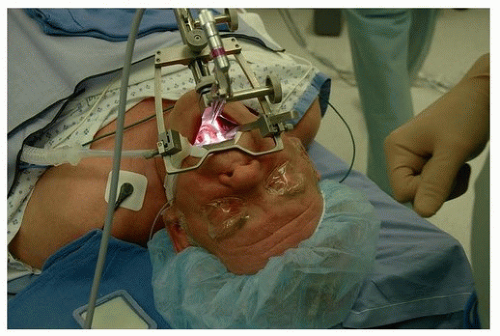
Anterior skull base CSF leaks were among the earliest advanced procedures performed transnasally with an endoscope.2 Creating safe techniques to separate the intracranial and sinonasal spaces was paramount in creating a safe and low-risk procedure. Initially, this involved the use of free mucosal grafts, adipose tissue, and nasal packing. In the 1990s, several authors described the use of the endoscope to assist in removal of tumors of the sinonasal and anterior cranial base, often with the combination of open and endoscopic techniques.3 In 2001, Casiano et al.4 reviewed the first series of purely endoscopic resection of esthesioneuroblastoma. Their success and the success of other authors led to a continued interest in the expansion of the use of this technique. Creation of hemostatic agents, finer and more angulated instrumentation, and techniques for closure of skull base defects were all results of these early surgical endeavors. Free mucosal grafting, inlay grafting techniques, and pedicled flaps have all played a role in the evolution of our surgical capabilities in this area. In addition, fine-cut CT and MRI scans, intraoperative navigation, and high-definition endoscopes have all allowed surgeons to address complex lesions more safely. Since these early studies, many surgical teams have gone on to describe large series of patients undergoing endoscopic resection of sinonasal malignancies.5,6 Most recently, the endoscopic endonasal approach (EEA) has been expanded to include options for management of tumors in areas other than the sinonasal cavity. This includes intracranial tumors, as well as tumors in the infratemporal fossa and pterygopalatine space. The sinonasal cavity has now, in many cases, become for the endoscopic approved corridor to the region of primary concern.


History of Laryngopharyngeal Endoscopy

At the same time as the early descriptions of endoscopy in the sinonasal cavity was first being described, Strong, Jako, Steiner, and others7,8 began to develop the field of endoscopic laser surgery (ELS) for the larynx. This led the way to transoral laser microsurgery (TLM). Since that time, techniques and instruments have evolved to the point where almost all regions of the upper aerodigestive tract are accessible for surgical endoscopy and resection of tumors.

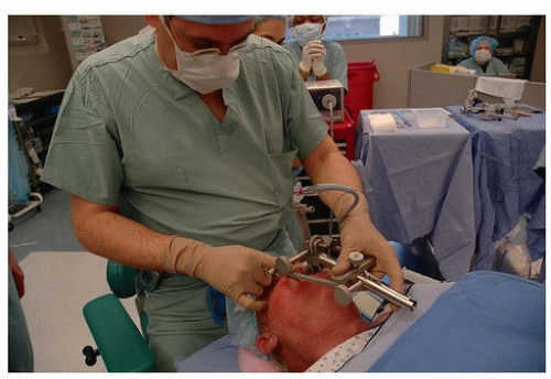
As is true with sinonasal endoscopy, endoscopy of the larynx required a continued expansion of the instruments, optics, and hemostatic methods to aid in its wider application. A variety of laryngoscopes, laryngeal microinstruments, specialized
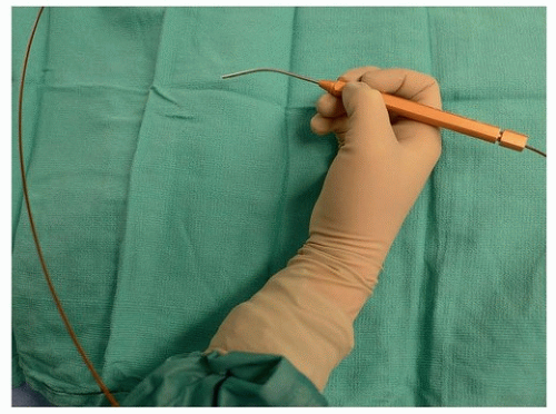
endotracheal tubes and suspension techniques have been designed to maximize the ability to visualize and safely operate in this tight space. The CO2 laser was an important tool for the safe performance of this surgery, as both an instrument of resection as well as a hemostatic tool. Its long wavelength (10,600 nm) and other physical properties originally required that it be delivered via a microscope-mounted beam splitter (or a direct mounted beam) into the surgical site. Until recently, the use of the CO2 laser has required a direct line of site from the laser emitter to the surgical site, often creating significant difficulty in both visualizing and manipulating the tumor. Recently, the invention of a hollow-core fiber to deliver the CO
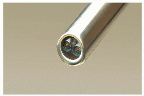
2 laser (OmniGuide system, OmniGuide Inc., Cambridge, MA) (Fig. 36.1) has broadened the indications for this surgery. It has removed the line of site necessary for management of the tumor and has brought the laser into the field on a flexible fiber. This, in combination with angled endoscopes, microinstruments and high-definition optics, has allowed for continued expansion of the use of this TLM.


Figure 36.1. One of many interchangeable handpieces for the OmniGuide flexible CO2 laser.


History of Robotics in Otolaryngology

In many ways, robotic surgery is the natural extension of endoscopic surgery. Although the endoscope has allowed our optical equipment to move into the field, it has also made distal control of instrumentation more challenging. Thus, the obvious target for surgical innovation as it relates to minimally invasive surgery is to explore the notion of improved distal control of instruments. This is one of the areas in which robotics excels.

The presence of robotics in surgery has been a relatively recent phenomenon. In fact, despite the presence of robotics in nonmedical industries for over 60 years, the first reported robotic surgical case was performed in 1985. The Puma 560 was used to accurately localize neurosurgical biopsies. The same device was then used to perform transurethral biopsies of the prostate.9 In 1992, the U.S. Food and Drug Administration (FDA) approved the first medical use of a robot. During the same time period, NASA and the Department of Defense became interested in the notion of remote battlefield surgery. Through DARPA, these agencies funded this technology. It was conceptualized that deploying a remote controllable (telepresence) robot into the front lines of battle would allow a wounded soldier to be stabilized during the “golden hour” of trauma. This robot would be controlled by surgeons in a safe zone, allowing them to perform surgery during this critical window for survival, but without putting the surgeon in harm’s way. The funding for this project helped catalyze the creation of the first commercially available robotic systems, the AESOP (Computer Motion of Santa Barbara, CA) and the da Vinci (Intuitive Surgical, Sunnyvale, CA). Variations of these systems are the backbones for the robotic systems we use today.

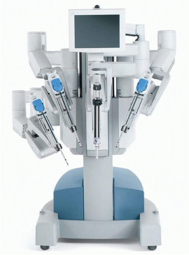
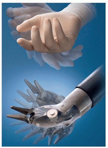
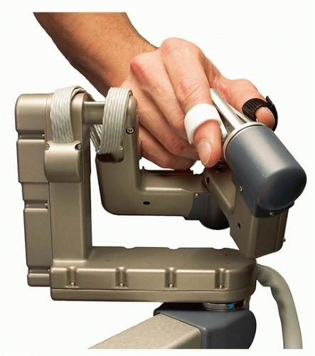
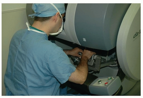
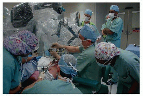
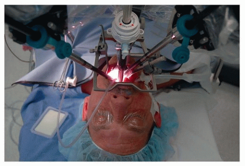
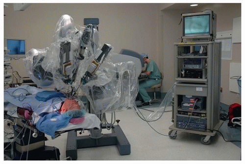
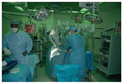
In otolaryngology (OTO-HNS), the primary robot in use today is the da Vinci system. In some ways, the word “robot” is a misnomer, in that full control of the instruments still rests in the hands of the operating surgeon. However, what sets it apart from other surgical tools is that the surgeon is controlling the instruments remote from the patient’s bedside. In its current form, the da Vinci robot consists primarily of two components. One is the surgeon console, where the operating surgeon sits. This consists of a stereoscopic (three-dimensional image) set of goggles, as well as controls for the robotic instruments (Figs. 36.2, 36.3, 36.4, 36.5, 36.6). The second component is the bedside patient cart, which consists of three separate instrument arms, with interchangeable instruments, and a camera arm all controlled by the operating surgeon. What makes these instruments different than most others is that they are designed with distally wristed function, so that they have the capacity to mimic or even exceed the natural range of motion of a human wrist. This gives the operating surgeon seven degrees of freedom with movement of the instruments. In combination with the stereoscopic view afforded by the dual optic rigid
camera arm, these small wristed instruments are well suited for surgery around corners or in tight spaces.


Figure 36.2. The Intuitive Surgical da Vinci bedside robotic console. Note the three instrument arms and the camera arm. (©2016 Intuitive Surgical, Inc.)


Figure 36.3. The end of the instrument arm on the da Vinci robot mimics the human wrist in creating seven degrees of freedom. (©2016 Intuitive Surgical, Inc.)


Figure 36.4. Close-up of the joystick-like controller used to move the robotic instruments. (©2016 Intuitive Surgical, Inc.)

The introduction of robotics to the field of OTO-HNS only occurred in the early 2000s. Initial reports described the use of the robot to help avoid neck incisions for surgery such as resection of a submandibular gland.10,11 The first use of the robot in a live patient was for the removal of a vallecular cyst, published in 2005.12 Within the next year, Drs. O’Malley and Hockstein described the first preclinical experiments that helped to establish transoral robotic surgery (TORS).13 Building upon these original experiments, Drs. Hockstein, Weinstein, and O’Malley at the University of Pennsylvania developed preclinical models to demonstrate the feasibility, safety, and efficacy of TORS. This included application of the technology to cadaver, and live canine procedures, prior to performing human studies.
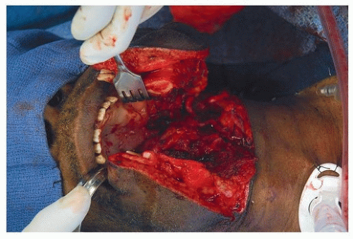
14,15,16,17


Figure 36.5. Surgeon at the console, using the joystick-like controls. (©2016 Intuitive Surgical, Inc.)


Figure 36.6. Close-up of the end of the camera arm, which houses two separate high-definition cameras and a light source.

Initial experiments surrounded the appropriate instrumentation for use with the robotic console. Most conventional laryngoscopes have a narrow inlet. This only allows for small, relatively parallel instruments to be inserted and used in the field. However, the robotic arms required broader access to the field. This obstacle was overcome by the incorporation of several different retractors. Early use of the Dingman, Crow Davis, and FK-WO laryngoscope provided enough room for the arms of the robot to access the pharynx (Figs. 36.7 and 36.8).

After appropriate access to the surgical site was accomplished, creating a safe set of procedures with low-risk profile was the next priority. Additional studies were performed to look at these parameters. We concluded that transoral robotics
surgery was safe and effective. This set the stage for human clinical trials.


Figure 36.7. Dr. Gregory Weinstein in an early case of TORS, placing a retractor into the patient’s mouth.


Figure 36.8. FK retractor in place, giving exposure of the surgical site and allowing adequate movement of the surgical arms.

Weinstein and O’Malley established a prospective human trial and conducted the first human experiments for TORS. The experiments included patients with various pathologies, including cancers of the oropharynx, larynx, and a variety or other benign and malignant conditions in the head and neck. These data,16 pooled with the data from several other authors,18 led to the approval from the FDA in December of 2009 of TORS for use in benign and select malignant lesions of the head and neck.


CURRENT INDICATIONS FOR ENDOSCOPY AND ROBOTICS


Oropharynx


Endoscopy

Due to the somewhat restricted access to tumors of the oropharynx, open surgery to this area is particularly complex. This approach often involves a mandibulotomy for access, a tracheostomy to secure the airway, and often a feeding tube to allow for healing of the surgical site postoperatively (Fig. 36.9). In addition, just achieving adequate exposure can result in one or more cranial neuropathies. Fortunately, alternatives to the open approach began to show promise. Beginning in the 1970s, radiation began to play a role in the postoperative setting and then as primary therapy for cancer of the oropharynx. As the results began to demonstrate efficacy, many centers began to favor a radiation-based approach to these cancers. In many cases, a completely nonsurgical approach to this area was gaining acceptance. The combination of chemotherapy and radiation has now become one of the standard options for the management of cancer of the oropharynx. The oncologic outcomes are generally good, leading to 3-year overall survival rates between 23% and 88%.19,20 However, posttreatment sequelae can be significant with gastrostomy tube dependence rates ranging from 0% to 26% in the same literature.


Figure 36.9. Traditional approach to tumors of the oropharynx. Note the mandibulotomy and tracheostomy.

The disease significantly increased in incidence over the same time period that the nonsurgical management of oropharyngeal cancer continued to gain acceptance. What was once an uncommon cancer primarily associated with smoking and drinking, largely affecting men in their 60s or older, has now become predominantly a virally induced (HPV) cancer, affecting many nonsmokers, often in their 40s and 50s. This change in the etiology of the cancer has been associated with an improved response to therapy and overall survival of patients with HPV positive tumors. Concurrent with our enhanced understanding of the biology and clinical behavior of HPV positive oropharyngeal cancers, the technology used to treat them has also evolved. The options for surgical management of primary cancer of the oropharynx have developed considerably and will be discussed below. In addition, even the management of the neck, once limited to a radical neck dissection, has now been shown to be amenable to more limited selective neck dissections.

At the same time that many centers were advocating a nonsurgical approach to cancers in the oropharynx, surgeons in Europe including Professor Wolfgang Steiner are credited with expanding the nascent field of TLM.7 This surgery is an alternative to primary radiation and at present is also an alternative to robotic surgery for the oropharynx. TLM, much like TORS, has benefitted from the evolution of optics and instrumentation and has the potential to give excellent outcomes without the need for open surgery. Steiner’s approach involves the use of distending bivalve laryngoscopes. This allows the surgeon to apply tension to the cancer and resect using the CO2 laser at
the appropriate distance from the tumor. This approach often requires a “piece-meal” transection of the cancer in order to continuously evaluate the depth of the tumor and to obtain clear its margins. Although the adoption of this technique has been slow, it has resulted in excellent outcomes in repeated studies, from multiple institutions. Several studies have demonstrated an excellent overall survival for patients using this technique, with rates from 52% to 87%.21,22,23,24


Robotics

The anatomic site that has benefitted most from the use of robotics in the field of head and neck surgery is the oropharynx. Although some of the earliest robotics experiments were performed in the larynx and neck, the robotic technology appeared best suited for the management of cancer in the oropharynx.25 The combination of improved visualization, four-handed management of the cancer, and relatively easy instrument access to the surgical site made this an obvious choice. Cancers in the tonsil and base of tongue are the primary targets for TORS (Fig. 36.10).

Prior to the creation of TORS, many of the patients with cancer of the oropharynx had limited surgical options. Open approaches are associated with significant morbidity and in the current era are often reserved for recurrent cancer after nonsurgical treatment. Although TLM has clearly demonstrated the feasibility of a nonopen approach to cancer of the oropharynx, the technical challenges of this approach have limited its acceptance.26 This combination of feasibility and technical challenges of TLM paved the way to the introduction of TORS for management of cancer of the oropharynx. Several of the obstacles facing a surgeon with TLM are removed with TORS. Reduction of the need for line of sight, the ability to use four-handed technique, improved hemostasis, a wider field of view, and wristed distal dexterity instruments have all made the resection of tumors in this area less challenging (Fig. 36.11). Although still less than a decade since its inception, the early results are good. Several studies have reported 2- and 3-year overall survival results from 85% to 92% in selected patients.27,28,29

TORS is performed using a variety of laryngeal and oropharyngeal retractors. Once adequate visualization of the cancer is obtained, the robotic arms are brought into place in the oral cavity. The primary surgeon sits at the console, and the assistant at the patient bedside, at the head of the patient (Figs. 36.12 and 36.13). This two-surgeon approach allows for excellent suctioning, retraction, and hemostasis, as four hands are in the field at once. Because this exposure generally allows for a view of the entire tumor, an “en-bloc” approach is often possible and favored.


Figure 36.10. Early photo demonstrating the technique for surgical setup for TORS.


Figure 36.11. Surgical setup, demonstrating the position of the instrument arms and the camera arm.


Figure 36.12. Setup of the room and the robotic instruments in an early surgical case. The bedside console, the vision cart, and the surgeon console are all visible.


Figure 36.13. Operating room setup for TORS.


Table 36.1 Indications and Contraindications for TORS


Indications


1. Adequate visualization of the tumor


2. Adequate exposure for resection


During staged laryngoscopy, the appropriate retractors may need to be inserted to determine if the tumor will be amenable to negative-margin resection with TORS techniques


Characteristics such as trismus, anterior positioned larynx, large tongue, or morbid obesity often make placement of the pharyngeal retractors impossible


Contraindications


Unresectability of involved neck nodes


Invasion of the mandible


Involvement of the base of the tongue requiring resection of >50% of the tongue base


Involvement of the pharyngeal wall requiring resection of >50% of the posterior pharyngeal wall


Radiographic confirmation of carotid artery involvement


Fixation of the tumor to the prevertebral fascia


Clear indications, tumor characteristics, and contraindications have now been set out for TORS. They include features listed in Table 36.1.9

The timing of management of the neck in patients undergoing TORS continues to evolve, as does the procedure itself. Initially, the neck dissection was routinely staged after the primary surgery, to reduce postoperative pharyngeal edema as well as to reduce the incidence of fistulization. Recently, many surgeons have begun to stage the neck component of the surgery before or at the same time as the resection of the primary. This may help to reduce the incidence of postoperative bleeding.


Larynx


Endoscopy

TLM (the history of which is described above) is an accepted standard in the treatment of well-selected patients with cancer of the larynx. There is an extensive literature demonstrating the safety, efficacy, and outcomes of this technique.30,31,32,33,34 It is most often used in the management of previously untreated cancers, but results in the face of recurrence following radiation have also been quite good. Because good exposure is one of the keys to success in this surgery, a combination of closed or distending laryngoscopes is used to visualize the cancer.31 The technique, especially for larger cancers, often involves a stepwise resection of the cancer by cutting through the cancer with the laser in order to create a three-dimensional map of the depth and extent of the cancer. Specific blocks of cancer are then removed until the entire cancer is resected with frozen section evaluation of the margins. The laryngoscopes are repositioned as necessary during surgery in order to optimize exposure.35 TLM has been advocated for use in many earlystaged glottis cancers, but has also been shown to be effective in the treatment of selected patients with more advanced, staged T3 and T4 cancers36 (Fig. 36.14). In select cases, it may also be an option for treating recurrent cancers after surgery or radiation-based therapies.37

Only gold members can continue reading. Log In or Register to continue

Stay updated, free articles. Join our Telegram channel

Dec 18, 2016 | Posted by in ONCOLOGY | Comments Off on Application of Advances in Endoscopic and Robot-Assisted Approaches to the Treatment of Head and Neck Cancer

Full access? Get Clinical Tree

Get Clinical Tree app for offline access