Antimetabolites
Sheila Shaigany
Paul I. Schneiderman
Antimetabolites represent a broad class of chemotherapy agents that halt tumor growth by disrupting cellular division in the S phase of the cell cycle. By structurally resembling normal cell components, they work to inhibit metabolites required for DNA and RNA synthesis. Antimetabolites can be divided into three categories:
Purine analogs (ie, 6-mercaptopurine, thioguanine, fludarabine, cladribine)
Pyrimidine analogs (ie, capecitabine, gemcitabine, cytarabine, fluorouracil)
Folate antagonists (ie, methotrexate (MTX), pemetrexed)
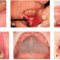
As with other classes of cytotoxic agents, antimetabolites target rapidly dividing cells of the hair, skin, nails, and mucosal surfaces. Therefore, almost all drugs within this class may lead to varying degrees of reversible alopecia, stomatitis/mucositis, and nail dystrophy.
TOXIC ERYTHEMA OF CHEMOTHERAPY
Toxic erythema of chemotherapy (TEC), an umbrella term used to describe a range of cutaneous reactions to cytotoxic chemotherapies, is frequently seen with antimetabolites such as cytarabine, 5-FU, capecitabine, and MTX.1 TEC includes reactions such as handfoot syndrome (HFS), epidermal dysmaturation, and intertriginous eruptions (ie, “malignant intertrigo”). These reactions can sometimes be differentiated based on clinical and histological findings, but frequently exhibit significant overlap.
HFS (also referred to as acral erythema or palmo-plantar erythrodysesthesia [PPE]) is a commonly observed cutaneous adverse event, most frequently seen with capecitabine (50%-60%), cytarabine, and 5-fluorouracil (5-FU).2 Concomitant use of additional chemotherapies (ie, anthracyclines such as doxorubicin) may also increase the risk of HFS up to 89%.3 It typically presents within 2 to 21 days after starting therapy, but may occur up to 10 months later, particularly with dose increases. HFS is both dose and time dependent, with more severe reactions occurring from prolonged infusions or oral therapy (ie, capecitabine).
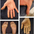
Patients initially experience prodromal tingling and burning, followed by symmetric, tender edema and erythema of the palms>soles. HFS has a predilection for the distal phalangeal fat pads, thenar and hypothenar eminence, and lateral aspect of the fingers, but it may also extend to the dorsal surface. Vesicles and blisters can erupt and progress to
erosions and desquamation (Figure 23.1). A severe bullous variant causing epidermal necrosis may occur with both cytarabine and MTX (Figure 23.2). In Fitzpatrick skin types V and VI, HFS may present with more hyperpigmentation rather than erythema, especially in patients taking capecitabine.2
erosions and desquamation (Figure 23.1). A severe bullous variant causing epidermal necrosis may occur with both cytarabine and MTX (Figure 23.2). In Fitzpatrick skin types V and VI, HFS may present with more hyperpigmentation rather than erythema, especially in patients taking capecitabine.2
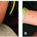
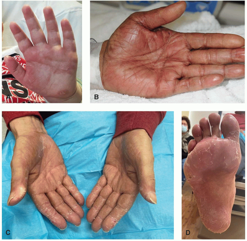 Figure 23.1. Hand-foot syndrome due to cytarabine (A and B) and capecitabine (C and D) with palmar erythema, edema, and desquamation, favoring the distal fat pads and thenar eminences, with relative sparing of the flexural finger creases. |
Management Pearls
HFS resolves with cessation of therapy in an average of 1 to 2 weeks. Treatment is mostly supportive. Manage symptoms with daily emollients, cold compresses, pain control, local wound care, and high-potency topical steroids. Systemic corticosteroids can be considered for severe, debilitating reactions.
CTCAE Grade 3 reactions (blistering, peeling, bleeding, and/or edema along with pain limiting self-care activities of daily living) generally require chemotherapy interruption and
either subsequent dose reduction or switch to an alternative agent. HFS is often more severe with rechallenge if the dose is not reduced.
either subsequent dose reduction or switch to an alternative agent. HFS is often more severe with rechallenge if the dose is not reduced.
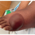
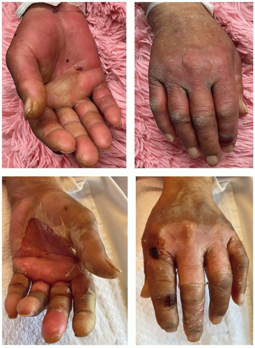 Figure 23.2. Severe, painful bullous variant of hand-foot syndrome due to high-dose cytarabine involving both the palmar and dorsal hand, with progressive evolution of edema to bullae formation and ulcerations over the course of several days. |
Daily application of 10% urea cream as prophylaxis can be considered, given the low side effect profile, although studies examining its efficacy demonstrate conflicting findings. Regional cooling with ice packs or ice water immersion while receiving intravenous therapy may also be considered, as it has shown to be helpful in preventing anthracycline and taxane-induced HFS. We do not recommend daily oral pyridoxine or celecoxib for the prevention of HFS for all patients due to a paucity of strong evidence and the potential for adverse events with celecoxib (cardiovascular toxicity, GI bleed); evidence best supports the use of celecoxib for capecitabine-induced HFS, when it should be considered to maintain the use of this therapeutic agent.
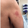
Non-HFS TEC presents as symmetric, erythematous to dusky patches often accompanied by edema, purpura, and desquamation. As with HFS, pathogenesis is thought to involve a direct toxic effect on keratinocytes and eccrine glands as the drug is excreted through sweat. Rashes often localize to regions containing a high proportion of sweat glands and/or areas under occlusion such as the axilla, inframammary folds, inguinal folds, palms, and soles (Figure 23.3A and B). A distinctive papular purpuric eruption with violaceous erythema of the groin, axillae, back of the neck, scalp, ears, and intertriginous areas is a recognizable pattern of cytarabine toxicity.4 Blisters may develop, forming large erosions in the skin folds (Figure 23.4C). A rare type of acral erythema involving the ears due to cytarabine, termed “Ara-C ears,” may also develop with bilateral ear swelling and erythema.
DRUG-INDUCED NEUTROPHILIC ECCRINE HIDRADENITIS
Drug-induced neutrophilic eccrine hidradenitis (NEH), a neutrophilic dermatosis classically associated with cytarabine, presents 7 to 14 days after starting therapy, with tender red or purpuric macules and papules on the trunk, distal extremities, and head/neck. The histology is distinct, demonstrating perieccrine neutrophils (may be absent in neutropenic patients receiving cytotoxic chemotherapy) along with eccrine vacuolar interface and eccrine gland necrosis. As with HFS and TEC, it is thought to result from a direct cytotoxic effect of drug within the eccrine glands. Eccrine squamous syringometaplasia is a noninflammatory version of NEH diagnosed exclusively by skin biopsy.
Management Pearls
It is important to rule out acute graft-versus-host disease (GVHD) in stem cell transplant patients as skin findings may be similar to NEH. The presence of gastrointestinal signs and symptoms (ie, diarrhea, hepatitis) favors a diagnosis of GVHD.
Treatment is supportive and involves local wound care to erosions to prevent infection and for analgesia. Topical steroids may also be used, but their utility is not entirely clear. Rash improves with reduction of dose and/or frequency of administration. Severe cases warrant discontinuation of drug as the rash recurs with rechallenge.
While NEH resolves spontaneously within a few weeks, it usually recurs with rechallenge. The rash can be treated with high-potency topical steroids or steroid sparing agents such as dapsone and colchicine. Oral steroids are used as a last resort as patients are often neutropenic.
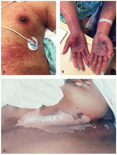 Figure 23.3. Toxic erythema of chemotherapy due to fludarabine presenting as coalescing erythematous and dusky macules that localize to sweat gland-rich areas such as the axilla, areola, umbilicus (A), palms, and soles (B), with prominent edema, and hemorrhagic bullae formation on the palms. A flexural bullous intertriginous eruption in a patient taking cytarabine, idarubicin, and etoposide (C).
Related posts:Stay updated, free articles. Join our Telegram channel
Full access? Get Clinical Tree
 Get Clinical Tree app for offline access

|