18
Antigen Processing and Presentation
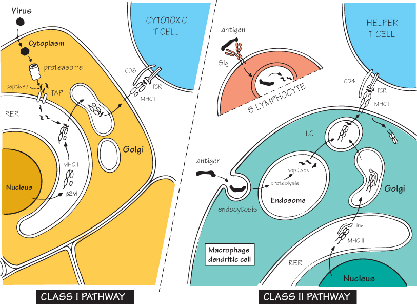
In discussing the MHC and the T-cell receptor (see Figs 11 and 12), frequent allusion has been made to the foreign peptides that bind to the former and are recognized by the latter. These peptides become associated with MHC molecules through two quite separate pathways (see figure), usually known for convenience as the class I pathway (left) and the class II pathway (right).
The first evidence that the MHC was involved in presenting antigens to T cells was the demonstration of ‘MHC restriction’ – the fact that T cells are specific for both antigen and MHC molecule. Then it was discovered that cytotoxic T cells could respond to viral nuclear antigens, which are not displayed on the surface of the virus! How could a T cell ‘see’ such a well-concealed antigen? The answer is shown in the left-hand figure above: virus-derived peptides become bound to class I MHC molecules inside the cell and are then transported to the surface, where T cells can recognize the peptide–MHC combination and, under appropriate circumstances, kill the virus-infected cell (for more details see Fig. 21).
At the same time, it was shown that a rather similar process occurs in the ‘antigen-presenting’ cells, mainly macrophages and dendritic cells, which activate helper T cells, with the difference that here it is the class II MHC molecules that transport the peptides to the surface (right-hand figure). This process is kept separate from the class I pathway by occurring in the endosomal/lysosomal vacuoles in which foreign material is normally digested (see Fig. 9). B lymphocytes can also process and present antigen, but only when they are able to bind it via their surface immunoglobulin (top right). Presentation by B cells to T cells is an essential step in T-cell help (see Fig. 19).
One can now appreciate that the real role of the MHC system is to transport samples of intracellular proteins to the cell surface for T cells to inspect them and react if necessary – by proliferating into clones and then helping macrophages or B cells or killing virus-infected cells, as described in the following pages.
The Class I Pathway
Virus
Because they are synthesized in the cell, viral proteins are available in the cytoplasm, alongside self-proteins.
RER
Rough endoplasmic reticulum, where proteins, including those of the MHC, are synthesized.
MHC I
The single three-domain α chain associates with β2-microglobulin to make a class I MHC molecule, whose structure is not fully stable until a peptide has been bound (see below). The efficient folding of MHC molecules around an antigen peptide requires a set of other ‘chaperone’ molecules found within the RER.
Proteasome
Related posts:

Stay updated, free articles. Join our Telegram channel

Full access? Get Clinical Tree


