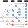
Considering that the antibody in serum is a mixture of perhaps 100 million slightly different types of molecule, the unravelling of its structure was no mean feat. Early work depended on separation into fragments by chemical treatment (top left in figure); the fine details have come from amino acid sequencing and X-ray crystallography, both of which require the use of completely homogeneous (monoclonal) antibody. This was originally available only in the form of myeloma proteins, the product of malignant B lymphocytes, but is produced nowadays by the hybridoma method (see Fig. 15) or by genetic engineering in bacteria, yeast or a variety of mammalian cell types.
A typical antibody molecule (IgG, centre) has 12 domains, arranged in two heavy and two light (H and L) chains, linked through cysteine residues by disulphide bonds so that the domains lie together in pairs, the whole molecule having the shape of a flexible Y. In each chain the N-terminal domain is the most variable, the rest being relatively constant. Within the variable (V) regions, the maximum variation in amino acid sequence is seen in the six hypervariable regions (three per chain) which come together to form the antigen-binding site (bottom left in figure). The constant (C) regions vary mainly in those portions that interact with complement or various cell-surface receptors; the right-hand part of the figure shows the different features of the C region in the five classes of antibody: M, G, A, E and D. The result is a huge variety of molecules able to bring any antigen into contact with any one of several effective disposal mechanisms. The basic structure (MW about 160 000) can form dimers (IgA, MW 400 000) or pentamers (IgM, MW 900 000) (see right-hand side of figure).
There are species differences, especially in the heavy chain subclasses, which have evolved comparatively recently; the examples shown here illustrate human antibodies. Interestingly, camels and llamas also have antibodies with only heavy chains. These antibodies may be able to attach to some targets not accessible to conventional antibodies, and examples are being tested as possible new ways of preventing infection by viruses such as HIV (see Fig. 28). The carbohydrate side-chains (shown here in black) may constitute up to 12% of the whole molecule.
Note: The illustration shows an IgG molecule with its 12 domains stylized. The actual three-dimensional structure is more like the molecule shown binding to antigen in Fig. 13 (extreme right).
Fragments
produced by chemical treatment: these fragments were of great importance in elucidating the chemical structure of antibody. Fab and F(ab)2 fragments allow binding to specific antigen in the absence of secondary interactions with other cells mediated via the constant region.
- H, L: heavy and light chains which, being only disulphide-linked, separate under reducing conditions.
- Fab: antigen-binding fragment (papain digestion).
- Fc: crystallizable (because relatively homogeneous) fragment (papain digestion).
- F(ab)2: two Fab fragments united by disulphide bonds (pepsin digestion).
Affinity and Avidity
Related posts:


Stay updated, free articles. Join our Telegram channel

Full access? Get Clinical Tree


