Anorexia/Weight Loss


Anorexia/Weight Loss


Egidio Del Fabbro

Eduardo Bruera


CACHEXIA

Cancer cachexia is defined as a multifactorial syndrome characterized by involuntary loss of skeletal muscle (with or without fat loss) that leads to progressive, impaired function and cannot be fully reversed by conventional nutritional support (1). Cancer cachexia is common, reported to occur in up to 90% of patients with advanced cancer, and is often accompanied by a pro-inflammatory response and a cluster of symptoms that include fatigue and poor appetite. The clinical implications of cancer cachexia are profound, since weight loss is associated with more chemotherapy-related side effects; fewer completed cycles of chemotherapy; and decreased survival (2,3). Furthermore, severe involuntary weight loss impairs cancer patients’ quality of life (QOL) (4) and sense of dignity (5).


EPIDEMIOLOGY

An autopsy series of 816 patients with solid tumors at MD Anderson Cancer Center found that infection was the commonest cause of death, followed by organ failure; however, in 10% of patients no cause could be attributed other than severe emaciation and/or electrolyte imbalance (6). Subsequently, a landmark study by Dewys et al. (3) of more than 3,000 patients reported a shorter median survival in patients with weight loss compared with those without weight loss. Except in patients with pancreatic or gastric cancer, decreased weight correlated with poor performance status and within each performance status category or tumor stage, weight loss was associated with decreased survival. Up to 70% of patients with breast cancer or acute lymphoblastic leukemia had no evidence of weight loss, but about half of prostate cancer, colon cancer, and lung cancer patients experienced weight loss and 85% of gastric and pancreatic patients lost weight (one-third >10%). The study also suggested that any weight loss (0% to 5%) was associated with a poorer prognosis, and the prognostic effect of weight loss was greater in patients with a more favorable prognosis (good performance status or early tumor stage).

A prospective study carried out in 1979 at the National Cancer Institute in Milan of 280 patients with a variety of tumors (7) reported that specific cancers and advanced stage were more likely to be associated with weight loss. Patients with upper gastrointestinal tumors (esophagus and stomach) had significantly decreased weight, serum albumin, and triceps skinfold thickness, and treatment with chemotherapy or radiotherapy was associated with an additional decrease in arm muscle circumference. Malnutrition also became progressively more severe as the disease advanced, except in patients with breast or cervix cancer.


PROGNOSIS

A number of studies have reported that weight loss is associated with a poor prognosis in patients with advanced cancer. A systematic review of prognostic factors in patients with recently diagnosed incurable cancer showed (8) that weight loss, fatigue, anorexia, nausea, dyspnea, pain, multiple comorbidities, and poor performance status were all associated with decreased survival.

In 1,555 consecutive patients with locally advanced or metastatic gastrointestinal carcinomas (esophagus, stomach, pancreas, and colorectal), weight loss at presentation was more common in men than in women and correlated with decreased tumor response, QOL, and performance status (9). Patients with weight loss received lower chemotherapy doses, but they developed more severe dose-limiting toxicity (plantar-palmar syndrome and stomatitis) and received 1 month less treatment. Another prospective study of 770 lung cancer patients found that weight loss was reported by 59%, 58%, and 76% of patients with small cell lung cancer, non-small cell lung cancer (NSCLC), and mesothelioma, respectively (10), and those patients with weight loss had increased treatment toxicity and decreased survival.


MECHANISMS OF CACHEXIA


Starvation versus Cachexia

Unlike starvation, which is characterized by a loss of fat primarily, weight loss due to cachexia is characterized by a loss of both muscle and fat (Table 10.1). Cachexia will occur despite caloric supplementation and can be differentiated from starvation by endocrine abnormalities such as insulin and ghrelin resistance and frequently also by increased
resting energy expenditure. Unfortunately, most patients with cancer cachexia will also have a significant component of “starvation” that exacerbates muscle wasting through either a decline in appetite or other symptoms that affect oral intake (e.g., nausea and dysgeusia).


TABLE 10.1 Starvation versus cachexia


Starvation


Cachexia


Caloric intake


↓↓


Resting energy expenditure


↓↑ or ↔


Body fat


↓↓


Lean body mass


↓↓


Inflammatory markers


↑ or ↔


Insulin


↔, unchanged; ↓, reduced; ↓↓, markedly reduced; ↓↑, increased or reduced; ↑, increased.


Inflammation

An aberrant pro-inflammatory response as a result of the tumor-host interaction has detrimental effects on cell metabolism, protein synthesis, hormone action, and the autonomic nervous system (ANS). Although increased activity of proinflammatory cytokines is proposed as the principle unifying mechanism causing both anorexia (11) and muscle wasting (12), the pathophysiology of cachexia is complex and multifactorial (Figure 10.1), so none of the interrelated mechanisms that contribute to this syndrome should be viewed in isolation. The primary sites of dysregulation are known to be the hypothalamus centrally (producing anorexia) and skeletal muscle and adipose tissue peripherally (increased catabolism and decreased anabolism).


Peripheral

In cancer cachexia, pro-inflammatory cytokines appear to induce muscle wasting by targeting only certain muscle gene products. Skeletal muscle is composed of core myofibrillar proteins, including myosin heavy chain (MyHC), actin, troponin, and tropomyosin. Transcription of the myosin heavy chain gene and many other muscle genes is regulated in part by the nuclear transcription factor MyoD. Myogenic cell cultures and animal models of tumor-induced cachexia indicate that MyHC is a selective target for pro-cachectic inflammatory cytokines (TNF-α and IFN-γ) by inhibiting MyoD and increasing the degradation of MyHC via the ligase-dependent ubiquitin-proteasome pathway (by IL-6) (13). The ubiquitin ligase-dependent proteasome pathway is a major cellular mechanism that degrades proteins and regulates skeletal muscle wasting in cancer and other disease states.

These basic science studies demonstrating the dual requirement of inflammatory cytokines for muscle wasting are supported by recent clinical trials showing interventions (etanercept (14) and infliximab (15)) against a single cytokine (TNF-α) produced no improvement in cachexia-related clinical outcomes.

More recently, animal models indicated that the muscle wasting induced by circulating cytokines may be modulated by genetic ablation of adipose triglyceride lipase (ATL) (16). Animals with and without ATL activity were injected with tumor cells, which resulted in high circulating levels of inflammatory cytokines such as IL-6, TNF-α, and lipidmobilizing factor (zinc-α2-glycoprotein). However, only the animals with ATL activity experienced adipose tissue wasting and subsequent muscle loss accompanied by an increased expression of the ubiquitin-proteasome pathway. These studies suggest that lipolysis plays an important role in cancer cachexia and that lipases may be important therapeutic targets for the prevention of muscle wasting.

Finally, the identification of specific factors produced by the tumor, e.g., PIF (proteolysis-inducing factor), may be important for future therapeutic targets (17).


Central

Systemic pro-inflammatory cytokines stimulate the production of cytokines within the hypothalamus. Proinflammatory cytokines are implicated in causing anorexia by stimulating neural pathways within the arcuate nucleus of the hypothalamus to secrete anorexigenic peptides such as α-melanocyte-stimulating hormone (α-MSH), which is derived from proopiomelanocortin. α-MSH inhibits feeding and increases energy expenditure by activating melanocortin receptors, primarily the type 4 melanocortin receptor (MC4-R). In healthy animals, orally administered selective MC4-R antagonists (18) that penetrate the blood-brain barrier increase food intake, and in mice with C26 adenocarcinoma, selective MC4-R antagonists prevent tumorinduced loss of body weight, fat mass, and lean body mass. Inflammatory cytokines also inhibit the release of orexigenic neuropeptides such as agouti-related protein from another set of neurons within the hypothalamus that stimulate feeding.


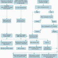
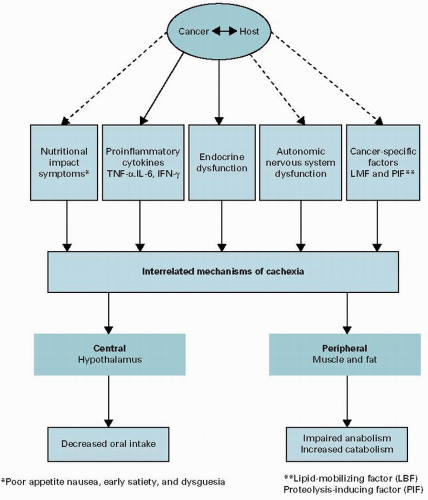
Figure 10.1. Mechanisms of cancer cachexia.

Cytokines also have other central effects that decrease the oral intake indirectly by producing symptoms such as early satiety (via IL-1) and depression (IL-6) (19).

Although animal models have shown a compelling association between pro-inflammatory cytokines and cachexia, the evidence in human studies is less consistent with some studies demonstrating a correlation between serum cytokines and clinical outcomes, e.g., weight loss (20,21) or performance status (22,23), and others showing no significant relationship (24,25,26). It might be that pro-inflammatory cytokines such as TNF-α act in a paracrine rather than in an endocrine fashion, so that serum levels do not accurately reflect tissue concentrations (27) or that single-nucleotide polymorphisms (28,29,30) determine an individual’s susceptibility to developing cachexia. In addition, as described earlier in this chapter, animal models of cachexia suggest that downstream targets such as ATL may be key determinants of susceptibility to circulating inflammatory cytokines.


Neuroendocrine

The neural pathways within the hypothalamus are also influenced by orexigenic anti-inflammatory hormones such as ghrelin and testosterone. Ghrelin and insulin levels are elevated in patients with cachexia, suggesting resistance to these hormones, possibly mediated by inflammatory cytokines. In addition, the ANS may play a role in producing the syndrome of cachexia. Since many patients with advanced cancer have a dysfunctional ANS (31), other chronic sympathetic activation (32,33) or abnormalities of parasympathetic nerves such as the vagus, which has a role in ghrelin activity (34) and an acute anti-inflammatory effect (35), may amplify the mechanisms causing cachexia (Figure 10.2).


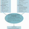
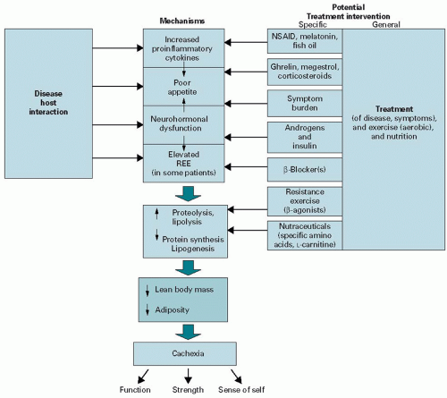
Figure 10.2. A multimodal treatment model. REE, resting energy expenditure; NSAID, non-steroidal anti-inflammatory drug.


DIAGNOSIS OF CACHEXIA

The diagnosis of cachexia is obvious in those patients who present with temporal wasting, anorexia, poor performance status, and a markedly underweight body mass index (BMI). The challenge is to diagnose patients earlier in the disease trajectory so as to initiate effective therapy before cachexia becomes “refractory” to intervention or patients are unable to comply with anti-cachexia treatment because of disease progression, frailty, or delirium. A history of weight loss >5% within the previous 6 months in the absence of simple starvation is most consistently used for the diagnosis of cachexia in clinical trials. The history of weight loss is important to obtain, since an underweight BMI at an initial visit is the exception in most outpatients because of the increased prevalence of obesity in the general population; 95% of patients referred to a cachexia clinic at a comprehensive cancer center had a normal or even overweight BMI (36) despite having metastatic cancer and a palliative care outpatient center reported marked weight loss in 71% of patients (30% with significant muscle mass reduction) in spite of a normal or increased BMI (37).


CLASSIFICATION

Patients considered for intervention trials should be enrolled at similar points along their illness trajectory. Unfortunately, patients with cachexia may be enrolled in clinical trials at different inception points, making it very difficult to compare outcomes between participants and between clinical trials. Also, many participants in cachexia intervention trials are frail and often unable to complete the full duration of study treatment. To address these challenges a classification of cachexia has been proposed by an International Consensus
Group that divides the syndrome into cachexia, pre-cachexia, and refractory cachexia (Figure 10.3). Pre-cachexia is characterized by early clinical and metabolic signs (e.g., anorexia, impaired glucose tolerance, or elevated C-reactive protein [CRP]) that may precede an involuntary weight loss of ≤5%. Patients who have >5% loss of body weight over the last 6 months, or a BMI <20 kg/m2 and ongoing weight loss of >2%, or sarcopenia (skeletal muscle index of males <7.26 kg/m2 and females <5.45 kg/m2) and ongoing weight loss of >2% (but have not entered the refractory stage) are classified as having cachexia. The last stage, refractory cachexia, attempts to identify those patients with weight loss that are thought to be “refractory” to anti-cachexia therapy. Patients with cachexia who have a poor performance status (WHO 3 or 4) and a life expectancy of <3 months are placed within this category. In future, criteria need to be determined for patients that will be predictive of resistance to therapeutic interventions.


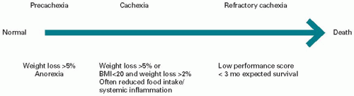
Figure 10.3. Classification of cachexia.


CLINICAL ASSESSMENT

The clinical assessment includes a careful history that is focused on nutrition (quantity and composition), symptoms contributing to poor oral intake, weight and body composition, a physical examination, and identification of any reversible metabolic abnormalities.

A clinical approach to cachexia in terms of “primary” and “secondary” cachexia is a useful framework for approaching the clinical management of patients with cancer and involuntary weight loss. Primary cachexia denotes the syndrome (discussed earlier in this chapter) that is characterized by muscle loss (with or without loss of fat). Secondary cachexia includes potentially treatable contributors to the weight loss of primary cachexia such as nutritional impact symptoms (NISs) and comorbid metabolic abnormalities (e.g., hypogonadism, thyroid dysfunction, and vitamin B12 and vitamin D deficiency). Other causes of weight loss that have a predominant starvation component such as gastrointestinal obstruction may also be included in this category, especially if they respond to endoscopic or surgical treatment (e.g., stent placement or endoscopic dilatation for esophageal obstruction).


Nutritional Impact Symptoms

Advanced cancer patients with cachexia may have multiple concurrent symptoms that compromise oral intake and contribute to nutritional decline (Table 10.2). These NISs have been included in the Patient-Generated Subjective Global Assessment (PG-SGA) of Nutritional Status, a validated screening tool that includes a patient report of weight and weight change, food intake, symptoms, activities, function, and a physician evaluation of the disease and its related nutritional requirements. NISs listed in the PG-SGA include poor appetite, pain, nausea, vomiting, constipation, altered smell and taste, mouth sores, dry mouth, dysphagia, depression, and diarrhea. The NIS section of the PG-SGA questionnaire is completed by patients and it provides the clinician with important information since many NISs can be treated effectively with inexpensive therapies. Although NIS can be assessed without completion of the entire PG-SGA, the screening tool has other advantages in terms of identifying patients at risk for malnutrition and facilitating quantitative outcome data collection for research purposes. The PG-SGA uses a numerical score as well as provides a global rating of well-nourished, moderately, or suspected of being malnourished or severely malnourished. The higher the score, the greater is the risk for malnutrition. A score of >9 indicates a critical need for nutrition intervention. Nutrition triage recommendations include patient and family education, symptom management, and nutrition intervention such as additional food, oral nutrition supplements, and enteral or parenteral nutrition (PN). In clinical practice, the scored PG-SGA has been shown to be a quick, valid, and reliable nutrition assessment tool that enables malnourished patients with cancer to be identified and triaged for nutritional support (38) and has been accepted by the Oncology Nutrition Dietetic Practice Group of the American Dietetic Association as the standard for nutrition assessment of patients with cancer. A retrospective study at a comprehensive cancer center found that most patients with involuntary weight loss had three or more uncontrolled NISs Table 10.2 shows the interventions commonly used to treat NIS.

The Edmonton Symptom Assessment Scale is a valid, commonly used questionnaire in cancer patients that can
identify the presence as well as the severity of some NISs such as nausea, depression, and fatigue, but unfortunately it does not include many other NISs such as early satiety, constipation, and dysgeusia.


TABLE 10.2 Findings and interventions for secondary nutritional impact in 151 cancer patients referred to a cachexia clinic


Nutritional Impact Symptoms


Number (%)


Corresponding Interventions


Number (% Treated Among Affected Individuals)


Early satiety


94 (62%)


Metoclopramide


74 (79%)


Constipation


78 (52%)


Laxatives


68 (87%)


Nausea or vomiting


67 (44%)


Antiemetics (mostly metoclopramide)


54 (81%)


Depressed mood


63 (42%)


Antidepressant (mostly mirtazapine)


51 (81%)


Dysgeusia


42 (28%)


Zinc supplement


20 (48%)


Dysphagia


21 (14%)


GI or speech therapy evaluation


5 (24%)


Dry mouth


14 (9%)


Artificial saliva


2 (14%)


Mucositis


11 (7%)


Opioids and topical mouthwash


3 (27%)


Dental pain


8 (5%)


Dental referral


2 (25%)


GI, gastrointestinal.


From Del Fabbro E, Hui D, Dalal S, et al. Clinical outcomes and contributors to weight loss in a cancer cachexia clinic. J Palliat Med. 2011 September;14(9):1004-1008.


Body Composition

Although both muscle and fat are usually depleted in patients with cachexia, the body composition in patients with the same BMI could vary considerably. Most patients reporting weight loss have normal or elevated BMIs and a significant proportion of obese cancer patients (20%) are sarcopenic. Muscle wasting may therefore be underrecognized and masked by adipose tissue. Patients with the combination of sarcopenia and obesity have a worse prognosis and a higher risk of chemotherapy-related adverse effects (39).

The least invasive method to assess body composition is the midarm muscle area which requires measuring the triceps skinfold thickness using calipers (in millimeters) and the midarm circumference (in centimeters) and entering them into the following equation: midarm muscle area = (midarm circumference in centimeters) – π × tricipital skinfold thickness in millimeters)2/(4 × π) – a correction factor of 10 for men and 6.5 for women.

Bioimpedance analysis (BIA) relies on the different electrical properties of fat and muscle (increased water content). The bioimpedance devices are relatively easy to use and not burdensome to patients, but they may provide falsely elevated results of muscle mass when patients have edema. Additional information such as the ratio of resistance and reactance (phase angle) provided by BIA may also be useful for prognostication (40).

Dual-energy X-ray absorptiometry (DEXA) scans are based on the three-component model of body composition. DEXA uses two x-ray energies to measure body fat, muscle, and bone mineral. DEXA is more burdensome to patients (must be in supine position while the image is taken) than BIA and is more expensive. The results may be viewed as whole body estimates of body fat, muscle, and bone mineral as well as regional body estimates.

Computed tomography (CT) scan is expensive and not practical for many palliative care patients with cachexia. However for those patients who are being routinely evaluated for the purpose of restaging and follow-up, CT is useful to distinguish between muscle and adipose tissue (41) and can be used to assess body composition changes longitudinally.


Indirect Calorimetry

Handheld indirect calorimetry (IC) provides more detailed information regarding patients’ caloric requirements and identifies the presence of hypermetabolism. Since more than 80% of caloric daily requirements are due to basal energy expenditure (BEE), IC will provide an accurate measured BEE for an individual rather than a “predicted estimate,” e.g., by using a formula such as the Harris Benedict Equation (HBE). About half of patients with cachexia will be hypermetabolic (BEE > 110% of predicted). The assessment of BEE by IC allows clinicians to recommend more accurate, individualized daily caloric goals. In addition there is evidence that elevated metabolic rates may be modified pharmacologically, so that IC may be useful in future to determine therapeutic options. Preliminary studies using β-blockers (42) have demonstrated some success in decreasing the resting energy expenditure of hypermetabolic patients and maintaining lean body mass.


When IC is not practical, energy expenditure can be estimated by using equations such as the HBE or a general estimate of 30 to 35 kcal/kg body weight. These estimates should be used with caution since there are inconsistencies and variations in predicted estimates for energy requirements (43).

Harris Benedict Equation (kcal/day):

Males = 66.5 + (13.7 × W) + (5.0 × H) – (6.8 × A)

Females = 655 + (9.6 × W) + (1.7 × H) – (4.7 × A)

W = usual or adjusted weight in kilograms, H = height in centimeters, and A = age in years.

When using HBE, the total caloric requirements (TCRs) can be estimated by multiplying the BEE by the sum of the stress and activity factors, i.e., TCR = BEE × activity factor × stress factor. The stress factor for cancer is controversial, but depending on the clinical condition of the patient it ranges from 1.1 to 1.3 (44). The activity factors for sedentary patients are usually reported as 1.2.


Laboratory Assessment

Laboratory tests for levels of hormones and vitamins, such as testosterone, thyroid-stimulating hormone, vitamin B12, and vitamin D, may be helpful to identify secondary causes of weight loss or muscle weakness. A retrospective study of patients with cancer referred to a cachexia clinic found that 73% of males were hypogonadic, while 4% of patients were hypothyroid and only 3% were vitamin B12 deficient (36). Another study of 100 consecutive patients complaining of moderate fatigue or poor appetite demonstrated a high frequency of low vitamin D levels (70%) (45). Although these vitamin and testosterone deficiencies are associated with muscle weakness and weight loss, their clinical relevance in cancer patients is unclear. Randomized placebo-controlled trials of replacement doses will be required to assess their impact on symptoms, function, and QOL. An elevated serum CRP level is helpful (but not essential) for the diagnosis of cachexia (and pre-cachexia) and useful for directing therapy with an immune modulator (e.g., non-steroidal anti-inflammatory drugs [NSAIDs]). Other laboratory tests that provide indirect evidence of the pro-inflammatory state and metabolic dysregulation include a decreased hemoglobin, elevated white blood cell count, and hypoalbuminemia.


MANAGEMENT

In the past, cachexia was seen as an inevitable consequence of cancer’s progression, with no effective therapeutic interventions. While there have been advances in the management of cancer cachexia, with several trials (see Table 10.3) showing improved clinical outcomes including lean body mass, appetite, and function, it must be emphasized that there is still no standard treatment for cachexia. Because the mechanisms of the cachexia syndrome are multifactorial, a comprehensive multidimensional approach using pharmacologic and nonpharmacologic interventions is most likely to be effective in reversing or stabilizing weight loss and muscle wasting (46).

Ideally, treatment should be individualized, taking into account the patients’ overall condition, the principal mechanisms of their weight loss, and their goals of care. Although many patients and their families perceive poor appetite as a significant burden, patients with cachexia may have very different priorities and so the therapeutic options may vary quite considerably. For many, maintaining lean body mass and function may be important, while for others the ability to preserve their appetite in order to enjoy meals with family and friends may be the primary goal. Some patients may be very concerned about their body image and the obvious, visible external manifestation of their illness.


Symptom Management

The foundation of cachexia management should begin with the identification and management of NISs contributing to decreased nutritional intake (Table 10.2). Early satiety is the most common gastrointestinal symptom in cachectic patients and can be treated effectively with metoclopramide (10 mg every 4 hours orally) (69). Metoclopramide enables the stomach to accommodate more food and improves motility. Patients should be made aware of the risk for extrapyramidal symptoms (particularly within the first 48 hours), which are usually reversible. Tardive dyskinesia, however, is often irreversible and the duration of treatment and total cumulative dose are associated with an increased risk. Metoclopramide treatment considered beyond 3 months should be discussed with patients regarding the risk versus benefit. Constipation may also contribute to early satiety and can be effectively managed with laxatives.

Depressed mood can also lead to decreased oral intake and should be managed with counseling and antidepressants if indicated. Mirtazapine and olanzapine are useful agents for both depression and nausea (70). Mirtazapine improves gastric accommodation and like metoclopramide has 5HT4 agonist activity that promotes gastric (71) emptying and intestinal secretions.

In animal models, orally administered zinc increases appetite and appears to mediate its effects through the vagus to increase expression of orexigenic hypothalamic neuropeptides (72). Zinc supplementation has objectively improved dysgeusia in a study of patients with advanced cancer complaining of taste alteration (73), but it was not effective in another placebo-controlled study (74) when administered prior to radiation therapy. In the absence of other effective agents, a trial of zinc sulfate is warranted in patients with dysgeusia since this supplement has few side effects.

Only gold members can continue reading. Log In or Register to continue

Stay updated, free articles. Join our Telegram channel

Aug 25, 2016 | Posted by in ONCOLOGY | Comments Off on Anorexia/Weight Loss

Full access? Get Clinical Tree

Get Clinical Tree app for offline access