Fig. 1
External compression was one of the first techniques used to address tumor motion in lung SBRT

Fig. 2
Image guidance with CBCT prior to delivery was sufficient to provide treatment that allowed for a 5 mm PTV margin
Audiovisual biofeedback (AVB) allows for patients to be an active participant in managing their tumor motion. Specialized eyewear can display a particular breathing pattern that is customized to the patient, which the patient can then follow during simulation, pre-treatment image guidance, and therapy. Lee et al. compared the consistency of displacement (or amplitude), and periodicity of breathing patterns as seen on MRI, in patients receiving AVB versus free breathing (Lee et al. 2016). They showed a significantly higher level of consistency in the AVB cohort, for both inter-fraction and intra-fraction breathing. AVB had the strongest benefit with periodicity (70% improvement compared to free-breathing) compared to displacement. These results have spawned the development of a phase II multi-institutional randomized trial in Australia comparing AVB versus the free-breathing approach (Pollock et al. 2015).
Even when controlling or restricting the motion trajectory, it is still quite common for tumors to have displacements of more than 1 cm, especially those located in the lower lobes. In these cases, delivering dose during a limited range in the trajectory, or gating, can facilitate using narrower PTV margins and also expose less normal lung tissue (Jang et al. 2014). In addition, the process of gating itself has not been shown to impact tumor motion variability, highlighting the reproducibility of this approach (Saito et al. 2014). Advances have been made to use fiducial marker motion data generated from on-board kilo-voltage (kV) imaging (Ali et al. 2011; Wan et al. 2016). This has led to development of emerging technologies such as gated-CBCT and tumor-tracking treatment delivery.
The implantation of small inert metal markers near or within a tumor target to guide setup accuracy is not a novel concept. Before the advent of CBCT, this was the main approach for localizing the prostate gland and helped foster the coupling of dose-escalation with narrower PTV margins. Techniques of Implanting such markers in the lung have dramatically improved over the past 10 years, with advances in electromagnetic navigational bronchoscopy. A recent report by Minnich et al. indicated marker retention rates exceeding 90% (Minnich et al. 2015). Others have shown similar outcomes, with very low rates of complications and minimal intra-fraction migration (Nabavizadeh et al. 2014; Rong et al. 2015). These markers can be used for localization on the CBCT, and are also seen on intra-fraction kV images during arc-IMRT delivery. This allows for opportunity to correct for shifts that can occur during longer treatment delivery sessions.
One limitation of inert markers is the reliance of obtaining serial imaging repeatedly during the delivery fraction, and the inevitable inherent time lag in receiving the marker positional data and the ability for the therapist to intervene if necessary. With this mind, the feasibility of placing electro-magnetic transponder fiducials (Calypso Inc, Seattle, WA) in the lung were first reported in a pilot study of 7 patients (Shah et al. 2013). Two markers were placed per patient using bronchoscopic guidance. Placement into the lung itself was difficult, and therefore markers were placed into the most distal bronchus that was closest to the tumor. Thirteen of the 14 markers remained stable and were able to be tracked by the system. Based on this data, the Calypso system is now approved for intra-fraction motion monitoring and gating in lung cancer patients.
Active tumor tracking is the ability of the linear accelerator to shape the radiation beam to match the contour of the lung tumor, but treating it during the entire trajectory. The benefit to this approach is a shorter treatment time compared to gating, which can minimize risk for intra-fraction positional changes in the tumor and/or patient. One phantom study has demonstrated feasibility to reconstruct motion of the fiducial marker data to improve imaging artifact of CBCT due to patient breathing (Ali et al. 2011). This is an important development that can provide real-time motion data to the linear accelerator to assist with tracking. A new linear accelerator platform has been developed with a gimble-pivoting mechanism to permit simultaneous tracking and treatment of the lung tumor throughout the respiratory cycle (Vero Inc) The commissioning and quality assurance report is presented by Solberg et al. (2014). Clinical outcome data in the United States are still pending.
3 Prostate Cancer—Intact Gland
There have been remarkable advances in the technology of radiation treatment delivery for prostate cancer over the past 20 years. The advent of intensity modulated radiation therapy (IMRT), with more conformal dose distributions and steeper dose gradients next to normal tissue, enabled clinicians to employ narrower PTV margins. This also enabled the ability to increase the potency of treatment by increasing the prescription dose. Doses as high as 86 Gy are now used in the definitive setting with conventional fractionation, with excellent outcomes and acceptable toxicity (Spratt et al. 2013). Hypofractionated dosing schedules have also been studied to increase patient convenience. The Radiation Therapy Oncology Group (RTOG) protocol 0415 was recently published by Lee et al., indicating that 70 Gy in 28 fractions is not inferior to conventionally fractionated treatment (73.8 Gy in 41 fractions) (Lee et al. 2016). SBRT has also been studied for low-risk and intermediate-risk prostate cancer, with greater than five year follow-up (Hannan et al. 2016; Katz et al. 2013). With dose escalation to 50 Gy in 5 fractions, Hannan et al. report biochemical control rates of 100% at five years (Hannan et al. 2016). As clinical outcomes from more potent dose schedules continue to emerge, there have also been a parallel of advancements in image-guidance to monitor and limit intra-fraction motion.
The only commercially available wireless radiotransponder fiducial system (Calypso Inc, Seattle, WA) was originally pioneered in patients with prostate cancer (Willoughby et al. 2006). Kupelian et al. reported multi-insitutional intra-fraction motion data on 35 patients (Kupelian et al. 2007). They found that displacement of the beacons exceeded 3 mm in more than 40% of treatment sessions. Motion trajectory was unpredictable in majority of cases. Radiotransponder beacons were used in the SBRT trial by Hannan et al., although intra-fraction motion data have not been reported. The majority of the clinical experience comes from patients treated using the Cyberknife platform, which uses orthogonal kV images to assess implanted marker motion at multiple time-points during delivery. A report of pooled outcomes using the Cyberknife system has been recently published by King et al. (2013).
Alternatives to wireless transponders are also being explored, given several limitations with this system, most notably imaging artifact on MRI. Keal et al. report on a novel approach known as kilovoltage intrafraction monitoring (KIM), using inert metal fiducial markers (Keall et al. 2016). A major advantage with KIM is it uses the standard kV-imager already built into the standard modern linear accelerator without necessity to purchase any additional hardware. In a preliminary study of 6 patients, they assessed the impact of KIM as a method for reducing gating events using a 3 mm/5 s action threshold, compared to patients without KIM. Out of 200 delivered fractions, 15% had a gating event. Percentage of beam-on time with the prostate being >3 mm away from isocenter was reduced in patients who had KIM (24% vs. 73%). The accuracy of KIM was also measured as <0.3 mm in all 3 dimensions by comparing it to simultaneously acquired kv/MV triangulation data. Given that the majority of published prostate SBRT studies did not use Calypso, this approach to intra-fraction motion management may have far-reaching clinical impact.
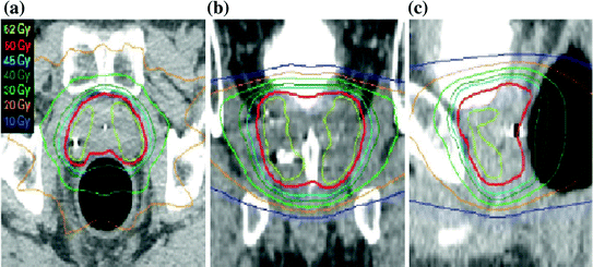
The use of an endo-rectal balloon (ERB) may overcome daily variation in rectal distention and peristalsis. This physiologic motion is the dominating contributor to intra-fraction motion of the prostate gland. Langen et al. demonstrated that the magnitude of intra-fraction motion using Calypso was largest in the anterior-posterior direction, with both positional drift and transient pulsatile motion (Langen et al. 2008). The total elapsed treatment time also had a significant impact on the motion, with larger movements seen with longer treatment times. In the setting of SBRT, such displacement of the target organ can result in under-dosing the PTV. To assess the potential benefit of ERB, Wang et al. compared the motion between 30 patients who were treated with and without ERB (Wang et al. 2012). They report that the ERB group had significant decreases in the motion in all dimensions, especially the anterior-posterior direction. In the University of Texas phase I prostate SBRT trial, daily endorectal balloon was used for simulation and treatment (Hannan et al. 2016). The rectal catheter was filled a pre-determined quantity of air, thereby fixing the interface between the anterior rectal wall and the prostate itself (Fig. 3). Another purpose of the ERB is to also displace the lateral and posterior rectal wall away from the PTV, facilitating lower doses received to these areas. The lack of any grade 2 or higher late gastro-intestinal toxicities in the 45 Gy arm, with a median follow-up of 74 months, illustrates the benefit with this technique (Hannan et al. 2016). The 45 Gy starting dose was the highest 5-fraction dose reported in the literature to date. Intra-fraction motion data has not been reported for this trial.


Fig. 3
Rectal balloon placement for prostate SBRT (Boike et al., Journal of Clinical Oncology © 2011). Reprinted with permission
4 Prostate Cancer Following Prostatectomy
Salvage XRT is a standard treatment recommendation to treat biochemical recurrence of prostate cancer following radical prostatectomy. IMRT is now considered the preferred technique to optimize sparing of adjacent rectal tissues. Given the lack of a solid tumor target, radiation delivery in this setting presents multiple challenges. As IMRT inherently results in sharper dose gradients away from the target volume, intra-fraction data on the location of the tumor bed is critical. The definition of the CTV itself is fundamentally based on the relationship between the bladder and rectum. After multiple reports of successful implantation of fiducial markers in the intact-gland, a similar approach was started in the prostate bed.
Inter- and Intra-fraction motion data from 20 patients who received Calypso implantation was presented by Klayton et al. (2012). Prostate bed displacement was measured after aligning to bony landmarks. The shift in the superior-inferior direction exceeded 5 mm in more than 21% of delivered fractions. During delivery, motion was predominant in the posterior direction toward the rectum. Approximately 15% of all treatments were interrupted due to motion threshold being exceeded. It is possible that ERB may be useful to minimize motion of the prostate bed. In the absence of markers, soft tissue imaging with CBCT is essential to visualize the rectal wall. Besides traditional x, y, and z translation movements, yaw, pitch, and roll changes have also been shown to be contributors to intra-fractional target changes using Calypso (Zhu et al. 2013). Real-time adaptive planning strategies may be important in order maximize target coverage. It is proposed by Zhu et al. that intra-fraction data obtained early in the treatment course can be helpful in the decision making process to modify the existing treatment plan (Zhu et al. 2013).
Stay updated, free articles. Join our Telegram channel

Full access? Get Clinical Tree


