Inflammation and Current HPV Status in Head and Neck Malignancy
Roman Carlos Zamora1, *, Jose Gutiérrez Jodas2, Norhafiza Mat Lazim3
Abstract
Head and neck malignancy is on the rise, where the majority of the tumors are squamous cell carcinoma (HNSCC). Previously, alcohol and tobacco are reported to be the well-established risk factors for HNSCC development. Currently, the HPV driven HNSCC has shown an increase in incidence globally, with oropharyngeal and oral cavity carcinoma predominating at certain geographic locations. HPV associated oropharyngeal squamous cell carcinoma commonly occurs in Europe and certain Western countries. They have different biological profiles compared to HPV-negative HNSCC. HPV-positive HNSCC patients have different characteristics and prognosis, which remarkably affect the management of this subset of patients. HPV is a significant inflammatory agent that can promote carcinogenesis via multiple critical mechanisms that are discussed in the chapters. Targeting HPV for future research is a great promising avenue for the discovery of novel screening, diagnostic, and therapeutic targets.
* Corresponding author Roman Carlos Zamora: Department of Otorhinolaryngology-Head and Neck Surgery Hospital Universitario Reina Sofia, Cordoba Spain. Direction: Avenida Menendez Pidal, s/n, Postal code 14004, Cordoba, Spain; Tel: +34634219251; E-mail: romanchos@hotmail.com
INTRODUCTION
This chapter focuses on chronic inflammation, HPV, and its relation to head and neck malignancy, as well as its various treatment and preventive options. It is well documented that chronic inflammation in the complex world of the immune system plays an important role in malignancy. Currently, the tendency of
treatment options for HNSCC patients is becoming less invasive, more conservative, and aims to have a more molecular approach. This can be ideally achieved by incorporating inflammation markers as promising therapeutic agents in the near future. Prevention plays an important role in combatting HNSCC.
As early as the 1800s, the perception of inflammation associated with cancer has been thought of but not demonstrated. In recent years, molecular studies have been able to show that inflammation contributes to the survival and proliferation of malignant cells [1]. Many of the well-established risk factors for head and neck malignancy are associated with inflammation. For instance, the environmental carcinogens such as sharp tooth and periodontitis for oral cavity carcinoma, Epstein Barr virus (EBV) for nasopharyngeal carcinoma (NPC), wood dust exposure for sinonasal malignancy and human papillomavirus (HPV) for oropharyngeal carcinoma. HPV-related HNSCC is significant as this tumor group has a different biological profile and treatment outcomes compared to HPV-negative tumors.
CHRONIC INFLAMMATION AND HEAD AND NECK ORAL CARCINOMA
Squamous cell carcinoma is the most common malignant lesion in the oral cavity. There is a significant increase in oral cancer incidence in young people, with 350,000-400,000 new cases worldwide per year. Bacteria, viruses, and fungi have been implicated and highly related to certain cancers [2]. For these reasons alone, it is important to gather more research and investigations about the risk factors of head and neck squamous cell carcinoma (HNSCC) in patients under 45 years old [3]. The oral cavity contains many bacterial species, with some of them being able to produce oral pathology. In recent years, the relationship between oral flora and head and neck cancer has been a subject of interest. Emerging evidence suggests that chronic inflammation could play an important role in the development of cancer. Importantly, studies have shown that periodontitis can promote carcinogenesis and lead to oral cavity cancers.
There is a strong association between head and neck carcinoma risk and oral leukoplakia, oral fibrosis, and repetitive dental ulcer injury [4]. There is a complex inflammatory process involved in the progression of premalignant lesions to squamous cell carcinoma. The role of T-cells (Tregs) and tumor-associated macrophages in immunohistochemical staining of cytokines showed that there was an increase in disease progression in premalignant oral lesions. In the early stages of premalignant lesions, IL-10 was seen to be increasing [5]. All of these inflammatory markers play a crucial role in the pathogenesis of head and
neck malignancy, and some have been discussed in the other chapters in this book.
Generally, during the progression of oral dysplasia, IL-4+ macrophages were seen from premalignant lesions. However, TGF-β1+ macrophages were seen in oral squamous cell carcinoma (OSCC) in less quantity than premalignant lesions as well as the expression of IFN-γ. These findings suggest that chronic inflammation promotes tumorigenesis in OSCC, rather than initializing it [5]. Overpopulation of pathogenic oral bacteria may be secondary to poor oral hygiene. This, in turn, can switch the chronic inflammation process and progression into OSCC. Three species of oral bacteria associated with an increased risk of oral squamous carcinoma were Fusobacterium nucleatum, Prevotella tannerae, and Prevotella intermedia. Additionally, it was also seen that alcohol, cigarettes, and poor oral hygiene were associated with an increase in oral pathogenic bacteria. Salivary IL1β was associated with a rise in periodontal-pathogenic bacteria and OSCC risk, which in turn can be influenced by genetic factors and lifestyle. Critically, all these results suggested that good oral hygiene may reduce OSCC risk and should be part of a prevention campaign [6].
The risk of cancer can potentially be predicted through alterations seen in the oral microbiota. Further understanding can be achieved by molecular advances in monitoring the role of oral microbiota and oral carcinogenesis [7]. For example, the formation of 8-oxo-7,8-dihydro-2′-deoxyguanosine and 8-nitroguanine are mutagenic DNA lesions associated with inflammation-related cancers. The formation of these mutagenic DNA has been seen in precancerous lesions due to infection and pro-inflammatory factors. Several studies have suggested that cancer development is triggered by inflammation associated-DNA damage in cancer stem-like cells. An increase in oxidative stress due to dysfunction of anti-oxidative proteins, DNA methylation, and microRNA dysregulation can lead to carcinogenesis. One example is Epstein-Barr virus-related nasopharyngeal carcinoma, in which quantitative RT-PCR analysis confirmed the downregulation of miR-497 in cancer tissues and plasma. These findings can be useful biomarkers in liquid biopsy for prevention and early detection [8] of HNSCC.
C-reactive protein (CRP) is an acute-phase protein that serves as a marker for inflammation and the progression of various cancers. A study by Metgud et al. compared CRP in saliva and serum in 20 normal individuals, 20 patients with OSCC, and 20 patients with oral premalignant lesions to assess as a prognostic indicator for OSCC. Mean CRP levels were more elevated in patients with premalignant lesions and OSCC compared to controls [9]. Oral cancer has become an important problem in many parts of the world, with more cases seen in developing countries. This is the reason why molecularly targeted prevention of oral cancer and the link to chronic inflammation is important. Other inflammatory mediators that play a role in oral cancer development, apart from the aforementioned, include VEGF, prostaglandin pathways, p53, inflammatory cytokines, reactive oxygen, and nitrogen species. Currently, the cytology and biopsy can make the diagnosis of HNSCC, and this can be combined with testing of inflammatory biomarkers that could be beneficial for early detection of HNSCC [10].
Alcohol and tobacco, human papillomavirus (HPV), or Epstein-Barr viruses (EBV) can start and maintain a chronic inflammation through genomic alterations or viral oncoproteins via phosphatidylinositol 3-kinase (PI3K) and transcription factor nuclear factor-kappa B (NF-κB). Various ongoing studies at molecular therapies targeting signaling in cancer cells are being developed to explore better ways of controlling cancer. One example is the immune checkpoint inhibitors in combination with inflammatory cells in the immune system [11]. It is important to understand cancer development comes from activated oncogenes or dysfunctional tumor suppressor genes. However, these factors alone are not sufficient for the development of carcinogenesis. Importantly, the infiltration of immune cells facilitates neoplasm development by enabling tumors to evade the host immune response. Structural support to developing tumors is provided by the alteration of the extracellular matrix in inflammation. Hypoxia induces DNA damage and is tumorigenic, while tissue vasculature is important for maintaining the microenvironment that supplies cell division and metastatic spread. Inflammation and its ecosystem provide support for tissue homeostasis and repair, which is important to be understood in order to produce a potential treatment target for head and neck cancers [12]. Chronic inflammatory mediators have pleiotropic effects, i.e., they can favor carcinogenesis but can also limit tumor growth by stimulating immune effector mechanisms. The role of IL-1-signaling and stress protein involvement is an important mechanism in the development of anti-cancer immunity and anti-apoptotic functions. Chronic infection by various means, like reflux, viral or bacterial infection can cause up to 25% of human malignancies [13].
A study by Ambatipudi and colleagues assessed if DNA methylation derived systemic inflammation indices are associated with head and neck cancer development and survival. The multivariate logistic regression study showed that elevated Neutrophil-to-Lymphocyte ratio (NLR) is associated with increased odds of being an HNSCC case (OR = 3.25, 95% CI = 2.14-5.34, P = 4 × 10⁻⁷) while the contrary was observed with Lymphocyte-to-Monocyte ratio (LMR), OR = 0.88, 95% CI = 0.81-0.90, P = 2 × 10⁻³. It was seen that HPV16-E6 seropositive HNSCC cases had an elevated LMR and a lower NLR when compared to seronegative patients. Results showed that lower LMR but not lower levels of NLR were associated with an increased risk of death [14]. In another study by Wang et al. in inflammation-related DNA damage and cancer stem cell markers in nasopharyngeal carcinoma, several cancer stem/progenitor cell markers (CD44v6, CD24, and ALDH1A1) were studied. It was seen that CD44v6 and ALDH1A1 were significantly increased in cancer cells of primary NPC specimens in comparison to chronic nasopharyngitis. No significant difference was observed between chronic nasopharyngitis tissue and NPC in the case of CD24. The study concluded that CD44v6 and ALDH1A1 could be stem cell markers for NPC [15].
One study aimed to determine the expression of beta-defensins in nasal polyposis and chronic tonsillitis as well as to determine the relationship between the malignant process in tonsils and inflammation. In cases of chronic inflammation, the study showed large secretions of human beta defensins, while limited in malignant transformation. Endothelial nitric oxide synthase (eNOS) and nitric oxide molecule are involved in cell cycle regulation, cell proliferation and apoptosis results confirmed that eNOS is present in the upper airway in chronic inflammation and cancer [16]. It can be said that specific transcription factors enhance the expression of genes and have the ability to regulate the survival and proliferation of malignant cells. There is a link between inflammation and cancer. In the stroma of established cancers, there is a state of exaggerated inflammation and suppression of immune responses. There is no denying that precancerous lesions (oral submucous fibrosis, oral lichen planus), dento-gingival bacterial plaques, chronic periodontitis, chronic tonsillitis, etc., have inflammation as a common factor [17].
Oropharyngeal Carcinoma Associated with HPV
The malignancy of the aerodigestive tract represents about 600,000 new cases per year. It is well known that alcohol, tobacco and HPV are significant etiology factors. About 40-80% of head and neck malignancies are associated with HPV [18]. The risk of developing oral and oropharyngeal carcinoma is four times higher in patients with infected HPV. The relationship between HPV and oropharyngeal carcinoma has been recognized in 2009 [19]. The incidence of oropharyngeal carcinoma (OPSCC) associated with HPV has been increased in recent years. It is estimated that approximately 70-80% of OPSCCs in North America and Europe are related to HPV infection [20]. Oral HPV 16 detection was associated with incident HNSCC with a positive association for OPSCC [21]. Of note, the OPSCCs associated with HPV have higher survival than HPV negative. At present, the therapeutic approach is based on minimizing treatment, without decreasing overall survival, and conducting prevention campaigns through health education and vaccination campaigns.
Human Papilloma Virus (HPV)
The HPV virus belongs to the papillomaviridiae family. It is a DNA virus and more than 300 subtypes have been identified. The strains with the greatest oncogenic potential are HPV 16 and 18 [22]. It is well known that HPV is transmitted to the oropharynx through oral sex and profound tongue base kissing. The risk of infection increases with the number of couples and if there is an association with HIV. Most people eliminate HPV within one to two years but in some, the infection persists and becomes a chronic infection with the risk of developing carcinoma. The HPV infection occurs in the squamous epithelium of the oropharynx, in its basal stratum. A correct union and inhibition of certain proteins take place that allows the integration of viral DNA with the cellular DNA. Subsequently, this generates cellular changes that actívate cell proliferation.
HPV has a double-stranded DNA of 8000 base pairs being divided into three regions; E, L, and log control región, each of which will originate a certain group of proteins [23]. The E6 and E7 region encode oncoproteins that inactivate proteins that regulate the cell cycle, p16, p53, and pRB. These oncoproteins can be categorized into high and low risk. The low-risk oncoprotein does not have activity because they are inactivated mutations, this being the cause of the different oncogenicity among the different HPV [24]. Mutation of the p16 proteins inhibits the cell cycle and phosphorylation of pRB inactivity, which leads to a phase change of the cell cycle from G1 to S. The p53 protein is responsible for cell control, DNA repair and apoptosis control. The inactivation of p53 is caused by the E6AP proteins that maintain the binding between p53-E6, inactivating p53 results in chromosomal and genomic instability. The E7 oncoprotein ensures a correct replication of the viral DNA by inactivating the ‘pocket’ proteins which are the pRB, p100, and p130 [24].
HPV affects the basal layer of the stratified epithelium of the oropharynx. The viral DNA, E1 and E2 proteins are integrated into the cellular DNA. With times the proliferation takes place with genome amplification and multiplication occurs. This is carried out due to the activity of E6 and E7 proteins that inactivate p53 and pRB. This mechanism that HPV has to enter into the DNA of the host cells prevents an immune reaction to be eliminated. The elimination of the virus is carried out when the cells reach the most superficial stratum which is the regression phase. The infection can be perpetuated for years if the regression phase is not completed. This is called the abortion cycle. This interruption in the cycle is due to failure of control of the E2 protein on the E6 and E7 causing the viral DNA unable to complete its cycle, thus generating genetic alteration and DNA instability with the consequent cell proliferation.
EPIDEMIOLOGY OF THE HPV RELATED HNSCC
There have been different studies that showed an increasing trend of OPSCC related HPV. The majority of this study was conducted in the US and Western Europe. In Stockholm [25] there is an increase in the prevalence of 0.7/100,000 between 1970-1979 to 1.65/100,000 between 2000-2006. In the US [26], there is an increase from 2.8/100,000 to 3.6/100,000 between the years 1988-2004. Whereas in Denmark [27] an increase in the annual incidence of 2.7% between 2000 and 2010. Although at present, it is not considered an epidemic, it is evident that there is an increase in the incidence of related HPV with OPSCC. The etiology of this increase is currently is unknown, although it could be justified by the greater number of sexual practices, the decrease in tonsillectomy in children or by a greater attempt to reach an etiological diagnosis in relation to HPV.
The association of HPV and OPSCC is considered a sexually transmitted disease, with oral sex being the main risk factor. The number of partners throughout life, early oral sex before the age of 18, deep tongue kissing, marijuana use, HPV infection of the anogenital tract and smoking are associated risk factors to the development of OPSCC and HPV [28, 29]. When comparing patients with OPSCC associated with HPV with non-associated HPV, it is observed that they are younger patients (<60 years), with lower consumption of tobacco and alcohol, higher sexual practices and higher socioeconomic status. With regard to gender, there is a greater proportion of men than women in the US and Europe. A population study of the National Cancer Database (NCDB) conducted during the period 2010 to 2014 showed a higher positive HPV in men than in women for both OPSCC (66% vs 50%) and OCSCC (16% vs 11%) [30]. HPV OPSCC positive in women appear at a later age and in an earlier stage than in men. Generally, sexual habits, lifestyle, tobacco and alcohol consumption cannot fully explain the differences of the prevalence of HPV-related HNSCC between the sexes. There should be other critical factors that are unknown. For instance, the hormonal factor, whereby the level of estrogen and progestogens fluctuation is inversely correlated with the development of OPSCC. Women who develop menopause before age 52 and those who become pregnant after age 35 have a higher risk of developing OPSCC associated HPV [30]. The race is another important factor to consider, although the most frequent HPV in all types of OPSCC is 16, African descent has been linked to HPV type 17. This difference is critical in relation to survival, since it is lower in the black race regardless of stage, applied treatment and sex. Racial disparity in the prognosis of cancer is strongly related to the genetic and epigenetic diversity, a greater metabolic propensity of obesity and the state of chronic inflammation of black people, as well as differences in innate immunity [31].
CLINICAL OVERVIEW OF HPV ASSOCIATED OROPHARYNGEAL CARCINOMA
Survival of OPSCC patients depends mainly on the stage at the time of diagnosis. In the earliest stages, symptoms are very scarce which is why this type of tumor is often diagnosed at late stages. Among the most frequent symptoms include odynophagia, dysphagia, otalgia, trismus and pharyngeal body sensation which are seen in advanced-stage diseases. The HPV positive OPSCC mainly harbor the virus within the palatine and lingual tonsils, which is a critical reservoir for HPV. As aforementioned, this group of patients has a specific profile. They are young and having multiple sexual partners, have a high socioeconomic status associated with tobacco and alcohol consumption.
It is common to find large cervical metastases and small tumors in the oropharynx which is difficult to spot in a routine examination of the oropharynx. In order to locate these small tumors, especially those smaller than 1 cm that is undetectable with white, the narrow band imaging (NBI) and autofluorescence based endoscopic imaging can be used. The NBI is currently used for the diagnosis of superficial carcinomas, assessing the hidden lesions, method of surveillance for patient post treatment and study of intraoperative resection margin [32]. In the Di Maia meta-analysis, the grouped sensitivity and specificity of NBI in patients with cervical metastases of HNSCC with unknown primary was 0.83 (99% CI, 0.54-0.95) and 0.88 (99% CI, 0.005-0.86). PET- CT is necessary for accurate staging of OPSCC. This will give information on the extent of the tumors, presence of cervical metastases, distant metastases and is highly useful for planning for radiotherapy and monitoring treatment response and evidence of recurrent diseases [33].
DIAGNOSTIC METHODS IN THE ASSESSMENT OF HPV
Of note, the diagnosis of HPV associated with oropharyngeal carcinoma is essential for its classification, treatment, and prognosis. There are currently more than 125 commercially available techniques for the detection of HPV. It is one of the most numerous diagnostic techniques available, but it is also less regulated. Generally, they can be broadly categorized into four types:
- Determination of p16 protein.
- DNA analysis by in situ hybridization
- DNA analysis by PCR
- Analysis of E6/E7 viral transcription RNA by reverse transcriptase PCR.
Classification HPV+ Oropharyngeal Carcinoma
The American Joint Committee on Cancer (AJCC) published the 8th edition on head and neck cancer staging in 2017 [34], introducing novel modifications with respect to the previous classification. The 7th edition classification system classified the majority of the OPSCC associated with HPV in stages III and IV. The introduction of the 8th edition has clearly differentiated between HPV positive and HPV negative.
The differentiation implies changes in stage, prognosis and treatment. The OPSCC associated or not with HPV have been classified based on overexpression of the p16 protein. The p16 is a widely available marker, inexpensive and easy to interpret. The cut-off point for overexpression of p16 is diffuse tumor expression (more than 75%) and with at least moderate staining intensity (+2/3). The new classification eliminates Tis (carcinoma in situ) in the OPSCC p16+, because there is no basement membrane in the epithelium of lingual and palatine tonsils. The OPSCC p16+ is the only carcinoma of the head and neck that maintain the T0 category. The other difference is in the classification of neck metastasis. For new classification, stage N1 encompasses ipsilateral neck metastasis less than 6cm, in contrast to the 7th edition classification which classifies N1 as single ipsilateral adenopathy less than 3cm. Additionally, presence of neck nodes in the OPSCC p16 positive cases larger than 6 cm are closely related to detrimental effects on survival [35]. The 8th edition classification system is more practical whereby it classifies the OPSCC HPV positive and OPSCC HPV negative based on the therapeutic and survival outcomes [34].
TREATMENT OF OROPHARYNGEAL CARCINOMA ASSOCIATED WITH HPV
In the past, the treatment of the OPSCC has been based on the use of radical radiotherapy and chemotherapy. This is because open transmandibular surgery carries high morbidities with mortality and swallowing deficit and lower survival compared to chemoradiation. Swallowing impairment secondary to treatment is one of the main sequelae that patients with OPSCC experienced. The chemoradiation, even though it has benefits, the local and systemic adverse effects are also substantial. It can also cause significant swallowing and mastication impairment due to fibrosis induced trismus. During period of radiotherapy, 62% of OPSCC patients require gastrostomy [36]. Interestingly, in order to reduce the morbidity associated with treatment without affecting survival, Trans Oral Ultrasonic (TOUSS), Transoral Laser Microsurgery (TLM) and Transoral Robotic Surgery (TORS) are being evaluated.
TOUSS was designed as a ‘no robot’ endoscopic transoral procedure, which is inspired by the laparoscopic approach of abdominal surgeons using a unique port, the mouth. TOUSS uses the Gyrus FK retractor, Olympus endoscope system and ultrasonic device such as the Thunderbeat (Fig. 1). This allows an endoscopic approach of the oropharynx at a much less cost than any other robotic system. This technique also gives an excellent oncological and functional result with minimal surgical complication. In a series by Fernandez et al. the majority of the OPSCC treated with TOUSS in stage T1 and T3 did not reveal any surgical complications. Only two patients in the series presented with post-surgical bleeding that was endoscopically controlled in the operation room. The resection margins were mostly negative for all surgical specimens [37]. TORS is another technique known for years through the robotic platform (Fig. 2). The best known is the Da Vinci Robot system. The indications of TORS for OPSCC have been extended to stage T3 and T4 without differentiating their association or not with HPV. The functional results are quite satisfactory. Choosing a candidate for transoral surgery will depend on T and N stage of the tumor that requires resection of the tongue base for more than 50% and the patient indicated for adjuvant chemoradiation [38]. In order to reduce the toxicity of chemoradiation and surgery, different therapeutic de-intensifications are being conducted. To evaluate whether lowering the dose of radiotherapy and substituting chemotherapeutic agents with lower toxicity and using minimally invasive techniques, the same oncological results are achieved as with the standard treatment. Another study evaluated the reduced dose of IMRT with or without cisplatin in the treatment of the patient with advanced oropharyngeal carcinoma [39].
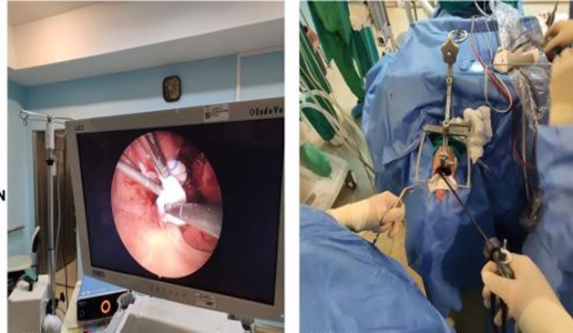
Images showing TOUSS procedure is being performed.
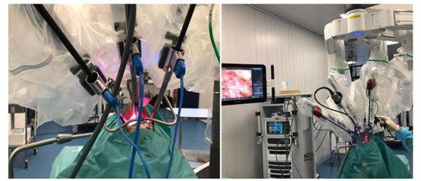
Images of TORS.
Chemotherapy De-Intensification Trials
The oncology RTOG group conducted a study which comprise of 182 centers in the USA and Canada incorporating HPV positive OPSCC patients. This study aims to evaluate whether cetuximab in combination with radiation can achieve greater survival and lower toxicity in stage III and IV OPSCC patients compared to the standard cisplatin treatment [40]. The result showed that the survival was lower with cetuximab and the toxicity was similar using both cetuximab and platinum regimes. It has been shown that a lower dose of IMRT produce favorable results with less late toxicities and a statistically significant improvement in swallowing solids can be achieved. This finding supports the hypothesis that a dose of 55 Gray or less is likely to affect swallowing function [41]. Avoiding the adjuvant chemotherapy in patients with extracapsular extension is another strategy to minimize the morbidity associated with the treatment. The Washington University group reported that the presence of extracapsular extension does not affect disease free survival. In their series of 151 patients with HPV positive OPSCC treated with TORS, the 3-year survival of patients with and without extracapsular extension was 89% (95% CI 84%-95% and 94% (95% CI 83%-100%) respectively [42].
The results of the trials should provide more information and adequate risk stratification and the selection of adjuvant treatment in the future. A proper selection of adjuvant treatment de-intensification is essential to obtain excellent results. Therefore, deintensification outside a clinical trial is currently not recommended. A better understanding of the molecular alterations underlying the HPV associated disease should be elucidated to provide more opportunities for targeted therapies with less toxicity. If these approaches are adequate, the quality of life of patients should improve significantly [43].
Related posts:
 of Head and Neck Cancer and Relation to Inflammation
Inflammation: Window for Therapeutic Strategy in Head and Neck Malignancy
the Screen: The Emergence of New Evidence
Diagnostic Tools for Head and Neck Cancer
Emergence of New Inflammatory Markers of Head and Neck Cancer and their Potentials
Managing Oncology Patients with Comorbid Autoimmune Disorders
of Head and Neck Cancer and Relation to Inflammation
Inflammation: Window for Therapeutic Strategy in Head and Neck Malignancy
the Screen: The Emergence of New Evidence
Diagnostic Tools for Head and Neck Cancer
Emergence of New Inflammatory Markers of Head and Neck Cancer and their Potentials
Managing Oncology Patients with Comorbid Autoimmune Disorders
Stay updated, free articles. Join our Telegram channel

Full access? Get Clinical Tree


