Fig. 1
Decreased Cath V (a), increased endostatin (b) and IL-18 (c) in BALF of Besnier-Boeck-Schaumann disease (BBS) patients as compared with healthy volunteers
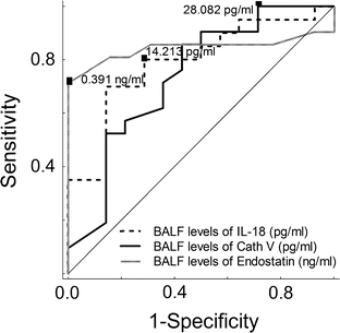
ROC curves for Cath V, endostatin, and IL-18 in BALF were applied to determine the cut-off values. Sensitivity and specificity of Cath V levels in the BBS patients relative to the healthy group were 90 % and 50 %, respectively, at a cut-off value of 28.08 pg/ml. Sensitivity and specificity of endostatin levels in the BBS patients relative to the healthy subjects were 71 % and 8 %, respectively, at a cut-off value of 0.39 ng/ml. Sensitivity and specificity of IL-18 levels in the BBS patients relative to the healthy subjects were 81 % and 28 %, respectively, at a cut-off value of 14.21 pg/ml. The areas under the curve for Cath V, endostatin, and IL-18 in BALF were 0.74, 0.84 and 0.79, respectively (Fig. 2).


Fig. 2
Receiver operating characteristic (ROC) curve for Cath V, endostatin, and IL-18 in BALF differentiating BBS and healthy subjects (AUC 0.74, 0.84, and 0.79, respectively)
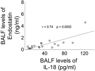
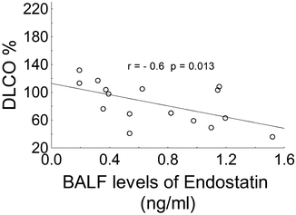
In the BBS group, a positive correlation was found between the BALF levels of endostatin and IL-18 (r = 0.74, p < 0.001) (Fig. 3). We observed a negative correlation between BALF levels of endostatin and %DLCO in the BBS group (Fig. 4). Moreover, in the BBS group, BALF concentrations of endostatin correlated with following parameters: %lymphocytes (r = 0.52, p = 0.019), %macrophages (r = 0.52, p = 0.018) and CD4+/CD8+ (r = 0.86, p = 0.013). Cath V concentration negatively correlated with CD4+/CD8+ in BALF of BBS patients (r = −0.83, p = 0.003).


Fig. 3
Correlation between concentrations of endostatin, and IL-18 in BALF of Besnier-Boeck-Schaumann disease (BBS) patients

Fig. 4
Correlation between endostatin and DL CO (diffusing capacity for carbon monoxide) in BALF of Besnier-Boeck-Schaumann disease (BBS) patients
4 Discussion
In the present study we revealed that patients with BBS had a lower level of Cath V in BALF than healthy people. To our knowledge, this study is the first concerning the cathepsin concentration in BALF of humans. Our study is consistent with the findings of Samokhin et al. (2011) in the mouse lungs. That study, which focused on the cathepsin K, L, and S, showed that lack of cathepsin activity prevents the development of lung granulomas. These cysteine proteases degrade major extracellular proteins (Shi et al. 1992) and are involved in immune responses (Hsing and Rudensky 2005; Honey and Rudensky 2003). Cathepsins L and S play a significant role in the antigen presentation and T cell selection (Honey and Rudensky 2003; Nakagawa et al. 1998), and the formation of granulomas has been linked to T cell activation (Gerke and Hunninghake 2008; Grunewald and Eklund 2007; Noor and Knox 2007). The role of Cath V in humans has not yet been clarified, but in vitro experiments have demonstrated that it has similar physiological properties to cathepsin S (Berdowska 2004). Possible explanation for the decreased Cath V level, in the present study, is proteolytic degradation. Pulmonary sarcoidosis is associated with chronic lung inflammation; thus, it is possible that pro-inflammatory protease may degrade Cath V and lead to its reduced level in BALF. We presume that a low level of Cath V in BBS patients is due to its being used up during the destruction of the extracellular matrix. This process is essential to the lung granuloma formation (Gerke and Hunninghake 2008; Grunewald and Eklund 2007; Noor and Knox 2007). A negative correlation between the BALF level of Cath V and CD4+/CD8+ is consistent with the hypothesis above outlined. Lymphocytic alveolitis with predominance of CD4+Th cells and macrophages is typical in sarcoidosis (Grunewald and Eklund 2007).
Several studies on cathepsins have demonstrated that these enzymes participate in signaling pathways leading to apoptosis (Turk et al. 2002). Apoptosis plays an important role in the resolution of granulomas (van Maarsseveen et al. 2009). Recently, it has been demonstrated that BBS lymphocytes CD4+ present resistance to apoptosis (Dubaniewicz et al. 2006). Petzmann et al. (2006) described decreased apoptosis of antigen-primed T cells in BALF of BBS patients. These findings are in accord with our study because BBS patients had lower levels of Cath V than healthy subjects.
Shi et al. (2003) described that cathepsins are involved in the controlled extracellular matrix degradation, enabling endothelial cells to penetrate the vascular basement membranes to form new vessels. Several investigators confirmed that in pulmonary BBS the angiogenic/angiostatic balance is also disturbed. Fireman et al. (2009) reported that VEGF in the alveolar space is lower in BBS than in healthy subjects. The authors also revealed that the level of VEGF in patients at Stage 3–4 of disease is significantly lower than that at Stage 1–2, indicating a less fibrotic parenchymal disorder in the latter.
Stay updated, free articles. Join our Telegram channel

Full access? Get Clinical Tree


