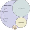
Philip P. Smith, George A. Kuchel Although traditional classification considers the upper and lower urinary tracts as part of one system, each serves a distinct function. In this edition, upper and lower urinary tract components will be considered, emphasizing the known effects of aging on each system. Nevertheless, a number of potentially pertinent topics will not be discussed in this chapter. For example, age-related changes in the renal handling of water and electrolytes are addressed in Chapter 82, and diseases that commonly affect the aged kidney, prostate, and gynecologic structures are discussed in Chapters 81, 83, and 85, respectively. Given the multifactorial systemic complexity inherent to aging and common geriatric syndromes (Chapter 15),1 the discussion will need to cross traditional organ-based boundaries. Therefore, we will also discuss the ability of age-related declines in renal function to influence key geriatric measures, such as cognitive function and mobility performance. Conversely, given growing evidence that oxidative stress, inflammation, and nutrition can influence aging- and disease-related processes across many different organs, the ability of these systemic factors to modify urinary tract aging will also be considered. Finally, the contribution of lower and upper urinary tract dysfunction to urinary incontinence, a major geriatric syndrome, is discussed in Chapter 106. Declines in renal function represent one of the best documented and most dramatic physiologic alterations in human aging. In spite of great progress, important issues remain. For example, it has been difficult to explain why renal aging can be so variable between seemingly “normal” individuals and to establish which of these changes may potentially be reversible. Nevertheless, developments and continuing research in this area offer unique opportunities for improving the lives of older adults.2–5 Age-related declines in glomerular filtration rate (GFR) are well-established, yet contrary to general belief, GFR does not inevitably decrease with age. Among Baltimore Longitudinal Study of Aging participants, mean GFR declined approximately 8.0 mL/min per 1.73 m2 per decade from the middle of the fourth decade of life.6 However, these decrements were not universal, with approximately one third of these subjects showing no significant decrease in GFR over time.6 This high degree of interindividual variability among relatively healthy older adults has raised the hope that age-related declines in GFR may not be inevitable and could ultimately be preventable, even in the absence of an overt disease process. At the same time, clinicians wishing to prescribe renally excreted medications to healthy older adults clearly require reliable tools to estimate GFR accurately. The decrease in GFR with age is generally not accompanied by elevations in serum creatinine levels6 because age-related declines in muscle mass tend to parallel those observed for GFR, causing overall creatinine production also to fall with age. Thus, serum creatinine levels generally overestimate GFR with age, and in women and underweight individuals, the serum creatinine level is most insensitive to impaired kidney function.7 Although many formulas have been devised for estimating creatinine clearance based on normative data,8,9 their reliability in predicting individual renal function is poor.10,11 In frail and severely ill patients on multiple medications, where the need for accurate estimation is greatest, the reliability of such estimates may be the most questionable. In consequence, timed short-duration urine collections for creatinine clearance measurement are generally recommended.10,12 In contrast to the poor predictive ability of low creatinine levels, elevations in serum creatinine levels above 132 mmol/L (1.5 mg/dL) reflect declines in GFR greater than what would be typically expected with normal aging, representing likely underlying pathology. Ultimately, even creatinine clearance has limitations and may underestimate GFR.13 Cystatin C, a measure of kidney function that is independent of muscle mass, has been advocated as an improved marker of reduced GFR in older adults with creatinine levels within the normal range.14 Although U.S. Food and Drug Administration (FDA)–approved kits for its measurement have been available since 2001, and in spite of its potential attraction in the management of frail older adults, the precise role of cystatin C measurements in clinical decision making remains to be clearly defined. On average, aging is associated with a progressive decrease in renal plasma flow.15,16 Losses of 10% per decade have been described, with typical values declining from 600 mL/min in a young adult to 300 mL/min at 80 years of age.15,16 Perfusion of the renal medulla is maintained in the presence of lower blood flow to the cortex, which can be observed as patchy cortical defects on renal scans obtained in healthy older adults. Regional renal flow and GRF are determined by a balance between the vascular tone involving the afferent and efferent renal blood supply. Generally, renal vasoconstriction increases in old age, whereas the capacity of the vascular bed to dilate is decreased. Responsiveness to vasodilators (e.g., nitric oxide, prostacyclin) appears to be attenuated, whereas responsiveness to vasoconstrictors (e.g., angiotensin II) is enhanced.5 Basal renin and angiotensin II levels are significantly lower in older adults, and the ability of various different stimuli to activate the renin-angiotensin-aldosterone system (RAAS) is blunted. The ability of the tubules to excrete and reabsorb specific solutes plays a crucial role in maintaining normal fluid and electrolyte balance. The impact of aging and specific disease processes on the ability of tubules to handle specific solutes is discussed elsewhere (Chapter 82). Nevertheless, some overarching principles, are worthy of note2,5,17: For example, the ability to conserve and excrete sodium is impaired, with reduced salt resorption in the ascending loop of Henle, reduced serum aldosterone secretion, and a relative resistance to aldosterone and angiotensin II.2,5 As a result, older adults take longer to reduce their sodium excretion in response to a salt-restricted diet; conversely, older adults take longer to excrete a sodium load. Qualitatively similar changes have been described in regard to the tubular capacity to adjust to changes in water. The aged kidney is granular in appearance, with modest declines in parenchymal mass.2,5 The most impressive changes involve a reduction in the number and size of nephrons in the renal cortex, with a relative sparing of the medullary regions. Loss of parenchymal mass leads to a widening of interstitial spaces between the tubules and an increase in interstitial connective tissue. The numbers of visible glomeruli in aged kidneys decline in parallel with change in weight, with an increasing percentage of sclerotic glomeruli. Sclerosis is associated with lost lobulation of the glomerular tuft, increased mesangial cells, and decreased epithelial cells, resulting in decreased effective filtering surface. In response, remaining nonsclerotic glomeruli compensate by enlarging and hyperfiltering. Even in the absence of hypertension and other relevant diseases, important changes of the intrarenal vasculature can be observed in old age.2,5 Larger renal vessels may show sclerotic changes, whereas smaller vessels generally are spared. Nevertheless, arteriolar-glomerular units demonstrate distinctive changes in old age.2,5,18 Cortical changes are more profound, with hyalinization and collapse of glomerular tufts, luminal obliteration within preglomerular arterioles, and decreased blood flow. Structural changes within the medulla are less pronounced, and juxtamedullary regions demonstrate evidence of anatomic continuity and functional shunting between afferent and efferent arterioles. The hyperfiltration theory suggests that a loss of glomeruli results in increased capillary blood flow through the remaining glomeruli and a correspondingly high intracapillary pressure.2,5 Such age-related increases in intracapillary pressure (or shear stress) can also result in local endothelial cell damage and glomerular injury, contributing to the progressive glomerulosclerosis.2,5,19 Cytokines and other vasoactive humoral factors have been implicated in this type of pressure-mediated renal damage.2,5,20 Also in support of the hyperfiltration theory, restricted protein intake21 and antihypertensives that reduce single-nephron GFR (e.g., angiotensin-converting-enzyme [ACE] inhibitors and angiotensin II blockers)21 reduce glomerular capillary pressure and glomerular injury and prevent measurable declines in renal function. Other factors and mechanisms contribute to age-related declines in renal function. For example, individuals born with a reduced nephron mass could be more vulnerable to all categories of renal injury, including those associated with aging. A growing body of research has linked renal aging to the damaging effects of normal metabolism through the accumulation of toxins, such as reactive oxygen species (ROS), advanced glycosylation end products (AGEs), and advanced lipoxidation end products (ALEs).2,3,5,22,23 This toxin-mediated theory has many attractions: 1. These toxins accumulate with aging and can induce structural and functional changes. 2. They provide vital linkages between efforts to understand aging at the level of a single organ and traditional gerontologic research into longevity (see Chapter 5). 4. Such research has permitted the development of a pathophysiologic framework within which different risk factors (e.g., underlying genetic predisposition, renal progenitor cell behavior,24 gonadal hormone levels,25 diet,22 smoking,26 subclinical processes) can all influence how renal aging manifests in individuals.2,5,23 Renal aging cannot be viewed in isolation from aging at the systemic level. Not only are most patients with chronic kidney disease (CKD) older adults, but these patients are frail and at high risk of being disabled.4 Individuals with advanced CKD have an especially high risk of developing cardiovascular disease,27 cognitive declines,25–30 sarcopenia,31–33 and poor physical performance.27,34 It remains to be seen to what extent milder declines in renal function, more consistent with normal aging, may contribute to altered body composition and physiologic performance seen in generally healthy older adults. As discussed, creatinine-based estimates of GFR depend on skeletal muscle mass and tend to overestimate GFR in older adults. Thus, it is interesting that even mild declines in GFR, as measured using cystatin C, were associated with poorer physical function, whereas creatinine-based GFR estimates demonstrated a relationship only when less than 60 mL/min/1.73 m2.35 Ultimately, the development of an approach that places renal aging in a systems-based context, in which key functional issues are considered, may offer most exciting opportunities for developing interventions that will help maintain function and independence in late life. By storing and periodically releasing urine on a volitional basis, the lower urinary tract (LUT) serves to isolate the kidneys from the exterior environment while providing controlled elimination of metabolic byproducts. The anatomic arrangement of the nonrefluxing ureterovesical junction, fluid-tight urethral sphincteric mechanism, and interposed chamber—the bladder—create an effective barrier to the retrograde passage of infectious agents into the kidneys and from there into the bloodstream. Presumably, as the result of evolutionary pressures, the bladder and its outlet normally function as a urine storage structure sufficiently capacious to accept several hours’ volume of renal output while an efficient evacuation mechanism under voluntary permissive control can be quickly and voluntarily activated and then returned to storage status. Under normal circumstances, this process is under socially appropriate voluntary control in response to non-noxious perceptions related to bladder volume and voiding flow. The requirements for proper function of this system include normal sensory transduction of normal physiologic bladder filling, central transmission and subconscious processing, appropriate conscious recognition and processing, coordination of sphincteric relaxation and bladder pressurization via detrusor contraction, and normal biomechanical function of the bladder and its outflow, as well as intact urethral and bladder guarding and voiding reflexes. The individual experiences the perception of these processes. Biomechanical and functional changes as a result of the aging process per se involving the LUT and nervous system may alter an individual’s storage and evacuation capabilities. Bidirectional convergence of peripheral and central signaling pathways, including from the gut and skin,36 provide a physiologic basis for urinary symptoms arising from nonurinary sources. The association between mobility and cognition of urinary symptoms and dysfunction37–41 points to the centrality of integrative processes to effective urinary performance. In a broader perspective especially relevant to aging, the complexity of control and perception suggests that functional disturbances and urinary symptoms represent thresholds of failure of an integrative homeostatic system. Symptoms and objective dysfunctions thus should be regarded as syndromic, involving diverse nongenitourinary systems such as fluid balance and mobility, as well as sensory and decision making processes rather than as being reflective of merely isolated LUT pathology.42 Despite the nominal implications of current terminology, the relationships of LUT mechanistic capabilities, descriptive LUT physiology, and perceptions of urinary status (including the voluntary control of storage vs. voiding) are not reliable and are likely not fixed over the life span. Clinically measurable LUT function (e.g., flow rates, urodynamics, postvoid residual volumes) is the result of brain control over end-organ structures as controlled by cognitive (including perceptual) processes. The poor correlation between symptoms and objective function has long been recognized.43 A urodynamic study of continent older adults found that 63% were symptom-free, and 52% were both symptom-free and free of any potential confounding disease or medication use.44 Nevertheless, only 18% of these individuals were also free of any urodynamic abnormality.44 Moreover, nonvoiding bladder contractions during filling (so-called detrusor overactivity [DO]) unrelated to identifiable disease were observed in 53% of these individuals, with no correlation to gender or age.45 Variability in postvoid residual volumes also increases with aging, resulting in asymptomatic, elevated, postvoid residual volumes in some people.46,47 The perception of voiding difficulties (underactive bladder [UAB]) may relate more to abnormal bladder sensations than to a weak detrusor muscle contraction during voiding.48 Patient-perceived symptoms are clearly clinically important, especially when bothersome. Nevertheless, as a result of the complex syndromic nature of symptoms and dysfunction in older adults, and the related unreliable correlation of symptoms, dysfunction, and cause, the physiologic meaning of urinary symptoms and objective dysfunction in the older adult must be approached with caution. Relatively simplistic algorithmic care derived from studies of younger adults may represent a special case of a broader pathophysiologic model and therefore may not always be applicable in older adults. The mechanical interaction of the detrusor smooth muscle with nonmuscular components of the bladder wall gives the bladder its ability to distend compliantly (i.e., hold urine under low pressure) during storage and create expulsive force during voiding. The expression of bladder wall forces during voiding as a measurable detrusor pressure and/or urinary flow rate is dependent on the degree of urethral dispensability, which is itself the mechanical consequence of the interaction of urethral musculature and nonmuscular components. Furthermore, these wall forces relate to the sensitivity of afferent activity generated in response to volume and flow49,50 and thus to the LUT sensory information provided to brain control and perceptual processes. Finally, the smooth muscle of the detrusor and urethra are under autonomic control, potentially providing adjustability to this sensitivity in addition to the accepted importance of autonomic input in mediating urine storage and voiding. Although all these elements are subject to age-associated changes, the complex and centrifugal nature of urinary control by an integrative brain means that the functional impact of any individually changed parameter cannot always be reliably predicted. Even though the prevalence of LUT symptoms and dysfunction increases with aging, many older adults remain free of LUT problems despite harboring many age-related physiologic changes involving the LUT and associated structures. Much of the mechanistic research literature addressing LUT disorders in later life is based on animal modeling. This literature must be interpreted with caution for two reasons. First, unless at least three age groups are compared (young, mature, old), the biologic effects of maturation cannot be distinguished from those of aging. And, unless a fourth oldest-old group is included, effects observed in old animals may be more reflective of robust aging rather than late life frailty, thus limiting the translation of findings to the most problematic human clinical conditions. Second, animal model systems lack the human perceptual overlay and associated high-level cortical brain functions. Studies have suggested that cognitive processes related to perception have an active role in measurable function during filling and voiding, so the impact of mechanistic change on function should not be overinterpreted. Furthermore, animal models cannot provide direct information about symptom complexes such as overactive and underactive bladder because these symptoms are by definition perceptual. Aspects of cellular and structural contributors to detrusor muscular force creation demonstrate changes with aging, resulting in altered responsiveness of the detrusor muscle to neuropharmacologic stimulation. Structurally, aging is classically associated with a decrease in detrusor muscle–to–collagen ratio51 and nerve density in the bladder and urethra,52–54 but sensory neurons may be relatively spared.55 Quantitative assessment in a rat model demonstrated no diminution in nerve density at the bladder neck in aged compared to mature rats56,57 nor in the content of contractile proteins.58 Smooth and striated muscle thickness and fiber density in the bladder neck and urethra have been found to be diminished in older women relative to young women.59–62 Striated muscle changes are circumferentially uniform, although the decrease in smooth muscle is most pronounced on the dorsal-vaginal aspect of the urethra. The detrusor normally contracts in response to M3 muscarinic receptor activation via pelvic nerve efferent release of acetylcholine—M2 receptors are also present, but their precise role is not known.36 M3 receptor numbers decrease with age,63 and M3-stimulated activity is diminished, although the clinical importance of decreased contractile sensitivity is unclear.64 Against the decline in M3 responsiveness, other factors appear to become more important, including purinergic transmission,65–68 non-neuronal urothelial acetylcholine release,67and an increased contractile response to norepinephrine.60 Agonist-invoked mobilization of intracellular calcium is less in old mice, suggesting a reduced size of releasable calcium stores important for contraction.69 Rho kinase–mediated responses to carbachol correlate with age, whereas myosin light chain kinase–mediated contractions do not, indicating changes in the intracellular responses to stimulation.70 A 50% reduction in caveolae, specialized cell membrane regions important to detrusor muscle contraction, has been reported in a rat model.71 Diminished coordination and reactivity of autonomic discharge could contribute to inefficient use of available resources.72 Advances in functional neuroimaging have resulted in improved understanding of LUT control and the impact of aging and disease.73,74 Diminished activation in brain areas related to bladder sensory function and coordination are associated with aging.75 Some of these same regions are key to the ability to focus attention selectively on sensory input in preparation for conscious perception and action (attentional biasing).76–79 Frontal cortical areas monitor continuously increasing LUT afferent outflow during bladder filling, anticipating the threshold of afferent activity that requires action.80 Cognitive declines with aging and age-associated brain degenerative disorders such as white matter hyperintensities may interfere with the subconscious registration and transmission of LUT sensory information, precluding normal homeostatic control. Impaired sensory registration might also result in ill-prepared motor areas (bladder-sphincter and somatic-mobility centers), slowing responses and thus contributing to symptom severity and collateral dysfunctions. In view of all these considerations, geriatric incontinence may result from diminished capability of these individuals to sense, process, make decisions, and then execute decisions in the face of an unexpected bladder contraction, as opposed to being the result of the sensation of urgency developing in the first place.
Aging of the Urinary Tract
Introduction
Upper Urinary Tract: Kidneys and Ureters
Overview
Glomerular Filtration Rate
Renal Blood Flow
Tubular Function
Structural Changes
Mechanistic Considerations
System-Based Perspective
Lower Urinary Tract: Bladder and Outlet
Overview
Mechanistic Considerations
![]()
Stay updated, free articles. Join our Telegram channel

Full access? Get Clinical Tree


Aging of the Urinary Tract
22


