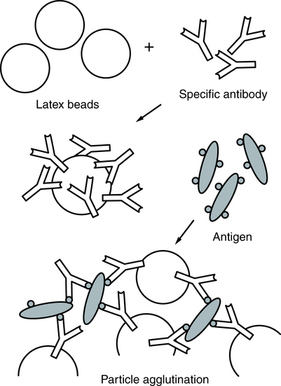
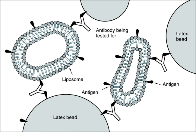
At the conclusion of this chapter, the reader should be able to: • Describe the principles of agglutination. • Identify and compare the characteristics of agglutination methods. • Explain methods for enhancing agglutination. • Describe the characteristics of graded agglutination reactions. • Discuss the principles of pregnancy testing, including sources of error. • Correctly answer case study related multiple choice questions. • Be prepared to participate in a discussion of critical thinking questions. • Explain agglutination reactions of the ABO blood group procedure. • Describe the principle and sources of error of the ABO blood group procedure. Precipitation and agglutination are the visible expression of the aggregation of antigens and antibodies through the formation of a framework in which antigen particles or molecules alternate with antibody molecules (Fig. 10-1). Precipitation is the term for the aggregation of soluble test antigens. Precipitation is the combination of soluble antigen with soluble antibody to produce a visible insoluble complex. Agglutination is the process whereby specific antigens (e.g., red blood cells) aggregate to form larger visible clumps when the corresponding specific antibody is present in the serum. Artificial carrier particles may be needed to indicate visibly that an antigen-antibody reaction has taken place; examples include latex particles and colloidal charcoal. Cells unrelated to the antigen, such as erythrocytes coated with antigen in a constant amount, can be used as biological carriers. Whole bacterial cells can contain an antigen that will bind with antibodies produced in response to that antigen when it is introduced into the host (Table 10-1). Table 10-1 The quality of test results depends on the following technical factors: • Time of incubation with the antibody source (e.g., patient serum) • Amount and avidity of an antigen conjugated to the carrier • Conditions of the test environment (e.g., pH, protein concentration) In latex agglutination procedures (Box 10-1), antibody molecules can be bound to the surface of latex beads. Many antibody molecules can be bound to each latex particle, increasing the potential number of exposed antigen-binding sites. If an antigen is present in a test specimen such as C-reactive protein, the antigen will bind to the combining sites of the antibody exposed on the surface of the latex beads, forming visible cross-linked aggregates of latex beads and antigen (Fig. 10-2). In some procedures (e.g., pregnancy testing, rubella antibody testing), latex particles can be coated with antigen. In the presence of serum antibodies, these particles agglutinate into large visible clumps. Procedures based on latex agglutination must be performed under standardized conditions. The amount of antigen-antibody binding is influenced by factors such as pH, osmolarity, and ionic concentration of the solution. A variety of conditions can produce false-positive or false-negative reactions in agglutination testing (see Table 10-4). Coagglutination and liposome-enhanced testing are variations of latex agglutination (Fig. 10-3). Coagglutination uses antibodies bound to a particle to enhance the visibility of agglutination. It is a highly specific method but may not be as sensitive as latex agglutination for detecting small quantities of antigen. See Latex agglutination slide tests have been replaced in many situations (e.g., home testing; see Chapter 9) by one-step chromatographic color-labeled immunoassays for the qualitative detection of hCG in urine (e.g., Clearview hCG II and Clearview hCG Easy, Wampole Laboratories, Princeton, NJ). Another variation is a one-step chromatographic color-labeled immunoassay for use with urine or serum (e.g., Wampole PreVue hCG Stick or Cassette, Status hCG). Flocculation testing can be used in syphilis serologic testing (see Chapter 18). These tests are the classic Venereal Disease Research Laboratories (VDRL) and rapid plasma reagin (RPR) tests. In the VDRL test, an antibody-like protein, reagin, binds to the test antigen, cardiolipin-lecithin–coated cholesterol particles, and produces the particles that flocculate. In the RPR test, the antigen, cardiolipin-lecithin–coated cholesterol with choline chloride, also contains charcoal particles that allow for macroscopically visible flocculation. By binding different antigens to the RBC surface in indirect hemagglutination or passive hemagglutination (PHA), the hemagglutination technique can be extended to detect antibodies to antigens other than those present on the cells (Box 10-2). Chemicals such as chromic chloride, tannic acid, and glutaraldehyde can be used to cross-link antigens to the cells. Some antibodies (e.g., immunoglobulin G [IgG]) do not directly agglutinate erythrocytes. This incomplete or blocking type of antibody may be detected by using an enhancement medium such as antihuman globulin (AHG) reagent (also known as Coombs reagent). If AHG reagent is added, this second antibody binds to the antibody present on the erythrocytes (see procedure in Chapter 26).
Agglutination Methods
Principles of Agglutination

A, Slide agglutination of bacteria with known antisera or known bacteria. Left, Positive reaction; right, negative reaction. B, Tube agglutination. Left, Positive reaction; right, negative reaction. (From Barrett JT: Textbook of immunology, ed 5, St Louis, 1988, Mosby.)
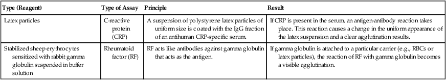
Type (Reagent)
Type of Assay
Principle
Result
Latex particles
C-reactive protein (CRP)
A suspension of polystyrene latex particles of uniform size is coated with the IgG fraction of an antihuman CRP-specific serum.
If CRP is present in the serum, an antigen-antibody reaction takes place. This reaction causes a change in the uniform appearance of the latex suspension and a clear agglutination results.
Stabilized sheep erythrocytes sensitized with rabbit gamma globulin suspended in buffer solution
Rheumatoid factor (RF)
RF acts like antibodies against gamma globulin that acts as the antigen.
If gamma globulin is attached to a particular carrier (e.g., RBCs or latex particles), the reaction of RF with gamma globulin becomes a visible agglutination.
Latex Agglutination
 Pregnancy Latex Slide Agglutination
Principle
 for the procedural protocol.
Alternate Procedural Protocols
Flocculation Tests
Hemagglutination
![]()
Stay updated, free articles. Join our Telegram channel

Full access? Get Clinical Tree


Oncohema Key
Fastest Oncology & Hematology Insight Engine