© Springer International Publishing Switzerland 2016
David S. Cooper and Cosimo Durante (eds.)Thyroid Cancer10.1007/978-3-319-22401-5_2121. A Patient with Postsurgical Recurrent Laryngeal Nerve Damage and Nerve Monitoring
(1)
Department of Otolaryngology – Head & Neck Surgery, University of Cincinnati College of Medicine, 231 Albert Sabin Way, MSB Room 6507, Cincinnati, OH 45267, USA
Keywords
Vocal cord paralysisRecurrent laryngeal nerveSuperior laryngeal nerveIntraoperative nerve monitoringInjection laryngoplastyNeurorrhaphyArytenoid adductionCase Presentation
A 54-year-old female presented to the clinic for evaluation of a known goiter with worsening compressive symptoms. She had no prior history of radiation exposure or a family history of thyroid cancer. She had been followed by her family physician with previous laboratory evaluation and was euthyroid. An ultrasound exam was performed showing a large goiter with mild substernal extension; no discrete nodules were visualized. A flexible laryngeal exam was performed which revealed symmetric, mobile vocal cords bilaterally. The patient desired a thyroidectomy for treatment of her compressive symptoms. The risks of postoperative hypocalcemia and vocal cord paralysis were explained, and she was scheduled for surgery.
Several weeks later, the patient underwent a total thyroidectomy . Intraoperative nerve monitoring (IONM) was performed using an endotracheal tube-based electrode. Her goiter was largest on the left and this was the initial side of surgery. The nerve stimulator was used to assist in finding the recurrent laryngeal nerve. After removal of the left lobe, the nerve stimulator confirmed the integrity of the recurrent laryngeal nerve. The right lobe was then dissected in a similar fashion. The nerve stimulator had a positive signal at the outset of dissection; however, after the thyroid was removed, there was a loss of nerve signal. The position of the endotracheal tube was checked as well as the setup of the remainder of the system. Despite troubleshooting, the nerve signal from the right recurrent laryngeal nerve (RLN) remained absent. Visually the nerve was grossly intact; however, palpation of the arytenoid failed to confirm nerve integrity with stimulation. The surgery was concluded and the patient was discharged home. She had no obvious voice disturbance immediately following surgery.
Assessment and Literature Review
RLN damage is a potential complication following thyroid surgery. Despite advances in surgical technique, anatomical knowledge, and intraoperative nerve monitoring, the rate of RLN injury remains relatively high. A recent systematic review reported an incidence of temporary RLN palsy in 9.8 % of thyroidectomies and permanent RLN injury in 2.3 % [1]. Injury rates are even higher for cases of secondary thyroidectomy, substernal goiter, and thyroid malignancy [2–5]. Surgeon experience has been shown to influence nerve injury with high-volume thyroid surgeons (those performing more than 100 thyroidectomies a year) having significantly lower nerve injury rates than lower-volume surgeons [6]. Vocal cord paralysis is known to negatively affect both quality of life and work activity with symptoms of breathy dysphonia and dysphagia with unilateral injury and stridor and airway obstruction with bilateral injury.
To accurately assess RLN function, a laryngeal exam is essential. Although some physicians reserve laryngeal exam for symptomatic patients, studies have shown a poor correlation between voice changes and vocal cord paralysis [7, 8]. Often patients will develop progressive paralysis over time with adequate contralateral vocal cord compensation to limit noticeable voice changes. In one study in particular, the authors performed routine laryngeal exam prior to thyroidectomy and noted that one third of patients with preoperative vocal fold motion impairment were asymptomatic [7]. Thus, to accurately determine the presence of iatrogenic vocal cord paralysis, a preoperative baseline exam is necessary. In cases of thyroid malignancy, preoperative RLN paralysis is highly predictive of invasive disease and thus alters surgical management and patient counseling [9]. Although there is variation among physician practices, an accurate assessment of preoperative laryngeal function is beneficial for both planning the extent of surgery and managing patients postoperatively.
Mechanism of Nerve Injury
The intimate relationship between the thyroid and RLN places the nerve at risk for injury during surgical removal. The majority of RLN injuries are secondary to compression, traction, devascularization, or thermal damage during thyroid removal wherein the structural integrity of the nerve remains intact but with damage of its underlying neural elements. The RLN innervates the intrinsic muscles of the larynx and consists of a complement of nerve fibers for both abduction and adduction of the vocal folds. Coordinated laryngeal function is highly dependent on each axon innervating its specific motor unit. When the RLN is damaged, the ultimate effect on laryngeal function, as well as prognosis for recovery, is dependent on the degree of nerve injury which was previously categorized by Sunderland [10]. Neuropraxic injuries are the least severe and are characterized by focal myelin damage without disruption of the axon leading to partial or temporary blockage of nerve conduction with a good prognosis for recovery. More severe damage results in disruption of the nerve axons, or axonotmesis , which leads to significant disruption of nerve conduction; however, the perineurium remains intact which improves the potential for functional recovery. Transection of the nerve, or neurotmesis, results in disruption of the axons, perineurium, and epineurium which is the most severe form of injury and carries the worst prognosis for recovery. The mechanism of injury is important when considering prognosis for recovery and postoperative management.
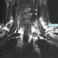
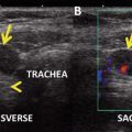
The superior laryngeal nerve and branches of the RLN must be considered during thyroidectomy. The external branch of the superior laryngeal nerve (EBSLN) traverses in an anteroinferior direction along the inferior constrictor muscle to innervate the cricothyroid muscle. Commonly the EBSLN is closely associated with the blood supply to the superior pole of the thyroid placing it at risk for injury during ligation of these vessels. Damage to the EBSLN often limits the ability to achieve higher pitches as well as increased vocal fatigue. Patients with injury to the EBSLN typically have normal laryngeal exams which limit accurate determination of the incidence of nerve damage. Few studies, all with limited patient populations, have utilized EMG to evaluate postoperative cricothyroid function with incidence of EBSLN injury ranging from 5 to 58 % [11, 12] (Figs. 21.1 and 21.2).
Branches of the RLN prior to entering the larynx are also subject to inadvertent injury. Typically these consist of an anterior and posterior branch and are present in 20–30 % of patients [13, 14]. The anterior branch is primarily responsible for laryngeal muscle function, while the posterior branch contains mainly sensory nerve fibers but may occasionally innervate the posterior cricoarytenoid muscle. If branching is not identified, the posterior branch may be mistaken for the primary RLN with inadvertent transection of the anterior branch resulting in vocal cord paralysis. When present, the branches typically occur distally and are often identifiable at the level of Berry’s ligament; however, variation does exist. The presence of these extralaryngeal branches doubles the likelihood of postoperative vocal cord paralysis and should be identified and preserved when present [15]. Branches of the RLN to the esophagus and inferior constrictor may also be present and when injured can impair swallowing postoperatively. Thus, surgeons must maintain a sound knowledge of anatomical variations to limit inadvertent injury to the various nerves in close approximation with the thyroid.
Evaluating Recurrent Laryngeal Nerve Function Intraoperatively
Injury to the RLN is oftentimes difficult to assess intraoperatively. Visually the nerve may appear intact while there can be significant functional damage. Given the inability to assess nerve function, surgeons poorly predict postoperative RLN function with one study reporting as few as 10 % of damaged nerves correctly identified intraoperatively [16]. IONM with nerve stimulation has several key benefits, but its most notable is the ability to assess neural integrity in the operating room and better predict postoperative laryngeal function. Most commonly, IONM is performed using an endotracheal tube-based surface electrode to continuously monitor RLN integrity during thyroidectomy. Manipulation or electrical stimulation of the RLN is recorded by either an audio only system or a system with both audio and visual waveform data to notify the surgeon of evoked neural activity. While continuous nerve monitoring can be useful, nerve stimulation has shown the most promise to improve surgical outcomes. Positive nerve stimulation is very sensitive for intact RLN function. However, a loss of signal is less specific for nerve damage, as this may be secondary to either technical issues or actual nerve damage. Efficient troubleshooting is necessary to limit inappropriate loss of signal. When used by surgeons familiar with IONM, high specificity has been shown to be obtainable [17]. This is particularly valuable when planning a total thyroidectomy to limit the frequency of bilateral RLN paralysis and its associated complications. When the RLN signal is lost on the initial side of surgery during a total thyroidectomy, some surgeons have started to postpone removal of the contralateral lobe. This change in surgical management resulted in a decrease in bilateral RLN paralysis from 17 % to zero percent when the initial side of dissection resulted in a loss of RLN signal [18]. Since most RLN injuries are temporary in nature, a completion thyroidectomy can be performed, if necessary, once nerve function returns.
Although nerve stimulation is a useful tool to protect RLN integrity, visual nerve identification remains the gold standard for nerve protection. Prior to the use of IONM, visual identification of the RLN was shown to decrease RLN injury and remains the gold standard for nerve protection [3, 19]. One of the main hopes of IONM was to further limit the incidence of RLN injury; however, this has yet to be clearly proven. Numerous studies have been published with varying results. A recent meta-analysis of 44 studies showed no statistical difference in RLN injury rates when comparing visual identification of the RLN to IONM [20]. Dralle et al. published a review of approximately 30,000 nerves at risk comparing visual identification versus nerve monitoring with no statistical decrease in RLN injury rates; however, this was thought secondary to lack of statistical power [2]. The only randomized clinical trial evaluating RLN injury rates and IONM did show a statistical benefit [21]. Other studies have shown benefit using IONM in more complex cases. Chan et al. showed decreased RLN injury rates when using IONM for secondary thyroidectomy and thyroidectomy for malignancy [4].
Despite the lack of evidence to support improved patient outcomes, the use of IONM has become increasingly popular over the past two decades. A recent survey of endocrine surgeons showed that approximately 40 % routinely use IONM, and this was most prevalent among high-volume surgeons (greater than 100 cases per year) and younger surgeons (age 35–44) [22].
Clinical Manifestations
A large majority of patients report voice and swallowing complaints after thyroidectomy regardless of RLN function. One study reported that 80 % of patients demonstrated hoarseness and 54 % reported dysphagia 1 week post-thyroidectomy despite normal RLN function [23]. These symptoms typically resolve over the first month postoperatively and are thought secondary to localized swelling, intubation trauma, or muscle injury. On the other hand, patients with RLN injury may not experience voice changes in the immediate postoperative period. Acute laryngeal edema from intubation trauma may create enough bulk for normal voice production; however, once swelling resolves, voice changes will become evident and often present as a “breathy” voice due to insufficient glottal closure. In contrast, bilateral RLN paralysis is often apparent in the immediate postoperative period with acute onset stridor and airway obstruction. Dysphagia is more common in elderly patients with few experiencing severe aspiration. Given the variety of symptoms, a routine laryngeal exam is required to definitively assess RLN function. This is typically performed between 1 and 8 weeks postoperatively and can be done by indirect mirror exam, flexible fiber-optic laryngeal exam, or video laryngoscopy. There can be large variability in laryngeal findings in the setting of RLN injury. Some patients may have persistent voice changes in the setting of normal vocal cord mobility, while other patients will have a flaccid hemilarynx with significant glottic incompetence. The degree of dysfunction is often related to the extent of nerve damage. The use of intraoperative steroids has shown mild benefit in postoperative voice outcomes [24, 25]. In particular, using steroids resulted in a shorter recovery time for temporary RLN paralysis when compared to placebo. The frequency of temporary RLN paralysis was also lower with intraoperative steroids; however, this was not statistically significant. Intraoperative findings should be correlated with laryngeal exam to assist with postoperative management, prognosis, and patient expectations.
Related posts:
 A Case of Multifocal Papillary Thyroid Microcarcinoma
A Patient with Papillary Thyroid Cancer and Biochemical Evidence of Disease at the One-Year Follow-Up Visit
Radioiodine Therapy in Lactating Women with Higher-Risk Differentiated Thyroid Cancer
A Case of Multifocal Papillary Thyroid Microcarcinoma
A Patient with Papillary Thyroid Cancer and Biochemical Evidence of Disease at the One-Year Follow-Up Visit
Radioiodine Therapy in Lactating Women with Higher-Risk Differentiated Thyroid Cancer
 A Patient with a Large Hürthle Cell Carcinoma of the Thyroid and Nodal Metastases
 Papillary Thyroid Carcinoma Diagnosed During Pregnancy
 Timing and Extent of Surgery for a Pediatric Patient with Hereditary MTC and Positive Screening for the S891A RET Mutation
Stay updated, free articles. Join our Telegram channel

Full access? Get Clinical Tree


